Miller Fisher syndrome (MFS) is a rare variant of Guillain–Barré syndrome characterized by ocular paralysis, ataxia, and insensitivity. This report describes the effect of Complex Korean Medicine Treatment (CKMT) on a patient previously diagnosed with MFS presenting with diplopia and facial palsy. The distance at which diplopia occurs, the diplopia questionnaire, the range of diplopia, the degree of facial paralysis, and the degree of ptosis were evaluated at the time of admission and weekly for 1 month. After receiving CKMT for 4 weeks the 62-year-old female had improved symptoms of diplopia, bilateral facial palsy and ptosis caused by MFS.
These results show the significant association of MFS with facial paralysis and the improvement achieved with CKMT.
©2021 Korean Acupuncture & Moxibustion Medicine Society. This is an open access article under the CC BY- NC-ND license (http://creativecommons.org/licenses/by-nc-nd/4.0/).
Article history:
Submitted: October 16, 2020 Revised: October 28, 2020 Accepted: November 05, 2020 Keywords:
diplopia, facial palsy, Miller Fisher syndrome
https://doi.org/10.13045/jar.2020.00367 pISSN 2586-288X eISSN 2586-2898
Case Report
A Case Study of a Patient with Diplopia and Bilateral Facial Palsy Due to Atypical Miller Fisher Syndrome:
Treatment with Complex Korean Medicine
Chae Hyun Park, Jae Hui Kang, Hwa Yeon Ryu, Ga Hyeon Jung, Yong Ho Ku, Hyun Lee*
Department of Acupuncture and Moxibustion Medicine, College of Korean Medicine, Daejeon University, Cheonan, Korea
ABSTRACT
Journal of Acupuncture Research
Journal homepage: http://www.e-jar.org
Introduction
Miller Fisher syndrome (MFS), a type of acute inflammatory polyneuropathy, was first described by Collier as a variant of Guillain–Barré syndrome (GBS) in 1932 [1]. The initial diagnosis of GBS is based on clinical presentation e.g., with numbness, pins and needles, muscle weakness, pain, problems with balance and coordination. In addition, presence of GQ1b antibody, lumbar puncture, and cerebral spinal fluid analysis are used to diagnose GBS [2]. However, not every MFS patient shows GQ1b antibody positivity, and this variant is called atypical MFS [3].
The treatment for MFS includes administration of steroids, intravenous immunoglobulins (IVIG), and plasma exchange [4].
In a recent study, there was no clear conclusion about the effect of immunoglobulin treatment, however the study was small [5].
Evaluation of the treatment for MFS entails an assessment of eye movement, the distance at which diplopia occurs, and use of the diplopia questionnaire. This report describes the improvement in symptoms of diplopia and facial palsy after receiving complex
Korean medical treatment (CKMT) at the Cheonan Korean Medicine Hospital of Daejeon University in a patient diagnosed with atypical MFS who had not shown improvement after IVIG and steroid treatment received previously at Dankook University Cheonan Hospital.
Case Report Patient
OOO (F/62) Chief complaint
Diplopia, bilateral facial palsy, sight problems, ptosis Medical history
Hospitalization due to norovirus gastroenteritis in March 2020.
On medication for osteoporosis since May 2020.
*
Corresponding author. Hyun LeeDepartment of Acupuncture and Moxibustion Medicine, Cheonan Korean Medicine Hospital of Daejeon University, 4, Notaesan-ro, Seobuk-gu, Cheonan, 331-958, Korea E-mail: [email protected]
ORCID: Chae Hyun Park https://orcid.org/0000-0001-5672-9026, Hyun Lee https://orcid.org/ 0000-0002-5160-7000, Jae Hui Kang https://orcid.org/0000-0003-4812-0557, Hwa Yeon Ryu https://orcid.org/0000-0002-9468-1227, Ga Hyeon Jung https://orcid.org/0000-0002-7759-0972, Yong Ho Ku https://orcid.org/0000-0001-7553-4144
©2021 Korean Acupuncture & Moxibustion Medicine Society. This is an open access article under the CC BY-NC-ND license (http://creativecommons.org/licenses/by-nc- nd/4.0/).
Family history
Mother has hypertension.
Present illness
During hospitalization due to syncope that occurred on May 12
th, 2020, the patient complained of diplopia and underwent a brain MRI, which showed normal findings. Soon after, the patient complained of ptosis and bilateral facial palsy. Cerebral spinal fluid examination showed albuminocytological dissociation, and the blood test was anti-GQ1b Ab negative. Thus, she was diagnosed with atypical MFS at Dankook University Cheonan Hospital, on June 6
th, 2020. She received IVIG 5 times during hospitalization at Cheonan Hospital but did not show any improvement. After steroid pulse therapy, the patient showed a slight improvement in diplopia and facial palsy.
The patient visited Cheonan Korean Medicine Hospital of Daejeon University. After hospitalization, steroid oral administration therapy was continued for 5 days.
Duration of treatment
The duration of treatment lasted from June 18
th, 2020, to July 17
th, 2020 (30 days of hospitalization).
To protect the patient’s personal information, medical records were obtained from the Institutional Review Board (no.: DJUMC- 2020-BM-13).
Treatment Acupuncture
The acupuncture needles used were 0.20 × 30 mm stainless steel standardized and disposable (Eastern acupuncture equipment
manufacturer, Boryung, Korea). Acupuncture was administered twice daily for 15 minutes: bilateral BL2, Ex-HN4, TE23, GB1, BL1, Ex-HN7, ST4, ST6, TE17, L14, ST36, and ST9. Electroacupuncture treatment was administered once daily for 15 min. The machine used for electroacupuncture was STN-110 (Stratek, Seoul, Korea), set with a stimulation frequency of 3 Hz (Table 1).
Pharmacopuncture
Pharmacopuncture was performed once daily on each day of hospitalization. The acupoints were TE17, ST6, and ST9. Hominis placenta pharmacopuncture (Korean Pharmacopuncture Institute, Seoul, Korea) was administered just before acupuncture therapy in the afternoon. Doses of 0.2 mL were inserted at each acupoint to a depth of 0.5-1.0 mm. The maximum total dose per treatment was 1.0 mL. This was administered using a 1.0 mL disposable syringe and needle (Jungrim Medical, Seoul, Korea, 30 G × 12.7 mm needle (Table 1).
Herbal medicine
The patient took herbal medication 3 times daily; Bojungikgitang- gamibang, Soyo-san, Samulgwibitang, and Kikukjihwangtang were given (Table 2).
Physiotherapy
Silver spike point and manual therapy were administered on the mimic muscles once daily.
Moxibustion treatment
Indirect electric moxibustion (Technoscience, Seoul, Korea) therapy was applied once daily at L14 and ST36.
Intervention Detail Details
Acupuncture rationale Reasoning Textbook of Acupuncture and Moxibustion Medicine
Variation Almost same
Needling details Points used Bilateral BL2, Ex-HN4, TE23, GB1, BL1, Ex-HN7, ST4, ST6, TE17, LI4, ST36 and sternocleidomastoid No. of needling 23 needles at 1 session
Depths of insertion 0.2-0.5 cm on acupoints Response sought Subjective De-qi sensation
Needle stimulation Manual stimulation of rotation, lifting and thrusting Needle retention time 15 min
Needle type 0.20 × 30 mm stainless steel standardized, and disposable
Treatment regimen Number of sessions 60 sessions
Frequency/ duration Twice a day for 30 d
Co-interventions Other components of treatment Electro-acupuncture, pharmacopuncture, herbal medicine, Moxibustion and physical therapy treatment.
Practitioner background Duration of clinical experience 2 Korean medicine doctors.
1) Resident trainee in acupuncture and moxibustion medicine with 1 year of experience 2) Specialist of acupuncture and moxibustion medicine with more than 25 years of experience.
Table 1. Treatment Intervention.
Bojungikgitang-gamibang
2020.06.18 - 2020.06.30 Soyo-san
2020.07.01 - 2020.07.05 Samulgwibitang
2020.07.06 - 2020.07.09
Kikukjihwangtang 2020.07.10 - 2020.07.17 2020.07.18 - 2020.07.25 Atractylodis Rhizoma Alba 16 Atractylodis Rhizoma
Alba 12 Longanae Arillus 16 Cistanches Herba 32
Paeoniae Radix Alba 12 Paeoniae Radix Alba 12 Angelicae Gigantis Radix 12 Rehmanniae Radix Preparat 20
Astragali Radix 12 Hoelen 8 Cnidii Rhizoma 12 Lycii Fructus 20
Crataegii Fructus 12 Bupleuri Radix 8 Paeoniae Radix Alba 12 Mori Fructus 20
Citri Pericarpium 12 Angelicae Gigantis Radix 8 Salviae Miltiorrhizae Radix 2 Corni Fructus 20
Raphani Semen 8 Liriopes Radix 8 Polygalae Radix 8 Gastrodiae Rhizoma
Poria cocos 8 Glycyrrhizae Radix 4 Atractylodis Rhizoma Alba 8 Chrysanthemi Flos 16
Ginseng Radix 8 Pinelliae Praeparatum 4 Hoelen cum Pini Radix 8 Phellodendri Cortex 16
Angelicae Gigantis Radix 8 Zingiberis Rhizoma 24 Coptidis Rhizoma 8 Alismatis Rhizoma 12
Platycodi Radix 6 Bambusae Caulis In Taeniam 8
Uncariae Ramulus 6 Crataegii Fructus 8
Perilla Herba 6 Amomi Fuctus 8
Osterici Radix 6 Lycii Fructus 8
Ledebouriella divaricata 6 Astragali Radix 8
Bombycis Corpus 6 Ginseng Radix 8
Angelica dahurica 6 Zizyphi Spinosae Semen 8
Bupleuri Radix 4 Aucklandiae Radix 8
Cimicifugae Rhizoma 4 Acori Graminei Rhizoma 8
Pinelliae Praeparatum cum
Zingiberis 4 Massa Medicata Fermentata 8
Aucklandiae Radix 4 Hordei Fructus Germiniatus 8
Massa Medicata Fermentata 4 Galli Stomachichum Corium 8
Amomi Fuctus 4 Glycyrrhizae Radix 4
Glycyrrhizae Radix 4 Zingiberis Rhizoma 24
Jujubae Fructus 16
Table 2. Herbal Composition of 4 Herbal Medications for Daily Dosage.
Evaluation
Diplopia questionnaire
In this case report the diplopia questionnaire was used to evaluate the severity of diplopia [6]. The questionnaire evaluation was conducted a total of 5 times, once a week, from June 18
th, 2020, to July 16
th, 2020 (Fig. 1; Appendix A).
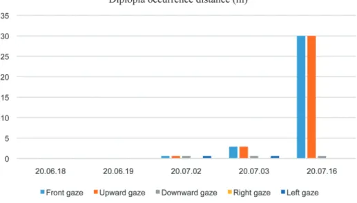
The distance at which diplopia occurs
The distance at which the patient’s diplopia appeared was measured from the patient’s glabella and expressed in meters. This method is often used to evaluate diplopia [7]. The maximum length that could be measured in the hospital room was 30 m (Fig. 2).
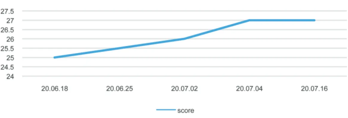
Fig. 1. Scores from the diplopia questionnaire over treatment time. The score
decreased to 13 points on July 16
th, 2020 from 25 points on June 18
th, 2020,
showing improvement in the diplopia the patient experienced.
week. Photographs of ptosis taken on July 16
th, 2020, in the front view showed that the pupils were clearly more visible compared with photographs taken on June 18
th, 2020 (Fig. 5).
Subjective state of the patient
The patient’s symptoms, such as dry eye syndrome, ocular fistula, and blurred vision were recorded electronically. Although subjective, these observations were used as indications of symptom improvement. On the day of admission, the patient complained that her eyes were easily tired and her vision was blurred. On Day 3, after admission, the patient said that her eyes were less watery and less tired. On Day 13, the patient reported that her eyes were not watering. On Day 38, dry eye syndrome had slightly improved. On September 4
th, 2020, normal findings were reported in the dry eye test and keratography. Blurred vision with cataract was diagnosed at another hospital after discharge (Fig. 6).
Fig. 2. Distance at which diplopia occurred over the treatment period. Diplopia was resolved within the measurable range in the forward and upward gaze. Diplopia was lost at 0.6 m in the downward and left gaze.
During the right gaze, diplopia was lost at 0.12 m, but appeared at a distance beyond that.
Fig. 3. Diplopia field. Compared with June 19
th, 2020, on July 16
th, 2020, when looking forward, upward, downward, and left, diplopia was resolved, and the range of diplopia on the right gaze was reduced by 2 cm.
Field of diplopia
To determine the degree of diplopia, the distance between the points was measured by marking the position where the center point was visible at a constant distance of 60 cm from the glabella of the patient. Since the patient complained of diplopia in all directions, this method was used to measure the degree and change in diplopia according to the direction (Fig. 3) [8].
Yanagihara system
To determine the degree of facial palsy, the Yanagihara scoring system [9] was used to measure 10 aspects of different facial function. The maximum score to grade the degree of nerve damage in facial nerve palsy is 30, and it was measured daily. (Fig. 4).
Photographs of ptosis
To evaluate the changes in ptosis, photos were taken once a
Fig. 4. Yanagihara score over the course of treatment. Compared with June 18
th, 2020, the score increased by 2 points on July 16
th, 2020.
Discussion
The male/female ratio of MFS is 2:1, with a mean age of 43.6 years at the onset of the disease [4]. The first symptoms of MFS are diplopia (38.6%) or ataxia (20.6%) [4]. In 57% of cases, the cranial nerves (other than the ocular motor nerve) such as the facial nerve (46%), glossary nerve and the vagus nerve (40%), and sublingual nerve (13%) may also be involved in MFS [4]. To diagnose MFS, the presence of a prior infection is reviewed, detection of anti- GQ1b antibodies, and cerebral spinal fluid analysis are used [2].
To treat MFS, steroid therapy, IVIG, or plasma exchange may be performed. However, more research is needed on their effects [5].
Acupuncture causes microscopic damage that increases local blood flow and promotes cell recovery [10]. You et al [11] treated ophthalmoplegia with acupuncture on acupoints GV20, BL2, Ex-HN4, TE23, ST2, TE17, LI04, LR03, and ST36. Wang et al
[12] reported using TE12, GB1, Ex-HN4, ST4, ST6, ST7, GB20 acupoints for patients with MFS, facial paralysis and diplopia.
In this current case, acupoints around the eyeball were selected (similar to other studies [8,11,12]) that facilitate the movement of eye muscles and affects the recovery of paralyzed muscles when stimulated with low-frequency electroacupuncture [13]. Although there are not many existing studies of MFS acupuncture treatment the most frequently used acupoints around the eyes were used to treat diplopia associated with MFS. In addition, since the patient also complained of facial paralysis acupoints used for facial paralysis were also used. Pharmacopuncture for facial nerve palsy may have the effect of supplementing qi and blood [14].
Bojungikgitang-gamibang is herbal medication prescribed for chronic facial palsy (where patients have passed the acute phase of the condition). In these patients, there has usually been more than 1 month since the onset of paralysis and there is often debility.
Fig. 6. Photographs of keratography. Normal findings in dry eye.
Fig. 5. Photographs of improvement in ptosis over the treatment period (June 18
th, 2020, June 25
th, 2020, July 2
nd, 2020, July 9
th, 2020, July 16
th, 2020).
(a) 20.06.18
(b) 20.06.25
(c) 20.07.02
(d) 20.07.09
(e) 20.07.16
Score
Gaze position Always Sometimes Never
Straight ahead into the distance 6 3 0
Up 2 1 0
Down 4 2 0
Right 4 2 0
Left 4 2 0
Reading 4 2 0
Any position 1 1 0
Total Appendix A. Diplopia Questionnaire.