대한임상신경생리학회지 13(1):44~47, 2011 ISSN 1229-6414
Copyright 2011 by The Korean Society of Clinical Neurophysiology 44
Original Article
밀러 피셔 증후군에서 보이는 지연성 안면마비의 임상양상과 전기생리학적 소견
대구가톨릭대학교 의과대학 신경과학교실
권두혁․석정임․한우호․이동국
Clinical and Electrophysiological Characteristics of Delayed Facial Palsy in Miller-Fisher Syndrome
Doo-Hyuk Kwon, M.D., Jung Im Seok, M.D., Woo Ho Han, M.D., Dong Kuck Lee, M.D.
Department of Neurology, School of Medicine, Catholic University of Daegu, Daegu, Korea
Received 10 December 2010; received in revised form 10 February 2011; accepted 17 March 2011.
Background: Miller-Fisher syndrome (MFS) is characterized by the clinical triad of ophthalmoplegia, ataxia, and areflexia, and is considered a variant form of Guillain-Barre syndrome. Although some cases of delayed-onset facial palsy in MFS have been reported, the characteristics of this facial palsy are poorly described in the literature. Methods: Between 2007 and 2010, six patients with MFS were seen at our hospital. Delayed facial palsy, defined as a facial palsy that developed while the other symptoms of MFS began to improve following intravenous immunoglobulin treatment, was confirmed in four patients. The clinical and electrophysiological characteristics of delayed facial palsy in MFS, as observed in these patients, are described here. Results: Four patients with delayed-onset facial palsy were included. Delayed facial palsy developed 8-16 days after initial symptom onset (5-9 days after treatment). Unilateral facial palsy occurred in three patients and asymmetric facial diplegia in one patient. The House-Brackmann score of facial palsy was grade III in one patient, IV in two patients, and V in one patient. None of the patients complained of posterior auricular pain. Facial nerve conduction studies revealed normal amplitude in all four patients. The blink reflex showed abnormal prolongation in two patients and the absence of action potential formation in two patients. Facial palsy resolved completely in all four patients within 3 months. Conclusions:
Delayed facial palsy is a frequent symptom in MFS and resolves completely without additional treatment. Thus, standard treatment and patient reassurance are sufficient in most cases.
Key Words: Miller-Fisher syndrome, Delayed facial palsy, Clinical characteristics, Electrophysiological characteristics
Address for correspondence;
Jung Im Seok, M.D.
Department of Neurology, Daegu Catholic University Hospital, 3056-6 Daemyeong 4-dong, Nam-gu, Daegu 705-718, Korea Tel: +82-53-650-3043 Fax: +82-53-654-9786
E-mail: [email protected]
Introduction
Miller-Fisher syndrome (MFS), a variant form of Guillain-Barre
syndrome (GBS), is characterized by the clinical triad of ophtha- lmoplegia, ataxia, and areflexia. Besides these three common symptoms, facial palsy is one of frequent symptoms of MFS.
Additionally, a few patients with MFS develop a facial palsy either during treatment or concomitant with a reduction in the severity of the neurological symptoms.1,2 However, in the literature, the clinical and electrophysiological characteristics of delayed facial palsy in MFS are poorly described.
밀러 피셔 증후군에서 보이는 지연성 안면마비의 임상양상과 전기생리학적 소견
Korean J Clin Neurophysiol / Volume 13 / June, 2011 45
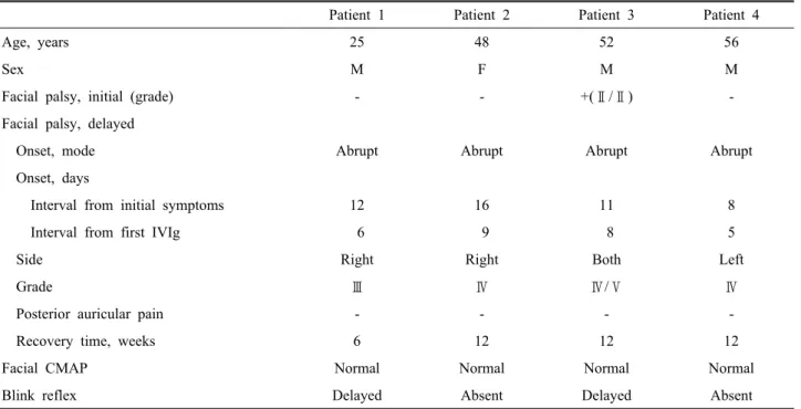
Table 1. Characteristics of demographic and clinical features
Patient 1 Patient 2 Patient 3 Patient 4
Age, years 25 48 52 56
Sex M F M M
Facial palsy, initial (grade) - - +(Ⅱ/Ⅱ) -
Facial palsy, delayed
Onset, mode Abrupt Abrupt Abrupt Abrupt
Onset, days
Interval from initial symptoms 12 16 11 8
Interval from first IVIg 6 9 8 5
Side Right Right Both Left
Grade Ⅲ Ⅳ Ⅳ/Ⅴ Ⅳ
Posterior auricular pain - - - -
Recovery time, weeks 6 12 12 12
Facial CMAP Normal Normal Normal Normal
Blink reflex Delayed Absent Delayed Absent
IVIg; intravenous immunoglobulin, CMAP; compound muscle action potential.
Methods
This retrospective study examined six consecutive patients with MFS who were seen at our hospital from January 2007 to June 2010. Patients with the clinical triad of ophthalmoplegia, ataxia, and areflexia with acute onset but without major limb weakness or other signs suggestive of central nervous system involvement were classified as having MFS.
Delayed facial palsy was defined as a facial palsy that developed while the other neurological symptoms of MFS began to improve following immunoglobulin treatment. Information on the patients’ demographic characteristics and the characteristics of the delayed facial palsy was collected for each patient with delayed facial palsy. Delayed facial palsy was characterized by the onset, severity, presence of concurrent posterior auricular pain, recovery time, and electrophysiological findings. The severity of facial palsy was graded by the House-Brackmann score.3 Nerve conduction studies (NCS) were performed on the bilateral median, ulnar, peroneal, posterior tibial, sural, and facial nerves using standard techniques.4 The blink reflex was recorded using the method described by Kimura.5 Facial NCS and blink reflex were recorded within 2 days of the onset of the delayed facial palsy.
Results
Delayed facial palsy was detected in four patients. Their demographic and clinical features are shown in Table 1. In three of them, facial palsy was absent initially and developed after treatment. In one patient, a delayed abrupt worsening of initial facial palsy was seen. The delayed facial palsy had developed abruptly 8-16 days after the initial symptoms (5-9 days after treatment). Unilateral facial palsy occurred in three patients and asymmetric facial diplegia in one patient. The House-Brackmann score of facial palsy was grade Ⅲ in one patient and Ⅳ in two patients with unilateral palsy. The score was grade Ⅳ on the right side and Ⅴ on the left side in one patient with asymmetric diplegia. No patient complained of posterior auricular pain.
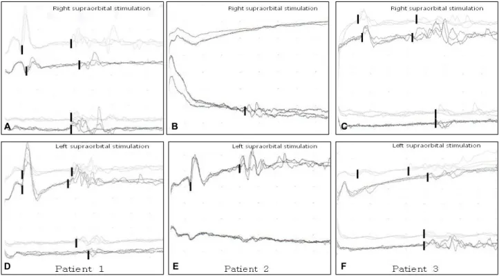
Facial NCS revealed normal compound muscle action potential (CMAP) amplitude in all four patients. In two patients (patient 1 and 3), the blink reflex showed abnormal prolongation and in two patients (patient 2 and 4), the absence of action potential formation. Figure 1 shows the blink wave recorded initially and at the time of delayed facial palsy. In all patients, delayed facial palsy resolves completely within 3 months without additional treatment.
권두혁․석정임․한우호․이동국
Korean J Clin Neurophysiol / Volume 13 / June 2011 46
Figure 1. Blink reflex in patients with delayed facial palsy. (A, B) Patient 1. Initial blink reflex (light line) reveals normal. Follow-up study (dark line) at the time of right facial palsy shows delayed R1 and R2 response on right supraorbital stimulation and delayed contralateral R2 on left stimulation. (C, D) Patient 2. Initial blink test was not done.
Follow-up study at the time of right facial palsy shows absent R1 and R2 responses on right stimulation and absent contralateral R2 on left stimulation. (E, F) Patient 3. This patient had mild facial diplegia initially. Initial blink test shows delayed R1, R2, and contralateral R2 on supraorbital stimulation on either side (more severe in left side). Follow-up study after worsening shows absent R1 response on left stimulation and more delayed responses. Blink wave that recorded initially (light line) and at the time of delayed facial palsy (dark line) were overlapped.
Discussion
MFS is a variant of GBS but has a better prognosis than GBS.
Although the incidence of GBS is about the same worldwide, that of MFS is higher in Asian countries than in Western countries. In Taiwan and Japan, for example, MFS reportedly accounts for 19% and 25% of the cases of GBS, respectively.1,6
Facial palsy, together with pupillary abnormalities and ptosis, is frequent in patients with MFS and is reported to occur in 30-45% of these cases.1,7 In one study, delayed worsening of facial palsy developed in six of the 50 patients with MFS,1 while in our study two-thirds of the patients with MFS developed delayed facial palsy.
The anti-GQ1b antibody has an important role in the pathogenesis of MFS. Immunochemical study with the anti-GQ1b antibody has shown prominent staining in the three ocular motor nerves, but not in the facial nerve.8 This evidence suggests that
unknown factors other than the anti-GQ1b antibody may contribute to the pathogenesis of facial palsy in MFS. Different antibody may be involved in the development of facial palsy.
Therefore, antibody testing would provide some clues to the mechanism of how facial palsy may develop in these cases. One report about delayed facial palsy during plasmapheresis suggests that various pathomechanisms are probably involved in different disease stages in MFS and plasmapheresis may not be effective in the prevention of facial palsy.9
Although MFS is usually associated with a good recovery, some patients experience worsening of neurological symptoms, which greatly distresses these patients. In this study, recovery in patients with delayed facial palsy was the same as in the other MFS patients, with the facial palsy as well as the other neurological symptoms resolving completely within 3 months.
Thus, standard treatment and reassurance are sufficient in patients with delayed facial palsy.
A B C
D E F
밀러 피셔 증후군에서 보이는 지연성 안면마비의 임상양상과 전기생리학적 소견
Korean J Clin Neurophysiol / Volume 13 / June, 2011 47
The most common cause of peripheral facial palsy is Bell’s palsy. The clinical characteristics of Bell’ palsy differed somewhat from those of the facial palsy in MFS. None of the MFS patients complained of posterior auricular pain, whereas it is a common associated symptom in Bell’s palsy. A study in our hospital showed that 77% of patients with Bell’s palsy had posterior auricular pain.10 These findings suggest that the pathogenesis of facial palsy in MFS differs from that of Bell’s palsy. In contrast, electrophysiological findings including facial NCS and blink reflex was similar to each other. A blink wave was present in two of the four patients with MFS and absent in other two patients.
Facial CMAP is usually normal and blink reflex is absent or delayed in patients with early-stage Bell’s palsy.11 EMG in facial innervated muscles did not performed in this study. It would be helpful to know the pathology of facial nerve, axonal damage or demyelination.
The results of our study should be interpreted with caution because of the very limited sample size and the fact that the data were derived from a single center. Thus, further studies on the incidence, characteristics, and electrophysiological findings of delayed facial palsy are warranted.
Delayed-onset facial palsy is common in MFS. As the neurologic deficits of MFS are transient, those with facial palsy can expect good recovery, without the need for additional treatment.
REFERENCES
1. Mori M, Kuwabara S, Fukutake T, Yuki N, Hattori T. Clinical features and prognosis of Miller Fisher syndrome. Neurology 2001;56:1104-1106.
2. Fernando MD, Samaranayake RA, Dissanayaka A. Unusual manifestations in Miller Fisher syndrome. Ceylon Med J 2007;
52:64-65.
3. House JW, Brackmann DE. Facial grading system. Otolaryngol Head Neck Surg 1985;93:146-147.
4. Oh SJ. Clinical electromyography: nerve conduction studies.
Baltimore: Williams & Wilkins, 1998.
5. Kimura J. The blink reflex. In: Kimura J. Electrodiagnosis in diseases of the nerve and muscle: principles and practice. 2nd ed.
Philadelphia, PA: Davis. 1989;307-331.
6. Lyu RK, Tang LM, Cheng SY, Hsu WC, Chen ST. Guillain-Barre syndrome in Taiwan: a clinical study of 167 patients. J Neurol Neurosurg Psychiatry 1997;63:494-500.
7. Li H, Yuan J. Miller Fisher syndrome toward a more comprehensive understanding. Chin Med J 2001;114:235-239.
8. Chiba A, Kusunoki S, Obata H, Machinami R, Kanazawa I. Serum anti-GQ1b IgG antibody is associated with ophthalmoplegia in Miller Fisher syndrome and Guillain-Barre syndrome: clinical and immunohistochemical studies. Neurolgy 1993;43:1911-1917.
9. Chida K, Takase S, Itoyama Y. Development of facial palsy during immunoadsorption plasmapheresis in Miller Fisher syndrome: a clinical report of two cases. J Neurol Neurosurg Psychiatry 1998;64:399-401.
10. Seok JI, Lee DK, Kim KJ. The usefulness of clinical findings in localising lesions in Bell's palsy: comparison with MRI. J Neurol Neurosurg Psychiatry 2008;79:418-420.
11. Valls-Sole J. Electrodiagnostic studies of the facial nerve in peripheral facial palsy and hemifacial spasm. Muscle Nerve 2007;36:14-20.