Effect of Hip Adduction Position on the Vastus Medialis Oblique and Vastus Lateralis During Closed Kinetic Chain Exercise in
Sitting Posture
Yong-su Cha1, MSc, PT, Hye-seon Jeon2,3, PhD, PT, Chung-hwi Yi2,3, PhD, PT, Oh-yun Kwon2,3, PhD, PT, Bo-ram Choi4, PhD, PT
1Dept. of Physical Therapy, Jeon Hospital
2Dept. of Physical Therapy, College of Health Science, Yonsei University
3Dept. of Ergonomic Therapy, The Graduate School of Health Science, Yonsei University
4Dept. of Physical Therapy, College of Health and Welfare, Silla University
Abstract
1)Background: Several studies have discussed diverse exercise methods considered to be useful for the selective contraction of the vastus medialis oblique (VMO) muscle for the treatment of patellofemoral pain syndrome. Some studies have reported that exercise methods, including hip adduction, in closed kinetic chain exercises are more effective in terms of the muscle activation of the VMO and the timing of the muscle’s initial contraction. We focused on isometric contraction during a closed kinetic chain exercise with hip adduction.
Objects: The purpose of this study was to examine muscle activation in the VMO and the vastus lateralis (VL) and the onset time difference between their initial contractions via closed kinetic chain isometric quadriceps femoris exercises including hip adduction.
Methods: In total, 36 healthy subjects adopted two hip positions during isometric contraction of the quadriceps femoris in a closed kinetic chain exercise (hip neutral and hip adduction position). Statistical analyses were conducted using a paired t-test (α=.05).
Results: Isometric contraction of the quadriceps femoris in a closed kinetic chain exercise caused a greater increase in VMO muscle activity in the hip adduction position [52.68±22.21 percentage of maximal voluntary isometric contraction (%MVIC)]than the hip neutral position (43.43±19.85 %MVIC). The onset time difference (VL-VMO) decreased more in the hip adduction position (-82.14±34.2 ㎳) than the hip neutral position (73.94±2.94 ㎳).
Conclusion: We recommend this exercise as a clinically useful therapeutic method for patients with patellofemoral pain syndrome due to weakening of the VMO muscle and lateral inclination of the patella.
Key Words: Closed kinetic chain exercise; Patellofemoral pain syndrome; Vastus medialis oblique.
Introduction
The quadriceps femoris group is the most im- portant muscle for maintaining normal knee function (Mellor and Hodges, 2005). This muscle is the first to move when knee extension is performed in the sagittal plane. Within this muscle, the vastus me- dialis oblique contributes greatly to the medial stabil- ity of the patella in knee extension movement
(Hubbard et al, 1997). However, the vastus medialis oblique, which controls the lateral inclination of the patella, is physiologically weakest and exhibits earlier weakening than the vastus lateralis (Francis and Scott, 1974). Thus, if the vastus medialis oblique be- comes weakened or subject to other problems, ex- ercises should be performed to strengthen it by re- cruiting a larger number of muscular fibers (Boling et al, 2006). One of the principal reasons for the ab-
Corresponding author: Bo-ram Choi [email protected]
normal lateral inclination of the patella is the onset time difference between the initial contractions of the vastus medialis oblique and the vastus lateralis (Voight and Wieder, 1991). Another cause is the superior force of the vastus lateralis, which simply has a larger cross-section than the vastus medialis oblique (Grabiner et al, 1991). As noted, unbalanced initial contractions of the vastus medialis oblique and vastus lateralis can lead to abnormal movement of the patella, which can ultimately result in patellofemoral joint pain syndrome.
Given the importance of the vastus medialis obli- que, various studies have recommended exercises to strengthen the quadriceps femoris group as a con- servative treatment method. Isometric quadriceps femoris exercises can be used from the initial stage of rehabilitation to prevent muscle weakening and maintain muscle strength. They also play a crucial role in protecting the patella by enhancing the sta- bility of the group (Arroll et al, 1997). Quadriceps femoris strengthening exercises that are commonly used in clinical practice include concentric patella extension exercises in a sitting posture on a Noland-Kickoff ta- ble (Monteiro-Pedro et al, 1999). Additionally, going up and down stairs, squat exercises, and open and closed kinetic chain exercises have been examined as exercise methods for strengthening the vastus me- dialis oblique (Laprade et al, 1998). However, such ex- ercises have several limitations for selectively train- ing the vastus medialis oblique and lateralis. Moreover, some studies have reported that the execution of quad- riceps femoris strengthening exercises did not result in any significant difference in muscle activation be- tween the vastus medialis oblique and lateralis.
To address these limitations, studies have been con- ducted on the selective enhancement of the strength of the vastus medialis oblique. When quadriceps femoris exercises were accompanied by hip adduction, pa- tients with patellofemoral joint pain syndrome re- ported pain reduction (Nakagawa et al, 2008). Choi et al (2011) compared the onset time for the vastus medialis oblique and lateralis during isometric patella extension exercises in neutral and adducted hip
postures. The onset time for the vastus medialis ob- lique was found to be advanced in the hip adduction posture. Lam and Ng (2001) emphasized that muscle activation in the vastus medialis oblique and lateralis was related to angle changes in the knee and hip.
Hanten and Schulthies (1990) discovered that in- dividuals who performed exercises while maintaining hip adduction registered an overall higher level of activation in the vastus medialis oblique than in the vastus lateralis. Previous studies maintained a 30°
flexion posture in the last stage of knee extension, performed as a selective exercise for the vastus me- dialis oblique. However, Steinkamp et al (1993) re- ported that the patella angle in quadriceps femoris ex- ercises should ideally be maintained at 45° or lower flexion angles while minimizing the repulsive force and pressure on the patellofemoral joint.
Many past studies have also focused on the varia- tions in the vastus medialis oblique and lateralis in open and closed kinetic chain exercises. Stensdotter et al (2007) carried out a comparison of open and closed kinetic chain exercises. The results revealed that the vastus medialis oblique showed a higher level of mus- cle activation than the vastus lateralis during closed kinetic chain exercises than in open chain exercises.
The onset time of the vastus medialis oblique was also found to be earlier during closed kinetic chain exercises.
Moreover, closed kinetic chain exercises serve to stabi- lize the movement of the center of the patella in the patellofemoral joint. Closed kinetic chain exercises gen- erate distributed forces by creating smaller levels of pressure between the tibiofemoral joints than open chain exercises, and generate larger forces than open chain exercises through multi-joint movements (Tang et al, 2001). Thus, patients with patellofemoral joint pain syndrome require closed kinetic chain quad- riceps-femoris-strengthening exercises to minimize re- pulsive force and pressure in the tibiofemoral joint.
As noted, some studies have reported that ex- ercises including hip adduction in closed kinetic chain exercises are more effective in terms of the muscle activation of the vastus medialis oblique and the
timing of the muscle’s initial contraction. However, there are few studies on exercise methods specifically for the vastus medialis oblique that involve hip ad- duction in closed kinetic chain isometric exercises. In the present study, we examined muscle activation in the vastus medialis oblique and the vastus lateralis and the onset time difference between their initial contractions via closed kinetic chain isometric quad- riceps femoris exercises including hip adduction.
The first hypothesis was that closed kinetic chain isometric patella exercises in neutral and adducted hip postures would result in different levels of mus- cle activation between the vastus medialis oblique and the vastus lateralis. The second hypothesis was that closed kinetic chain isometric patella exercises in neutral and adducted hip postures would lead to an onset time difference between the vastus medialis ob- lique and the vastus lateralis.
Methods
Subjects
We recruited 36 healthy subjects (30 males, 6 females) who had no lower extremity pain, were able to per- form voluntary knee extension exercises, and had no clinical problems. All the subjects consented volun- tarily to participate in the study.
Instrument
To analyze the surface muscle activity of the vastus medialis oblique and the vastus lateralis, surface elec- tromyography (TeleMyo 2400R, Noraxon Inc., Scottsdale, AZ, USA) was used. One channel electrode was at- tached to each vastus medialis oblique and vastus lat- eralis of the right (dominant) leg, and the ground elec- trode was attached to the protruding area of the tibia below the patella.
To measure the muscle activity of the vastus me- dialis oblique and the vastus lateralis, two channel double surface electrodes were attached to the right (dominant) leg. To minimize resistance against sur-
face electromyography signals, the electrodes attached to the surface of the skin were placed at a distance of 2 ㎝. After palpating the areas of the muscle to be measured, the locations of the muscles were identified.
To determine changes in the location of the muscle belly in parallel with the muscle fibers, resistance was applied when the muscles contracted. Regarding the vastus medialis oblique, the electrode was at- tached at a point 55° from the vertical line from the upper internal patella surface, 4 and 3 ㎝ internally.
For the vastus lateralis, the electrode was attached at a point 15° from the vertical line, 10 ㎝ up and 7
㎝ externally (Cowan et al, 2002). The single refer- ence electrode was attached to the protruding area of the tibia below the patella.
Hair was removed with a razor from the areas to which the electrodes were to be attached, and the areas were rubbed with fine sandpaper three or four times to rid the skin of keratinized layers. The skin was then disinfected with alcohol-soaked cotton. When the alcohol had evaporated, a small amount of a safe electrolyte gel was rubbed onto the skin, and the electrodes were attached. The experiment commenced when electromyography signals were detected. Prior to the experiment, maximal voluntary isometric contraction (MVIC) values were measured using manual re- sistances with the subjects in a sitting position. Three measurements were taken for 5 sec each. The values for the middle 3 sec, excluding the first and the last seconds, were averaged and used (Bowyer et al, 2008).
The collected surface electromyography data were analyzed using the MyoResearch software ver 1.06 XP (Noraxon Inc., Scottsdale, AZ, USA) on a per- sonal computer. The sampling rate of the electro- myography signals was set at 1000 ㎐, the frequency bandwidth at 20∼500 ㎐, and a 60 ㎐ notch filter was used. The collected electromyography signals were processed with the root-mean-square (RMS) method and quantified as percentage of MVIC (%MVIC). To analyze the onset time of muscle contraction, the col- lected electromyography signals were processed with the RMS method, using a computer-based onset de-
termination method. The standard deviation (SD) of the average value for a 100 ㎳ period prior to the on- set time of movements was calculated, and the onset time of muscle contraction was defined as the time taken to reach 3 SD (Choi et al, 2011).
Procedures
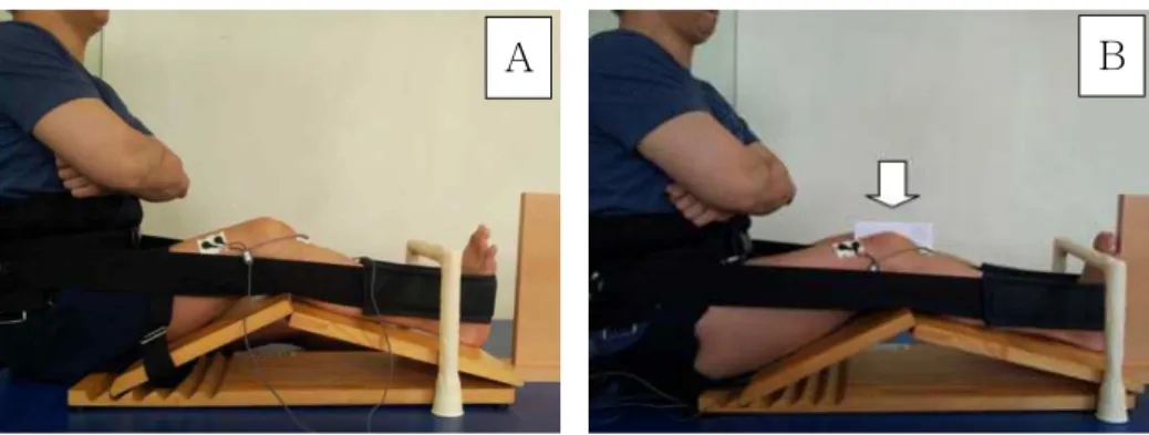
To maintain the knee joint bending angle at 45°, a triangle wooden plane (Figure 1) was used. For the isometric exercise, an inelastic strap, 7.5 ㎝ wide, was used. All the subjects who took part in the experiment put on knee pants and pushed their leg under the same conditions in a hip neutral position and in an adducted position against the strap, which was fixed with a belt placed on the lumbar region to the sole of the foot. Prior to the experiment, the subjects were given 10 min to become accustomed to the laboratory environment and the exercise method.
To ensure the collection of high-quality electro- myography signals, the experiment was not con- ducted on rainy days, wet days, or days when there was severe noise outside the laboratory. The subjects leaned their backs on the wall and placed their legs on the wooden plane in a sitting position. The belt was fixed to the lumbar region. To stop their legs moving, they were fixed from the soles of their feet to the belt with a strap. They conducted the ex- ercise by pushing their legs against the strap fixed to the soles of their feet. The measurement was per- formed five times for 5 sec each, with the quadriceps femoris muscle in a closed kinetic chain, neutral, and adduction positions. To minimize muscle fatigue, the subjects took a 1 min rest between measurements.
Position at the initiation of the experiment The subjects leaned their backs on the wall and placed their legs on the wooden plane in a comfort- able sitting position, folded their arms, and fixed their dominant leg using the inelastic strap designed for the experiment. The triangular wooden plane was placed below their legs, and their knee joint bending angle was maintained at 45°.
Hip neutral position
The hip neutral position was initiated with the dom- inant leg on the wooden plane and maintained in a straight line from the sole of the foot to the hip in the starting position. A target stick was placed at the center in front of the sole of the foot, and the experi- ment was conducted after ensuring that the sole of the foot had not deviated to the left or right side.
Additionally, a target stick designed for the foot not to be raised was placed to restrict the movement of that foot. The lumbar regions 1, 2, 3, 4, and 5 were fixed with a belt using an inelastic strap, which was fitted from the belt to the sole of the foot, and the subject pushed their legs forward against the strap. The measurement was performed five times for 5 sec each (Figure 1A).
Hip adduction position
In the hip adduction position, the dominant leg and the opposite leg were placed on the wooden plane, similarly to that in the neutral position. Thick paper was inserted between the knees, with the knees closed.
Fixation was achieved with the same strap used for the neutral position, and the dominant leg pushed the paper inside so that the paper between the knees did not fall and at the same time pushed the leg against the strap. The measurement was performed five times for 5 sec each (Figure 1B).
Statistical analysis
To compare the muscle activity of the vastus me- dialis oblique and the vastus lateralis and differences in the muscle contraction onset time, a paired t-test was used. The significance level was set at α=.05.
For statistical analyses, the SPSS software ver. 12.0 (SPSS Inc., Chicago, IL, USA) was used.
Results
Comparison of muscle activity between the hip neutral and hip adduction positions
In the neutral position, the muscle activity of the
Neutral position Adduction position p
VMOa (%MVICb) 43.43±19.85c 52.68±22.21 .002*
VLd (%MVIC) 51.60±20.98 49.60±22.21 .403
VMO-VLe (㎳) 73.94±2.94 -82.14±34.20 .013*
avastus medialis oblique,bpercentage of maximal voluntary isometric contraction,cmean±standard deviation,dvastus lateralis,
eonset time difference, *p<.05.
Table 1. Muscle activation and onset time difference of hip neutral and adduction position
A B
Figure 1. Hip neutral position (A), hip adduction position (B).
vastus medialis oblique was, on average, 43.43 %MVIC (SD=19.85). In the adduction position, the muscle ac- tivity of the vastus medialis oblique was 52.68
%MVIC (SD=22.21). However, in the neutral position, the muscle activity of the vastus lateralis was, on average, 51.60 %MVIC (SD=20.98). In the adduction position, the muscle activity of the vastus lateralis was 49.60 %MVIC (SD=22.21). For the vastus me- dialis oblique, the muscle activity in the hip adduction position was significantly higher (p<.05). However, for the vastus lateralis, there was no significant dif- ference between the muscle activities in the hip neu- tral and adduction positions (p>.05) (Table 1).
Comparison of the onset time difference (vastus medialis oblique-vastus laterails) between the hip neutral and hip adduction positions
When the subjects pushed their legs in the hip neu- tral position, the difference in the muscle contraction onset time between the vastus medialis oblique and the vastus lateralis was 73.94 ㎳ on average (SD=2.94
㎳). When the subjects pushed their legs in the hip ad-
duction position, the difference in muscle contraction onset time between the vastus medialis oblique and the vastus lateralis was -82.14 ㎳ on average (SD=34.2 ㎳).
According to the results of the statistical analysis, the onset time of the vastus medialis oblique in the hip adduction position was significantly more rapid than that of the vastus medialis oblique in the neutral po- sition (p<.05) (Table 1).
Discussion
The results of this study showed that the vastus medialis oblique exhibited a higher level of muscle activation in the adducted hip posture than in the neutral hip posture. However, the vastus lateralis re- vealed a higher level of muscle activation in the neutral hip posture than in the adducted hip posture.
This indicates that quadriceps femoris contraction ex- ercises accompanied by hip adduction can facilitate the selective contraction of the vastus medialis obli- que and reduce the lateral weight bearing of the patella. Anatomically, the vastus medialis oblique
originates from the distal adductor magnus, adductor longus, and medial intermuscular septum. Accordingly, the contraction of these adductors influences the contraction of the vastus medialis oblique by stabi- lizing the origin of the vastus medialis oblique.
Additionally, while the tilt angle of the vastus lateralis ranges from 30° to 40°, the tilt angle of the vastus medialis oblique is in the range 50° to 55°. Thus, the vastus medialis oblique provides a mechanical advant- age for an effective hip adduction action. When the vastus medialis oblique contracts simultaneously with hip adductors, a synergistic effect produces a differ- ence in muscle activation in the adducted posture.
Based on these facts, previous studies have meas- ured the selective activation of the vastus medialis oblique using electromyography after the execution of isomeric exercises, such as internal hip rotation, hip adduction, and knee extension. The results suggest that hip adduction exercises can be used to selectively strengthen the vastus medialis oblique. Hodges and Richardson (1993) noted that the combination of quadriceps femoris contraction and hip adduction was effective for the selective activation of the vastus medialis oblique.
During closed kinetic chain exercises, the quad- riceps femoris exhibits a relatively small force corre- sponding to knee extension, and this force increases steadily with knee flexion. The increase in force is distributed over a large contract area, which is en- larged according to the increase in knee flexion. The large contact area has been reported to prevent ex- cessive pressure on the tibiofemoral joint during the act of knee flexion. Steinkamp et al (1993) reported that, in comparison with knee extension against re- sistance, knee extension performed in the manner of a leg press showed a small degree of tension in the tibiofemoral joint at knee flexion angles of 0° to 48°, whereas in the range of 48° or above, the tibiofe- moral joint tension was higher during closed kinetic chain exercises than during open chain exercises. The authors recommended that closed kinetic chain ex- ercises should be performed at knee flexion angles of
45° or below by factoring in the pressure and load- ing on the tibiofemoral joint. They also suggested that quadriceps femoris strengthening exercises could be performed safely at knee flexion angles of 0° to 90° by means of closed kinetic chain exercises. They noted that increases in patellar flexion through closed kinetic chain exercises resulted in proportional in- creases in the electromyography activity of both the medialis and lateralis. Closed kinetic chain exercises are also known to reduce the displacement of the ti- biofemoral joint and facilitate proprioceptive functions by inducing simultaneous contractions of the quad- riceps femoris and hamstring and increased pressure on the tibiofemoral joint. Hodges and Richardson (1993) reported, in a study of open and closed kinetic chain isometric quadriceps femoris exercises, that on- ly closed kinetic chain isometric exercises resulted in increased activation of the vastus medialis oblique.
However, studies on activation of the vastus medialis oblique muscle in relation to closed kinetic chain ex- ercises are limited largely to squat exercises in a standing posture, and few relate to sitting postures.
In this regard, the present study shows that muscle activation differences between the vastus medialis oblique and the vastus lateralis in an adducted hip posture during closed kinetic chain exercises are caused not only in squatting positions but also in sitting positions.
Another outcome of this study is the onset time difference between the initial contractions of the vastus medialis oblique and vastus lateralis according to hip posture. During closed kinetic chain isometric exercises, the onset time difference showed a positive value in the neutral hip posture but a negative value in the adducted hip posture. This indicates that the adducted posture, rather than the neutral posture, advanced the timing of the initial contraction of the vastus medialis oblique. This suggests that during closed kinetic chain exercises, isometric quadriceps femoris exercises in an adducted hip posture are more closely associated with selective contraction of the vastus medialis oblique than in the neutral hip
posture. In particular, within the quadriceps femoris group, which provides the patellofemoral joint with dynamic stability, the vastus medialis oblique is re- garded as a major muscle for knee flexion and pa- tellar stability. As the vastus medialis oblique di- rectly and actively responds to lateral movement of the patella, the weakening of the vastus medialis ob- lique can cause excessive lateral movements of the patella by exhibiting a lower level of activation at the final angle of knee extension than the vastus lateralis. Such unbalanced contractions of the vastus medialis oblique and lateralis and the weakening of the vastus medialis oblique cause dysfunction in the patellofemoral joint and onset time differences in ini- tial muscle contractions (Grabiner et al, 1994). Clark et al (2000) induced the selective activation of the vastus medialis oblique using various methods such as patellar taping, isometric contraction of the vastus medialis oblique, contraction of the hip adductor, glu- teal strengthening, and going up and down stairs.
The results showed that the onset time difference between the initial contractions of the vastus me- dialis oblique and vastus lateralis was reduced and such exercises would enable the motor control of the vastus medialis oblique. An onset time difference between the initial contractions of the vastus me- dialis oblique and the vastus lateralis can give rise to an abnormal lateral inclination of the patella, and the control of unbalanced initial contractions between the two muscles is background knowledge that can be applied in the treatment of suitable patients (Cowan et al, 2001). Choi et al (2011) compared the time dif- ference in the initial contractions of the vastus me- dialis oblique and lateralis while the subjects were sitting in a chair or walking up and down stairs.
They observed that the timing of the initial con- traction of the vastus medialis oblique was earlier in an adducted hip posture than in a neutral hip posture. The timing of the initial contraction of the vastus medialis oblique in relation to the vastus lat- eralis plays an important role in reducing the lateral loading on the patella (Grabiner et al, 1991). When
the initial contraction timing of the vastus medialis oblique occurs earlier in relation to the vastus later- alis, the loading applied to the lateral patella is reduced. However, when the initial contraction timing of the vastus medialis oblique is delayed, the re- spective loading is increased (Neptune et al, 2000).
This study had several limitations. First, as the subjects were measured in a sitting position, differ- ences in their hamstring lengths might have influ- enced their muscle activation levels. Thus, further studies are required to address the effects of posture on the patellar flexion angle and pushing force by sufficient extension of the hamstring prior to the experiment. Second, all subjects in the study were healthy individuals, and 30 of the 36 subjects were males. Thus, the findings of the study cannot be gen- eralized to all patients or both genders. Finally, in this study, we were unable to obtain accurate kinematic information on the knee joint. Accordingly, additional studies should address these limitations. In particular, means of extracting accurate kinematic information on the knee joint based on a greater number of subjects who show patellofemoral joint pain syndrome or other types of patellar pain should be identified.
Conclusion
The present study analyzed differences in muscle activation and initial contraction timing for the vastus medialis oblique and vastus lateralis in neutral and adducted hip postures during closed kinetic chain iso- metric quadriceps femoris exercises. The results showed that during closed kinetic chain quadriceps femoris ex- ercises, the vastus medialis oblique had a higher level of muscle activation in an adducted hip posture than in a neutral hip posture. Moreover, during the ex- ercises, the timing of the initial contraction of the vastus medialis oblique was earlier in the adducted hip posture than in the neutral hip posture. In con- clusion, these exercises are considered a clinically use- ful therapeutic method for patients with patellofemoral
joint pain syndrome due to weakening of the vastus medialis oblique and lateral inclination of the patella, and for other patients who complain of pain in the knee joint due to abnormal movement of the patella, because of a muscular imbalance between the vastus medialis oblique and vastus lateralis.
References
Arroll B, Ellis-Pegler E, Edwards A, et al. Patellofemoral pain syndrome. A critical review of the clinical trials on nonoperative therapy. Am J Sports Med.
1997;25(2):207-212.
Boling M, Padua D, Blackburn JT, et al. Hip adduc- tion does not affect VMO EMG amplitude or VMO:
VL ratios during a dynamic squat exercise. J Sport Rehabil. 2006;15(3):195-205.
Bowyer D, Armstrong M, Dixon J, et al. The vastus medialis oblique: Vastus lateralis electromyo- graphic intensity ratio does not differ by gender in young participants without knee pathology.
Physiotherapy. 2008;94(2):168-173.
Choi B, Kim M, Jeon HS. The effects of an isometric knee extension with hip adduction (KEWHA) exercise on selective VMO muscle strengthening.
J Electromyogr Kinesiol. 2011;21(6):1011-1016.
http://dx.doi.org/10.1016/j.jelekin.2011.08.008
Clark DI, Downing N, Mitchell J, et al. Physiotherapy for anterior knee pain: A randomised controlled trial. Ann Rheum Dis. 2000;59(9):700-704.
Cowan SM, Bennell KL, Crossley KM, et al. Physical therapy alters recruitment of the vasti in pa- tellofemoral pain syndrome. Med Sci Sports Exerc.
2002;34(12):1879-1885.
Cowan SM, Bennell KL, Hodges PW, et al. Delayed onset of electromyographic activity of vastus medialis obliquus relative to vastus lateralis in subjects with patellofemoral pain syndrome.
Arch Phys Med Rehabil. 2001;82(2):183-189.
Francis RS, Scott DE. Hypertrophy of the vastus medialis in knee extension. Phys Ther. 1974;
54(10):1066-1070.
Grabiner MD, Koh TJ, Draganich LF. Neuromechanics of the patellofemoral joint. Med Sci Sports Exerc.
1994;26(1):10-21.
Grabiner MD, Koh TJ, Miller GF. Fatigue rates of vastus medialis oblique and vastus lateralis dur- ing static and dynamic knee extension. J Orthop Res. 1991;9(3):391-397.
Hanten WP, Schulthies SS. Exercise effect on elec- tromyographic activity of the vastus medialis oblique and vastus lateralis muscles. Phys Ther.
1990;70(9):561-565.
Hodges PW, Richardson CA. The influence of isometric hip adduction on quadriceps femoris activity.
Scand J Rehabil Med. 1993;25(2):57-62.
Hubbard JK, Sampson HW, Elledge JR. Prevalence and morphology of the vastus medialis oblique muscle in human cadavers. Anat Rec. 1997;249(1):
135-142.
Lam PL, Ng GY. Activation of the quadriceps mus- cle during semisquatting with different hip and knee positions in patients with anterior knee pain. Am J Phys Med Rehabil. 2001;80(11):804-808.
Laprade J, Culham E, Brouwer B. Comparison of five isometric exercises in the recruitment of the vastus medialis oblique in persons with and without patellofemoral pain syndrome. J Orthop Sports Phys Ther. 1998;27(3):197-204.
Mellor R, Hodges PW. Motor unit synchronization of the vasti muscles in closed and open chain tasks. Arch Phys Med Rehabil. 2005;86(4):716-721.
Monteiro-Pedro V, Vitti M, Berzin F, et al. The ef- fect of free isotonic and maximal isometric con- traction exercises of the hip adduction on vastus medialis oblique muscle: An electromyographic study. Electromyogr Clin Neurophysiol. 1999;39(7):
435-440.
Nakagawa TH, Muniz TB, Baldon Rde M, et al. The effect of additional strengthening of hip abductor and lateral rotator muscles in patellofemoral pain syndrome: A randomized controlled pilot study.
Clin Rehabil. 2008;22(12):1051-1060. http://dx.doi.org/
This article was received February 17, 2016, was reviewed February 17, 2016, and was accepted April 22, 2016.
10.1177/0269215508095357
Neptune RR, Wright IC, van den Bogert AJ. The in- fluence of orthotic devices and vastus medialis strength and timing on patellofemoral loads dur- ing running. Clin Biomech (Bristol, Avon). 2000;
15(8):611-618.
Steinkamp LA, Dillingham MF, Markel MD, et al.
Biomechanical considerations in patellofemoral joint rehabilitation. Am J Sports Med. 1993;21(3):
438-444.
Stensdotter AK, Hodges P, Ohberg F, et al.
Quadriceps EMG in open and closed kinetic chain tasks in women with patellofemoral pain.
J Mot Behav. 2007;39(3):194-202.
Tang SF, Chen CK, Hsu R, et al. Vastus medialis
obliquus and vastus lateralis activity in open and closed kinetic chain exercises in patients with patellofemoral pain syndrome: An electro- myographic study. Arch Phys Med Rehabil.
2001;82(10):1441-1445.
Voight ML, Wieder DL. Comparative reflex response times of vastus medialis obliquus and vastus lateralis in normal subjects and subjects with extensor mechanism dysfunction an electromyo- graphic study. Am J Sports Med. 1991;19(2):
131-137.