Inhibitory Effects of Water Extracts of Eucommiae Cortex and Psoraleae Semen Alone and in
Combination on Osteoclast Differentiation and Bone ※
Jin Soo Park
1, Ga Young Park
1, Han Gyul Choi
1, Seong Joung Kim
1, June Hyun Kim
1, Min Cheol park
2,3, Yun Kyung Kim
4,5, Sang Yong Han
4,5and Eun Heui Jo
1,3,*1
Department of Acupuncture & Moxibustion Medicine, College of Oriental Medicine, Wonkwang University
2
Department of Oriental Medical Ophthalmology & Otolaryngology & Der- matology, College of Oriental Medicine, Wonkwang University
3
Research Center of Traditional Korean Medicine, Wonkwang University
4
Department of Herbal Medicine, College of Pharmacy, Wonkwang Uni- versity
5
Wonkwang oriental Medicines Research Institute, Wonkwang University
[Abstract]
Objectives : The purpose of this study was to evaluate the effects of water extracts of Eucommiae cortex (EC), Psoraleae semen (PS), and their combination on receptor activator of nuclear fac- tor-kappa-B ligand (RANKL)-induced osteoclast differentiation.
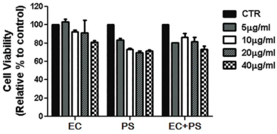
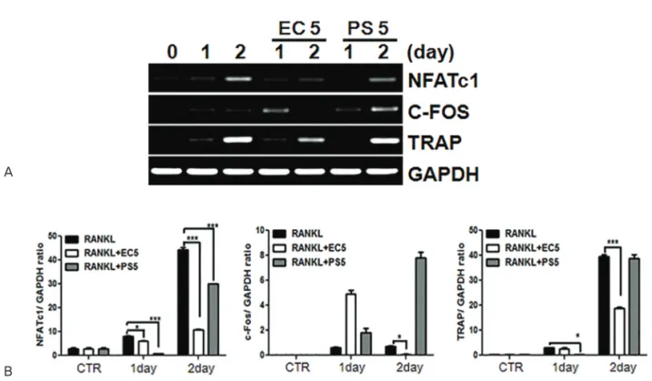
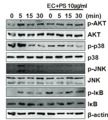
Methods : We assayed the protein expression levels of nuclear factor of activated T-cells, cy- toplasmic 1 (NFATc1), c-Fos, mitogen-activated protein kinases (MAPKs), and β-actin in cell lysates using western blotting. Similarly, mRNA expression levels of NFATc1, c-Fos, tartrate- resistant acid phosphate (TRAP), and glyceraldehyde-3-phosphate dehydrogenase, sper- matogeni (GAPDHS) from bone marrow macrophages (BMMs) were analyzed using reverse transcription-polymerase chain reaction (RT-PCR). Furthermore, we determined the anti-os- teoporotic effects of the water extracts of EC, PS, and their combination in a lipopolysaccharide (LPS)-induced bone-loss mouse model.
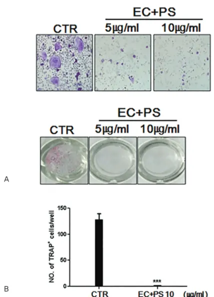
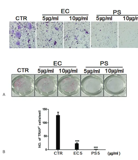
Results : The in vitro data revealed showed that the combination of EC and PS extract showed a more remarkable inhibition of osteoclast differentiation than each herb did alone. The com- bination downregulated the induction of c-Fos, NFATc1, and TRAP by suppressing the phos- phorylation of p38 and c-Jun N-terminal kinases (JNKs) and inhibiting nuclear factor kappa-light-chain-enhancer of activated B cells (NF-κB). Lastly, the in vivo data showed that PS reduced the LPS-induced bone erosion.
Conclusion : The result of this study suggests that EC and PS could be potential therapeutic agents for bone loss diseases such as osteoporosis.
※ This study was supported by academic research grant of Wonkwang University in 2017
✱ Corresponding author : Department of Acupuncture & Moxibustion Medicine, College of Oriental Medicine, Wonkwang University, 460, Iksandae-ro, Iksan-si, Jeollabuk-do, 54538, Republic of Korea
Tel : +82-63-270-1022 E-mail : [email protected]
Original Article
pISSN 1229-1137 eISSN 2287-7797http://dx.doi.org/10.13045/acupunct.2017079
Key words : Eucommiae cortex;
Psoraleae semen;
Osteoporosis;
Osteoclast
differentiation;
Bone resorption
Received : 2016. 11. 28.
Revised : 2017. 03. 16.
Accepted : 2017. 05. 02.
On-line : 2017. 05. 20.
This is an Open-Access article distributed under the terms of the Creative Commons Attribution Non-Commercial License (http://creativecommons.org/licenses/by- nc/3.0) which permits unrestricted non-commercial use, distribution, and reproduction in any medium, provided the original work is properly cited.