Pretreatment Lymphopenia, Poor Performance Status, and Early Courses of Therapy Are Risk Factors for Severe Bacterial Infection in Patients with Multiple Myeloma during Treatment with
Bortezomib-based Regimens
The aim of this study was to identify the risk factors associated with severe bacterial infection (SBI) in multiple myeloma (MM) patients during treatment with bortezomib- based regimens. A total of 98 patients with MM were evaluated during 427 treatment courses. SBI occurred in 57.1% (56/98) of the patients and during 19.0% (81/427) of the treatment courses. In the multivariate analysis for the factors associated with the development of SBI in each treatment course, poor performance status (Eastern Cooperative Oncology Group ≥ 2, P < 0.001), early course of therapy (≤ 2 courses, P < 0.001), and pretreatment lymphopenia (absolute lymphocyte count < 1.0 × 10⁹/L, P = 0.043) were confirmed as independent risk factors. The probability of developing SBI were 5.1%, 14.9%, 23.9%, and 59.5% in courses with 0, 1, 2, and 3 risk factors, respectively (P < 0.001). In conclusion, we identified three pretreatment risk factors associated with SBI in each course of bortezomib treatment. Therefore, MM patients with these risk factors should be more closely monitored for the development of SBI during bortezomib-based treatment.
Keywords: Multiple Myeloma; Infection; Bortezomib; Lymphopenia; Performance Status Shin Young Hyun,1* Sang Hoon Han,2
Soo-Jeong Kim,1 Ji Eun Jang,1 Yundeok Kim,1 Hyunsoo Cho,1 Jung Yeon Lee,1 June-Won Cheong,1 Yoo Hong Min,1 Jae-Woo Song,3 and Jin Seok Kim1
1Division of Hematology, Department of Internal Medicine, Severance Hospital, Yonsei University College of Medicine, Seoul, Korea; 2Division of Infection, Department of Internal Medicine, Severance Hospital, Yonsei University College of Medicine, Seoul, Korea; 3Department of Laboratory Medicine, Severance Hospital, Yonsei University College of Medicine, Seoul, Korea
*Present Address for Shin Young Hyun: Department of Internal Medicine, Yonsei University Wonju College of Medicine, Wonju, Korea, and Department of Medicine, The Graduate School, Yonsei University, Seoul, Korea
Received: 16 August 2015 Accepted: 23 December 2015 Address for Correspondence:
Jin Seok Kim, MD
Division of Hematology, Department of Internal Medicine, Severance Hospital, Yonsei University College of Medicine, 50-1 Yonsei-ro, Seodaemun-gu, Seoul 03722, Korea E-mail: [email protected]
http://dx.doi.org/10.3346/jkms.2016.31.4.510 • J Korean Med Sci 2016; 31: 510-518
INTRODUCTION
Infection is the leading cause of morbidity and mortality in mul- tiple myeloma (MM), in which 26%-45% of early deaths are at- tributable to infection (1,2). Infection is not only the direct cause of death but also affects disease progression due to the delaying of planned anti-myeloma therapy. The risk of bacterial infection increases 7-fold in patients with MM compared with matched controls (2), and the increased susceptibility to infection is due to disease-related immune deficits on innate and adaptive im- mune system including hypogammaglobulinemia (3,4), renal failure (3), treatment-related toxicities (4,5), and the use of high- dose corticosteroid therapy (6).
The treatment of MM has undergone a paradigm shift with the use of immunomodulatory drugs (thalidomide, lenalido-
mide, and pomalidomide) and proteasome inhibitors (bort- ezomib) (7). The effects of these drugs on the immune system are distinct from those of conventional anti-myeloma agents. In addition, these drugs are usually used in combination with high- dose dexamethasone, resulting in impairment of cell-mediated immunity. Therefore, the types and patterns of infections have changed by the administration of these novel agents. Bortezo- mib suppresses essential T-cell mediated immune responses by inhibiting the functions of human CD4+ T cells (8,9), reduc- ing antigen presentation by CD8+ T cells, and reducing the cy- totoxic T cell response (10). Consequently, the incidence of var- icella zoster virus infection increased in patients treated with bortezomib-based regimens compared with those treated with melphalan plus prednisone (11) or with high-dose dexametha- sone (12). In contrast to viral infections, the incidence of bacte-
rial infections was similar (10%-13%) (11,13) to that of regimens with melphalan plus prednisone (7%-10%) (14,15) but lower than that of regimens with intensive chemotherapeutic agents such as VAD (vincristine, doxorubicin, and dexamethasone) (14%-27%) (16,17) and DCEP (dexamethasone, cyclophospha- mide, etoposide, and cisplatin) (7.5%-22.5%) (18).
As the types and patterns of infections change, the risk factors for severe bacterial infection (SBI) in the MM patients treated with bortezomib-based regimens may be different from previ- ously known risk factors. The risk of bacterial infection increased in the early period of treatment with high tumor burden and decreased with a favorable response because of the restoration of the immune deficit (16). Therefore, a comprehensive pretreat- ment assessment of the risk factors for SBI in each treatment course can discriminate the patients at increased risk of SBI and allow the application of patient-oriented prophylactic and ther- apeutic approaches for SBI.
In this study, we analyzed the various clinical and laboratory parameters that were associated with infectious complications to determine the risk factors for SBI in 98 MM patients treated with bortezomib-based regimens during 427 treatment courses.
MATERIALS AND METHODS Patient eligibility
A total of 98 consecutive patients diagnosed with MM and who were treated with upfront or salvage bortezomib-based treat- ments in the Severance Hospital between January 2006 and De- cember 2012 were included. In the cases of patients treated with more than one bortezomib-based regimen as a different line of therapy, we used only the data related to the first bortezomib- based regimen. This study was approved by the Institutional Review Board of the Severance Hospital.
Treatment protocol and schedule
We retrospectively analyzed the data of 427 bortezomib treat- ment courses in 98 MM patients. Of the 98 MM patients, 58 pa- tients received bortezomib (1.3 mg/m2 intravenously [i.v.] on days 1, 4, 8, and 11) and dexamethasone (40 mg/day on days 1-4) every 3 weeks (VD regimen). Twenty-five patients received bortezomib (1.3 mg/m2 i.v. on days 1, 4, 8, 11, 22, 25, 29, and 32 during courses 1 to 4 and on days 1, 8, 22, and 29 during cours- es 5 to 9), melphalan (9 mg/m2 via oral route on days 1-4), and prednisolone (60 mg/m2 via oral route on days1-4) every 6 weeks (VMP regimen). To simplify the statistical analysis, one VMP regimen was divided into 2 courses, each with a 3-week sched- ule, and the VMP regimen variables presented are based on 1 course with a 3-week schedule. Fifteen patients received bort- ezomib (1.3 mg/m2 i.v. on days 1, 4, 8, and 11), doxorubicin (9 mg/m2 i.v. on days 1-4), and dexamethasone (40 mg on days 1-4) every 3 weeks (PAD regimen). Antibacterial prophylaxis
was not routinely administered but a prophylactic antibacterial agent (ciprofloxacin or levofloxacin) was used in 70.4% of the patients and a prophylactic antifungal agent (fluconazole) was used in 64.3% of the patients before or immediately after start- ing chemotherapy according to each physician’s decision. No patient received antiviral prophylaxis in this study.
Definitions
We investigated infectious complications and treatment-related toxicities within 1 month after each bortezomib course and the data from that period were analyzed. Chemotherapy-related toxicities were graded according to the National Cancer Insti- tute Common Terminology Criteria for Adverse Events (CTCAE) version 4.0. SBI was defined as grade 3-5 infection of each sys- tem or neutropenic fever. We divided patients into two groups:
1) the SBI group, composed of patients who developed SBI dur- ing the bortezomib treatment, 2) the non-SBI group, composed of patients who did not develop SBI during the bortezomib treat- ment. SBI was classified into three categories: 1) clinically doc- umented infection (CDI), for the cases in which clinical signs and symptoms of infection were present although the culture results were negative, 2) microbiologically documented infec- tion (MDI), for the cases in which pathogens were isolated from a blood sample or other cultures, 3) fever of unknown origin (FUO), for the cases in which fever was the only sign of infec- tion. In defining CDI and FUO, the episodes which were highly suspicious for bacterial infection were included and document- ed viral or fungal infections were excluded. Lymphopenia was defined as an absolute lymphocyte count (ALC) of < 1.0 × 10⁹/ L. Mortality was defined as infection-related death regardless of refractory or progressive disease.
Response to treatment was assessed according to the Inter- national Myeloma Working Group uniform response criteria (19). Progression-free survival (PFS) after bortezomib treatment was measured from the initial date of bortezomib treatment to the date of progression or the final follow-up visit without pro- gression or the date of death. Overall survival (OS) after bort- ezomib treatment was measured from the initial date of bort- ezomib treatment to the final follow-up date or the date of death from any cause.
Statistical analysis
To identify the risk factors for the development of SBI during bortezomib treatment, we analyzed the variables using two ap- proaches: 1) analysis of the patients who developed SBI during a specific period of treatment (total, 2 courses, or 3 months), or 2) analysis of each bortezomib treatment course.
Continuous variables were compared using Mann-Whitney U test and categorical variables were compared using χ2 test or Fisher’s exact test. Univariate analysis for the factors that affect- ed the development of SBI was performed using a logistic re-
gression method. The factors with P < 0.1 were selected and in- cluded in the multivariate logistic regression analysis. Potential confounding factors and multicollinearity were evaluated, and the factors strongly associated with other significant factors were excluded from the multivariate analysis. The survival curves were calculated using the Kaplan-Meier method and were com- pared using the log-rank test. A P value of < 0.05 was defined as statistically significant. All statistical calculations were performed with the PASW software version 20.0 (IBM Corp., Armonk, NY, USA).
Ethics statement
The protocol was approved by severance hospital’s institutional
review board (4-2013-0441). The data of this study was obtained from large numbers of patients for a retrospective chart review study so informed consent was waived.
RESULTS
Patient characteristics
The clinical characteristics of the 98 patients are summarized in Table 1. Ninety-eight patients received a median of 4 treatment courses (range, 1-15) of bortezomib-based chemotherapy and a total of 427 bortezomib courses. Most of the clinical charac- teristics were not significantly different between the SBI group and the non-SBI group except the performance status.
Table 1. Clinical characteristics of the 98 patients undergoing chemotherapy with the bortezomib-containing regimen
Variables No. (%) of patients by group
P value
Total SBI group Non-SBI group
At the time of diagnosis
Total number of patients 98 56 42
Age, yr 61 (39-80) 61 (39-76) 60 (42-80) 0.146
Gender Male
Female 53 (54.1)
45 (45.9) 26 (46.4)
30 (53.6) 27 (64.3)
15 (35.7)
0.060
Type of monoclonal component IgG
IgA IgD
Light chain only
57 (58.2) 19 (19.4) 4 (4.1) 18 (18.4)
35 (62.5) 11 (19.6) 1 (1.0) 9 (16.1)
22 (52.4) 8 (19.0) 3 (7.1) 9 (21.4)
0.477
Plasma cell (%) in bone marrow ≥ 50% 25 (45.5) 10 (35.7) 15 (55.6) 0.140
Durie-Salmon stage I
II III
3 (3.1) 9 (9.2) 86 (86.7)
2 (3.6) 4 (7.3) 49 (89.1)
1 (2.4) 5 (11.9) 36 (85.7)
0.704
Renal dysfunction (creatinine ≥ 2 mg/dL) 22 (22.4) 14 (25.5) 8 (19.0) 0.310
ISS stage*
I, II III
33 (33.6) 58 (59.2)
17 (32.7) 35 (67.3)
16 (41.0) 23 (59.0)
0.274
At the time of bortezomib treatment initiation
Age, yr 62 (39-80) 62 (39-78) 62 (45-80) 0.255
Disease status Newly diagnosed
Relapsed/refractory 30 (31.6)
68 (68.3) 16 (28.6)
40 (71.4) 14 (33.3)
28 (66.7)
0.461
Number of prior lines of therapy 0
1 2 ≥ 3
30 (31.6) 38 (38.8) 20 (20.4) 10 (10.1)
16 (28.6) 22 (39.3) 13 (23.2) 5 (9.0)
14 (33.3) 16 (38.1) 7 (16.7) 5 (11.9)
0.970
Performance status (ECOG)† ≥ 2 35 (36.8) 28 (50.0) 7 (16.7) < 0.001
Monoclonal protein ≥ 3.0 g/dL 19 (31.7) 10 (32.3) 9 (31.0) 0.570
Decrease of uninvolved immunoglobulins
≥ 1 immunoglobulin‡ 58/63 (92.1) 30/33 (90.9) 28/30 (93.3) 0.546
Regimen
bortezomib/dexamethasone
bortezomib/doxorubicin/dexamethasone bortezomib/melphalan/prednisone
58 (59.1) 15 (15.3) 25 (25.5)
34 (60.8) 7 (12.5) 15 (26.8)
24 (57.2) 8 (19.0) 10 (23.8)
0.668
Number of treatment courses 4.0 (1-15) 3.5 (1-12) 4.0 (1-15) 0.106
Follow up period after treatment, month 6.3 (0.5-57.1) 5.4 (0.5-57.6) 16.0 (0.8-55.8) 0.040
Continuous variables are presented as median (range). *Missed data in 7 patients; †Missed data in 3 patients; ‡Missed data in 35 patients. SBI, severe bacterial infection; ECOG, Eastern Cooperative Oncology Group; ISS, International Scoring System.
Infection profile
Infection development was assessed in each patient and for each treatment course. Of the 98 patients, SBI was observed in 56 (57.2%) patients, and 18 (18.3%) patients developed SBI more than once. Of the 427 treatment courses, 81 SBI cases occurred and the incidence of SBI per treatment course was 19.0%. Reac- tivation of herpes zoster viruses developed in 13 (13.3%) patients and in 14 (3.0%) treatment courses. Fungal infections, defined as proven or probable invasive fungal disease, developed in 13 (13.2%) patients and in 18 (4.2%) treatment courses. Most of the SBI episodes (79.0%) occurred in the first 3 courses and no SBI episode was observed after the 8th course (Fig. 1).
Of the 81 SBI episodes, 42 (52.5%) were CDI, 30 (37.0%) were MDI, and 9 (11.1%) were FUO (Table 2). Pneumonia was the most common type of infection in both CDI and MDI and ac- counted for 60.5% of the SBI episodes, and both bacteremia (n = 9) and urinary tract infection (n = 8) were frequently ob- served. In addition, non-neutropenic fever was frequently ob- served (n = 75) whereas neutropenic fever accounted for only 6 episodes (7.4%). Most of the SBI episodes (62 of 81) were appro- priately treated with systemic antibiotics although the subse- quent bortezomib courses were postponed in 31 episodes and interrupted in 9 episodes. Among total 19 patients died during the bortezomib-based treatment (19.3% of overall mortality rate), 12 patients died from infection with at least partial response to bortezomib and 7 patients died from infection in progressive or refractory status.
Variable pathogens were identified in 30 MDI cases. Gram- negative bacteria (n = 21) were more prevalent than gram-pos- itive bacteria (n = 9). Acinetobacter baumannii (n = 6), Esche- richia coli (n = 5), Klebsiella species (n = 4), coagulase-negative Staphylococci (n = 4), and Staphylococcus aureus (n = 3) were common pathogens.
Risk factors for developing SBI in 98 MM patients
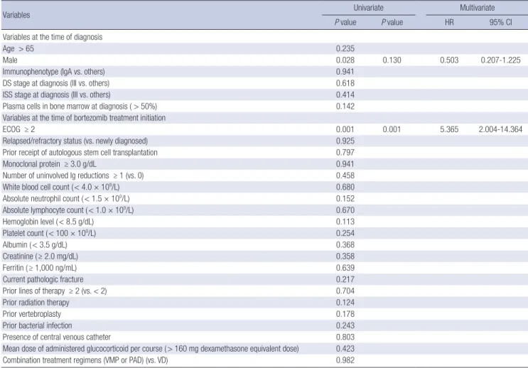
To identify the risk factors for developing SBI in 98 patients, three types of analysis were introduced in this study. An analysis of the risk factors was performed 1) during the total period of bort- ezomib-based treatment, 2) within 3 months from initiation of therapy, and 3) in the first 2 treatment courses. The clinical and myeloma-associated variables evaluated are presented in Table 3. Multivariate analysis showed that only poor performance sta- tus (Eastern Cooperative Oncology Group [ECOG] performance status ≥ 2) had statistical significance as a risk factor for devel- oping SBI during the total period of bortezomib-based treatment (hazard ratio [HR], 5.365; 95% confidence interval [CI], 2.004- 14.364, P = 0.001, Table 3), within 3 months (HR, 4.976; 95% CI, 1.817-13.624, P = 0.002), and within 2 courses (HR, 8.652; 95%
CI, 2.845-26.310, P < 0.001).
Risk factors for developing SBI during the 427 courses of bortezomib-based treatment
The risk factors that changed during each treatment course were evaluated. The incidence of SBI was higher in the treatment courses where poor performance status (ECOG ≥ 2, 34.2% vs.
10.6%, P < 0.001), pretreatment lymphocytopenia (ALC < 1.0 × 10⁹/L, 25.1% vs. 14.6%, P = 0.005), and pretreatment hypoalbu- minemia (serum albumin < 3.5 g/dL, 36.0% vs. 11.1%, P < 0.001) were reported, and early in the course of treatment (≤ 2 cours- es, 28.4% vs. 11.9%, P < 0.001) compared with other courses.
There was no significant change of the median ALC at each cy- cle during bortezomib-based treatment (P = 0.983). According to the univariate analysis, poor performance status (P < 0.001), pretreatment lymphocytopenia (P = 0.007), pretreatment hy- poalbuminemia (P < 0.001), and early course of treatment (P <
Cumulative number of SBI episodes
Courses
1 2 3 4 5 6 7 8 9 10 11 12 13 14 15 90
80 70 60 50 40 30 20 10 0
30 22
12 6
6 2 1 2
52 64 70 76 78 79 81 81 81 81 81 81 81 81
New SBI episodes at each course
Fig. 1. Number of severe bacterial infections (SBI) episodes during treatment with the bortezomib-based regimens. Cumulative numbers of SBI are presented at the bottom of the bars, and numbers of SBI episodes at each course are presented at the top of the bars.
Table 2. Eighty-one episodes of severe bacterial infections during chemotherapy with the bortezomib-containing regimens
Clinical conditions No. (%) of episodes
Classification of infection Clinically documented infection Pneumonia
Colitis Parotitis
Microbiologically documented infection Pneumonia
Bacteremia Urinary tract infection Colitis
Fever of unknown origin
42 (51.9) 37 4 1 30 (37.0)
12 9 8 1 9 (11.1) Type of fever
Neutropenic fever Grade 3 Grade 4 Grade 5
Non-neutropenic fever Grade 3
Grade 4 Grade 5
6 (7.4) 3 2 1 75 (92.6)
47 10 18
Table 3. Univariate and multivariate analysis for the risk of developing severe bacterial infection during bortezomib-based treatment in 98 patients (patient-based analysis)
Variables Univariate Multivariate
P value P value HR 95% CI
Variables at the time of diagnosis
Age > 65 0.235
Male 0.028 0.130 0.503 0.207-1.225
Immunophenotype (IgA vs. others) 0.941
DS stage at diagnosis (III vs. others) 0.618
ISS stage at diagnosis (III vs. others) 0.414
Plasma cells in bone marrow at diagnosis ( > 50%) 0.142
Variables at the time of bortezomib treatment initiation
ECOG ≥ 2 0.001 0.001 5.365 2.004-14.364
Relapsed/refractory status (vs. newly diagnosed) 0.925
Prior receipt of autologous stem cell transplantation 0.797
Monoclonal protein ≥ 3.0 g/dL 0.941
Number of uninvolved Ig reductions ≥ 1 (vs. 0) 0.458
White blood cell count ( < 4.0 × 10⁹/L) 0.680
Absolute neutrophil count ( < 1.5 × 10⁹/L) 0.152
Absolute lymphocyte count ( < 1.0 × 10⁹/L) 0.670
Hemoglobin level ( < 8.5 g/dL) 0.113
Platelet count ( < 100 × 109/L) 0.254
Albumin ( < 3.5 g/dL) 0.368
Creatinine ( ≥ 2.0 mg/dL) 0.358
Ferritin ( ≥ 1,000 ng/mL) 0.639
Current pathologic fracture 0.217
Prior lines of therapy ≥ 2 (vs. < 2) 0.704
Prior radiation therapy 0.124
Prior vertebroplasty 0.178
Prior bacterial infection 0.243
Presence of central venous catheter 0.803
Mean dose of administered glucocorticoid per course ( > 160 mg dexamethasone equivalent dose) 0.423
Combination treatment regimens (VMP or PAD) (vs. VD) 0.982
HR, hazard ratio; CI, confidence interval; ECOG, Eastern Cooperative Oncology Group; DS, Durie-Salmon; ISS, International Scoring System; VD, bortezomib-dexamethasone;
VMP, bortezomib melphalan prednisolone; PAD, bortezomib doxorubicin dexamethasone; WBC, white blood cell; ANC, absolute neutrophil count; ALC, absolute lymphocyte count;
Ig, immunoglobulin.
Table 4. Univariate and multivariate analysis for the risk of developing severe bacterial infections during the 427 courses of the bortezomib-based treatment according to the clinical and laboratory characteristic at the beginning of each course (course-based analysis)
Variables Univariate Multivariate
P value P value HR 95% CI
ECOG ≥ 2 < 0.001 < 0.001 3.920 2.305-6.666
Early courses of therapy ( ≤ 2 courses) < 0.001 < 0.001 2.782 1.633-4.740
Monoclonal protein ≥ 3.0 g/dL* 0.061
Number of uninvolved immunoglobulin reductions ( > 1) 0.268 Pretreatment white blood cell count ( < 4.0 × 10⁹/L) 0.362 Pretreatment absolute neutrophil count ( < 1.5 × 10⁹/L) 0.152
Pretreatment absolute lymphocyte count ( < 1.0 × 10⁹/L) 0.007 0.043 1.728 1.016-2.937
Pretreatment hemoglobin level ( < 8.5 g/dL)* 0.013
Pretreatment platelet count ( < 100 × 109/L) 0.754
Pretreatment albumin ( < 3.5 g/dL)* < 0.001
Pretreatment creatinine ( ≥ 2.0 mg/dL) 0.383
Pretreatment ferritin ( ≥ 1,000 ng/mL) 0.832
Dose of glucocorticoid used at each course ( >160 mg dexamethasone equivalent dose) 0.151
*These variables were excluded in the multivariate analysis because of collinearity with early courses of therapy. HR, hazard ratio; CI, confidence interval; ECOG, Eastern Coop- erative Oncology Group.
0.001) were significantly associated with the development of SBI in each treatment course (Table 4). In the multivariate logis-
tic analysis, poor performance status (HR, 3.920; 95% CI, 2.305- 6.666, P < 0.001), early course of treatment (HR, 2.782; 95% CI,
1.633-4.740, P < 0.001), and pretreatment lymphopenia (HR;
1.728; 95% CI, 1.016-2.937, P = 0.043) were risk factors for de- veloping SBI in each treatment course. The probability of devel- oping SBI was 5.1% in the absence of risk factors, 14.9% with 1 risk factor, 23.9% with 2 risk factors, and 59.5% with 3 risk fac- tors (P < 0.001, Fig. 2).
Treatment outcomes
The overall response rate (at least partial response) of the SBI group was 51.9%, which was lower than that of the non-SBI group (70.5%, P = 0.048). Among the 98 enrolled patients, 48 patients (49.0%) died during the median follow-up period from the ini- tiation of bortezomib treatment of 6.3 months (range, 0.5-57.1 months). The median OS was significantly higher in the non-SBI group compared with the SBI group (30.1 months vs. 6.1 months, P = 0.004) although PFS was not significantly different between
the two groups (21.9 months vs. 18.1 months, P = 0.418).
In the 57 patients aged less than 65 year, 14 of the 24 (58.3%) patients in the non-SBI group received autologous stem cell trans- plantation, but only 6 of 33 (18.2%) patients in the SBI group re- ceived autologous stem cell transplantation (P = 0.002). Among the patients aged below 65 year, the SBI group showed a signifi- cantly shorter median OS compared with the non-SBI group (11.2 months vs. not reached, P = 0.016) and a significantly short- er PFS compared with the non-SBI group (6.0 months vs. 24.4 months, P = 0.015, Fig. 3).
DISCUSSION
Herein we demonstrate that poor performance status (ECOG
≥ 2), early course of treatment (≤ 2 courses), and pretreatment lymphopenia (ALC < 1.0 × 10⁹/L) are risk factors for developing SBI in MM patients treated with bortezomib-based regimens.
More than half (59.5%) of the patients with all three risk factors developed SBI whereas only 5.1% of the patients without risk factors developed SBI. Therefore, we could expect the risk of developing SBI at the subsequent course of bortezomib-based treatment. It would be beneficial to manage MM patients sus- ceptible to SBI. Although we found the poor performance sta- tus at diagnosis as significant risk factor for developing SBI dur- ing the total period of bortezomib-based treatment, the assess- ment of the risk factors in each course of bortezomib-based treat- ment would be more suitable to predict the exact risk of devel- oping SBI in subsequent bortezomib-based treatments because the clinical and laboratory parameters changed in each treat- ment course.
The early course of treatment (≤ 2 courses) was defined as a Fig. 2. Probability of severe bacterial infection according to the number of risk factors
in each bortezomib course. Three risk factors are poor performance status (ECOG ≥ 2), early course of treatment ( ≤ 2 courses), and pretreatment lymphopenia (absolute lymphocyte count < 1.0 × 10⁹/L).
Number of risk factors
0 1 2 3
P < 0.001
5.1%
14.9%
23.9%
59.5%
Fig. 3. Overall survival (A) and progression-free survival (B) after the bortezomib-based treatment of 57 MM patients under 65 years of age according to the presence of severe bacterial infection (SBI). MM, multiple myeloma; SBI group, patients who developed SBI; Non-SBI group, patients who did not experience SBI.
Overall survival
Month
0 12 24 36 48 60 1.0
0.8
0.6
0.4
0.2
0.0
Non-SBI group
SBI group
P = 0.013
Progression-free survival
Month
0 12 24 36 48 60 1.0
0.8
0.6
0.4
0.2
0.0
Non-SBI group
SBI group P = 0.027
A B
risk factor for SBI in this analysis because of hypogammaglobu- linemia associated with high tumor burden in the early period of treatment (21). A decreased level of uninvolved immunoglob- ulins in MM serves as a risk factor for infection with encapsu- lated bacteria (e.g., Streptococcus pneumoniae or Haemophilus influenzae) (7). The degree of immunoglobulin suppression and disease stage are interrelated (20), and decreased levels of uninvolved immunoglobulin have been known as an indepen- dent prognostic factor for shorter OS (21). Many studies conduct- ed before the development of the novel agents demonstrated the increased risk of infections in the early period of treatment, particularly within 4-6 months following treatment, and the de- creased risk of infections after achieving a favorable response to treatment (16,22,23). Because of the serial reduction of tumor burden in subsequent courses of the bortezomib-based treat- ment, the risk of SBI was significantly higher in the first two cour- ses of bortezomib-based treatment in this study.
Poor performance status has been reported as a poor prog- nostic factor for survival (1,24), which increases treatment-re- lated toxicity and/or the risk of infection in MM (25). Because the bortezomib-based treatment for patients with poor perfor- mance status is usually performed in the inpatient setting, these patients can be easily exposed to a variety of nosocomial infec- tions. Cesana et al. reported a higher incidence of nosocomial infections in MM patients treated with VAD regimen in the in- patient setting compared with in the outpatient setting (6% vs.
0%) (16). Gram-positive encapsulated bacteria are known to be predominant pathogens in patients with MM. The incidence of infection with gram-negative bacilli increased after using alkyl- ating agents or novel antimyeloma agents (7,26). In this study, gram-negative bacteria accounted for 70% of MDI cases and the most common pathogen was Acinetobacter baumannii (n = 7), a major pathogen of hospital-acquired pneumonia (27).
Lymphopenia at the beginning of each course was a novel risk factor for SBI in this study. It is generally considered to be associated with viral and fungal infections (28). However, sev- eral studies reported lymphopenia as a marker of severe sepsis and septic shock (19,29). Lymphopenia indicates a preexisting immunosuppressed condition. Therefore, patients with lym- phopenia have inadequate immunologic reactions and conse- quently are vulnerable to bacterial infection. Until now, there is limited data about lymphopenia as a predictive factor for infec- tion in MM. Jung et al. analyzed the data from 139 patients treat- ed with bortezomib and observed that lymphopenia levels of
< 0.8 × 10⁹/L at diagnosis were a risk factor for SBI. Total 30 out of the 74 patients (40.5%) with lymphopenia developed SBI (30).
In this study, the incidence of SBI in treatment courses with lymphopenia (< 1.0 × 10⁹/L) was 42.0%, which was higher than the incidence of SBI in courses without lymphopenia (14.6%, P = 0.007). Considering the change of lymphocyte counts in each treatment course, the impact of lymphopenia at the be-
ginning of each course in this study would be more valuable in clinical application.
Corticosteroid therapy has been reported to increase the in- cidence of infections with intracellular pathogens such as Liste- ria monocytogenes, various fungal species, and herpes viruses (31). The risk for infection is associated with the dose and the duration of therapy, and doses exceeding 20 mg of prednisone equivalent per day or a cumulative dose of more than 700 mg of prednisone can increase the risk of infections (22). Large cumu- lative doses of corticosteroids are frequently used during MM treatments and contribute to increase the risk of infection. A re- cent clinical trial compared lenalidomide plus low-dose dexa- methasone (40 mg once a week, on days 1, 8, 15, and 22, every 28 days), lenalidomide plus high-dose dexamethasone (40 mg per day for four consecutive days on days 1-4, 9-12, and 17-20, every 28 days), and reported a higher incidence of infection in the high-dose dexamethasone arm (16%) compared with the low-dose dexamethasone arm (9%). The trial demonstrated a significantly decreased incidence of infectious complications and resultant improved OS in low-dose dexamethasone group (32). In this study, there was a trend to lower incidence of SBI in courses with lower doses of concomitant corticosteroid than higher doses of corticosteroid. The incidence of SBI was 18.0%
in course with lower doses of corticosteroid (≤ 160 mg of dexa- methasone equivalent dose) and 26.5% in course with higher doses of corticosteroid (> 160 mg of dexamethasone equivalent dose) (P = 0.151). However, we could not demonstrate a rela- tionship between corticosteroid doses and the incidence of SBI because most of our patients received relatively low doses of corticosteroids during bortezomib-based treatment. Of the pa- tients who received bortezomib combined with dexamethasone, the mean dose of dexamethasone was 157.45 ± 113.94 mg, which was very similar to the dose of low-dose dexamethasone, and only 6% of the patients received high-dose dexamethasone.
Therefore, our cohort would be considered as a homogenous population that is suitable for assessing the risk factors for SBI.
The prophylactic use of antibiotics for reducing the bacterial infections has been proposed in MM. However, evidence about the prophylactic use of antibiotics in non-neutropenic MM pa- tients is still inconclusive (33) whereas the prophylactic use of fluoroquinolone has been recommended by the National Com- prehensive Cancer Network (34). The emergence of resistance to antibiotics as well as their efficacy and safety are concerns with regard to the prophylactic antibiotics (35). Therefore, the prophylactic use of antibiotics should not be recommended to all MM patients but only to those at increased risk of bacterial infection. In addition, adjustments in the corticosteroid dose in patients with high risk of SBI in each treatment course can be a good strategy to decrease the incidence of treatment-related in- fectious complications. Therefore, further prospective studies are needed to elucidate the effectiveness of the prophylactic use
of antibiotics in MM patients and to assess the potential risks and benefits of using lower doses of corticosteroids.
One of the limitations of this study is its retrospective nature.
The prophylactic antibiotics and the corticosteroid dose were inconsistently administered depending on the patient’s situa- tion. Therefore, the assessment of the risk of SBI after removing these confounding factors was limited although these factors might affect the risk of SBI. Although the SBI rates were not sta- tistically different between the different regimens adopted (60.0%
for VMP, 46.7% for PAD, and 58.6% for VD regimen), the rate obtained in this study was somewhat higher than that of other studies (11,21,36). Because many of the patients in this study had a relatively poor performance status (36.8%) at the time of bortezomib initiation, the high incidence of SBI in this study may be attributed to this factor.
In conclusion, the three risk factors for developing SBI iden- tified in the present study were the early course of therapy, poor performance status, and lymphopenia at the beginning of each treatment course in MM patients treated with bortezomib-based regimen. Therefore, patients with these risk factors should be more closely monitored for developing bacterial infections dur- ing subsequent bortezomib-based treatments. Risk-adapted approaches such as the prophylactic use of antibiotics or modi- fication of the corticosteroid dose should be considered to re- duce the incidence of bacterial infection in those patients. Fur- ther prospective studies are needed to support the implemen- tation of effective preventive strategies against bacterial infection.
DISCLOSURE
The authors have no potential conflicts of interest to disclose.
AUTHOR CONTRIBUTION
Study design: Hyun SY, Kim JS. Data acquisition and patient care: Hyun SY, Kim JS, Han SH, Kim SJ, Jang JE, Kim YD, Cho H, Lee JY, Chung JW, Min YH, Song JW. Data analysis: Hyun SY, Kim JS. Writing the manuscript: Hyun SY, Kim JS. Review, revi- sion, and final approval: all authors.
ORCID
Shin Young Hyun http://orcid.org/0000-0002-8137-8675 Ji Eun Jang http://orcid.org/0000-0001-8832-1412 Jung Yeon Lee http://orcid.org/0000-0001-6162-5571 Hyunsoo Cho http://orcid.org/0000-0003-2651-6403 June-Won Cheong http://orcid.org/0000-0002-1744-0921 Yoo Hong Min http://orcid.org/0000-0001-8542-9583 Jae-Woo Song http://orcid.org/0000-0002-1877-5731 Jin Seok Kim http://orcid.org/0000-0001-8986-8436
REFERENCES
1. Augustson BM, Begum G, Dunn JA, Barth NJ, Davies F, Morgan G, Beh- rens J, Smith A, Child JA, Drayson MT. Early mortality after diagnosis of multiple myeloma: analysis of patients entered onto the United Kingdom Medical Research Council trials between 1980 and 2002--Medical Re- search Council Adult Leukaemia Working Party. J Clin Oncol 2005; 23:
9219-26.
2. Blimark C, Holmberg E, Mellqvist UH, Landgren O, Björkholm M, Hult- crantz M, Kjellander C, Turesson I, Kristinsson SY. Multiple myeloma and infections: a population-based study on 9253 multiple myeloma patients.
Haematologica 2015; 100: 107-13.
3. Kuroki Y, Tsuchida K, Go I, Aoyama M, Naganuma T, Takemoto Y, Naka- tani T. A study of innate immunity in patients with end-stage renal dis- ease: special reference to toll-like receptor-2 and -4 expression in periph- eral blood monocytes of hemodialysis patients. Int J Mol Med 2007; 19:
783-90.
4. Bodey GP, Buckley M, Sathe YS, Freireich EJ. Quantitative relationships between circulating leukocytes and infection in patients with acute leu- kemia. Ann Intern Med 1966; 64: 328-40.
5. Bow EJ. Infection in neutropenic patients with cancer. Crit Care Clin 2013;
29: 411-41.
6. Stuck AE, Minder CE, Frey FJ. Risk of infectious complications in patients taking glucocorticosteroids. Rev Infect Dis 1989; 11: 954-63.
7. Teh BW, Harrison SJ, Pellegrini M, Thursky KA, Worth LJ, Slavin MA. Chang- ing treatment paradigms for patients with plasma cell myeloma: impact upon immune determinants of infection. Blood Rev 2014; 28: 75-86.
8. Cerundolo V, Benham A, Braud V, Mukherjee S, Gould K, Macino B, Neefjes J, Townsend A. The proteasome-specific inhibitor lactacystin blocks pre- sentation of cytotoxic T lymphocyte epitopes in human and murine cells.
Eur J Immunol 1997; 27: 336-41.
9. Berges C, Haberstock H, Fuchs D, Miltz M, Sadeghi M, Opelz G, Daniel V, Naujokat C. Proteasome inhibition suppresses essential immune func- tions of human CD4+ T cells. Immunology 2008; 124: 234-46.
10. Basler M, Lauer C, Beck U, Groettrup M. The proteasome inhibitor bort- ezomib enhances the susceptibility to viral infection. J Immunol 2009;
183: 6145-50.
11. San Miguel JF, Schlag R, Khuageva NK, Dimopoulos MA, Shpilberg O, Kropff M, Spicka I, Petrucci MT, Palumbo A, Samoilova OS, et al. Bortezo- mib plus melphalan and prednisone for initial treatment of multiple my- eloma. N Engl J Med 2008; 359: 906-17.
12. Chanan-Khan A, Sonneveld P, Schuster MW, Stadtmauer EA, Facon T, Harousseau JL, Ben-Yehuda D, Lonial S, Goldschmidt H, Reece D, et al.
Analysis of herpes zoster events among bortezomib-treated patients in the phase III APEX study. J Clin Oncol 2008; 26: 4784-90.
13. Richardson PG, Sonneveld P, Schuster MW, Irwin D, Stadtmauer EA, Fa- con T, Harousseau JL, Ben-Yehuda D, Lonial S, Goldschmidt H, et al. Bort- ezomib or high-dose dexamethasone for relapsed multiple myeloma. N Engl J Med 2005; 352: 2487-98.
14. Facon T, Mary JY, Hulin C, Benboubker L, Attal M, Pegourie B, Renaud M, Harousseau JL, Guillerm G, Chaleteix C, et al. Melphalan and prednisone plus thalidomide versus melphalan and prednisone alone or reduced-in- tensity autologous stem cell transplantation in elderly patients with mul- tiple myeloma (IFM 99-06): a randomised trial. Lancet 2007; 370: 1209-18.
15. Facon T, Mary JY, Pégourie B, Attal M, Renaud M, Sadoun A, Voillat L, Dor- vaux V, Hulin C, Lepeu G, et al. Dexamethasone-based regimens versus melphalan-prednisone for elderly multiple myeloma patients ineligible for high-dose therapy. Blood 2006; 107: 1292-8.
16. Cesana C, Nosari AM, Klersy C, Miqueleiz S, Rossi V, Ferrando P, Valentini M, Barbarano L, Morra E. Risk factors for the development of bacterial infections in multiple myeloma treated with two different vincristine-adri- amycin-dexamethasone schedules. Haematologica 2003; 88: 1022-8.
17. Segeren CM, Sonneveld P, van der Holt B, Baars JW, Biesma DH, Cornellis- sen JJ, Croockewit AJ, Dekker AW, Fibbe WE, Löwenberg B, et al. Vincris- tine, doxorubicin and dexamethasone (VAD) administered as rapid in- travenous infusion for first-line treatment in untreated multiple myelo- ma. Br J Haematol 1999; 105: 127-30.
18. Corso A, Barbarano L, Zappasodi P, Cairoli R, Alessandrino EP, Mangia- cavalli S, Ferrari D, Fava S, Fiumanò M, Frigerio G, et al. The VAD-DCEP sequence is an effective pre-transplant therapy in untreated multiple my- eloma. Haematologica 2004; 89: 1124-7.
19. Durie BG, Harousseau JL, Miguel JS, Bladé J, Barlogie B, Anderson K, Gertz M, Dimopoulos M, Westin J, Sonneveld P, et al. International uniform re- sponse criteria for multiple myeloma. Leukemia 2006; 20: 1467-73.
20. Pruzanski W, Gidon MS, Roy A. Suppression of polyclonal immunoglob- ulins in multiple myeloma: relationship to the staging and other manifes- tations at diagnosis. Clin Immunol Immunopathol 1980; 17: 280-6.
21. Kastritis E, Zagouri F, Symeonidis A, Roussou M, Sioni A, Pouli A, Delim- pasi S, Katodritou E, Michalis E, Michael M, et al. Preserved levels of un- involved immunoglobulins are independently associated with favorable outcome in patients with symptomatic multiple myeloma. Leukemia 2014;
28: 2075-9.
22. Perri RT, Hebbel RP, Oken MM. Influence of treatment and response sta- tus on infection risk in multiple myeloma. Am J Med 1981; 71: 935-40.
23. Mainwaring CJ, Williams MA, Singer CR, Lush RJ, Smith JG, Haynes CL, Kelsey SM. Monocyte dysfunction in patients with multiple myeloma and lymphoplasmacytic disorders is related to serum paraprotein levels.
Br J Haematol 1999; 105: 948-54.
24. Takamatsu H, Honda S, Miyamoto T, Yokoyama K, Hagiwara S, Ito T, To- mita N, Iida S, Iwasaki T, Sakamaki H, et al. Changing trends in prognostic factors for patients with multiple myeloma after autologous stem cell trans- plantation during the immunomodulator drug/proteasome inhibitor era.
Cancer Sci 2015; 106: 179-85.
25. Yoshikawa TT. Epidemiology and unique aspects of aging and infectious diseases. Clin Infect Dis 2000; 30: 931-3.
26. Savage DG, Lindenbaum J, Garrett TJ. Biphasic pattern of bacterial infec- tion in multiple myeloma. Ann Intern Med 1982; 96: 47-50.
27. Howard A, O’Donoghue M, Feeney A, Sleator RD. Acinetobacter bauman- nii: an emerging opportunistic pathogen. Virulence 2012; 3: 243-50.
28. Yi YS, Chung JS, Song MK, Shin HJ, Seol YM, Choi YJ, Cho GJ, Lee GW, Moon JH, Hwang IH, et al. The risk factors for herpes zoster in bortezo- mib treatment in patients with multiple myeloma. Korean J Hematol 2010;
45: 188-92.
29. de Jager CP, van Wijk PT, Mathoera RB, de Jongh-Leuvenink J, van der Poll T, Wever PC. Lymphocytopenia and neutrophil-lymphocyte count ratio predict bacteremia better than conventional infection markers in an emergency care unit. Crit Care 2010; 14: R192.
30. Jung SH, Bae SY, Ahn JS, Kang SJ, Yang DH, Kim YK, Kim HJ, Lee JJ. Lym- phocytopenia is associated with an increased risk of severe infections in patients with multiple myeloma treated with bortezomib-based regimens.
Int J Hematol 2013; 97: 382-7.
31. Klein NC, Go CH, Cunha BA. Infections associated with steroid use. In- fect Dis Clin North Am 2001; 15: 423-32.
32. Rajkumar SV, Jacobus S, Callander NS, Fonseca R, Vesole DH, Williams ME, Abonour R, Siegel DS, Katz M, Greipp PR; Eastern Cooperative On- cology Group. Lenalidomide plus high-dose dexamethasone versus le- nalidomide plus low-dose dexamethasone as initial therapy for newly di- agnosed multiple myeloma: an open-label randomised controlled trial.
Lancet Oncol 2010; 11: 29-37.
33. Vesole DH, Oken MM, Heckler C, Greipp PR, Katz MS, Jacobus S, Morrow GR; University of Rochester Cancer Center and the Eastern Cooperative Oncology Group. Oral antibiotic prophylaxis of early infection in multiple myeloma: a URCC/ECOG randomized phase III study. Leukemia 2012;
26: 2517-20.
34. National Comprehensive Cancer Network (US). NCCN clinical practice guidelines in oncology: prevention and treatment of cancer-related infec- tions, version 2 [Internet]. Fort Washington, PA: National Comprehensive Cancer Network, 2015. Available at http://www.nccn.org/professionals/
physician_gls/pdf/infections.pdf [accessed on 10 August 2015].
35. Wittekamp BH, Bonten MJ. Antibiotic prophylaxis in the era of multidrug- resistant bacteria. Expert Opin Investig Drugs 2012; 21: 767-72.
36. Teh BW, Teng JC, Urbancic K, Grigg A, Harrison SJ, Worth LJ, Slavin MA, Thursky KA. Invasive fungal infections in patients with multiple myelo- ma: a multi-center study in the era of novel myeloma therapies. Haema- tologica 2015; 100: e28-31.