Comparison of Drug-Eluting Balloon Followed by Bare Metal Stent with Drug-Eluting Stent for Treatment of de Novo Lesions:
Randomized, Controlled, Single-Center Clinical Trial
The combined use of a drug-eluting balloon (DEB) and a bare metal stent (BMS) for the treatment of de novo non-small vessel coronary artery diseases (CAD) remains to be evaluated. We investigated the efficacy of a sequential treatment using a DEB together with a BMS implantation in comparison to a zotarolimus-eluting stent (ZES). This study was a prospective, randomized, open-label study. We designed it to demonstrate the non- inferiority of a sequential treatment using a DEB first followed by a BMS (DEB + BMS) compared with the use of a ZES. The primary endpoint was in-segment late loss (LL) at 9 months measured by quantitative coronary angiography (QCA). A total of 180 patients were enrolled in the study. The 9-month follow-up angiography was performed in 72 patients with DEB + BMS and 74 patients with ZES. When comparing the DEB + BMS results with the ZES ones, LL was 0.50 ± 0.46 mm in DEB + BMS patients vs. 0.21 ± 0.44 mm in ZES patients (P < 0.001). The mean difference of the LL was 0.31 mm, which was larger than the prespecified non-inferiority margin of 0.19 mm, and the 2-sided 95%
confidence interval was 0.15–0.48. The clinical outcomes were not significantly different.
In conclusion, the DEB + BMS strategy is inferior to the ZES one in terms of the LL result at 9 months. The DEB strategy for de novo coronary artery lesions needs to be improved for it to become an alternative treatment option. This was a clinical trial study and was registered at www.ClinicalTrials.gov (Identifier: NCT01539603; http://www.clinicaltrials.gov/ct2/
show/NCT01539603).
Keywords: Drug-eluting Balloon; Bare Metal Stent; Drug-eluting Stent; In-segment Late Loss; Coronary Artery Disease
In-Ho Chae, Chang-Hwan Yoon, Jin Joo Park, Il-Young Oh, Jung-Won Suh, Young-Seok Cho, Tae-Jin Youn, and Dong-Ju Choi Department of Internal Medicine, Cardiovascular Center, Seoul National University Bundang Hospital, Seongnam, Korea
Received: 10 November 2016 Accepted: 19 March 2017 Address for Correspondence:
Chang-Hwan Yoon, MD, PhD
Department of Internal Medicine, Cardiovascular Center, Seoul National University Bundang Hospital, 82 Gumi-ro 173-beon- gil, Bundang-gu, Seongnam 13620, Republic of Korea E-mail: [email protected]
Funding: This work was supported by the Industrial Strategic Technology Development program (No. 10052980) funded by the Ministry of Trade, Industry and Energy, Korea.
https://doi.org/10.3346/jkms.2017.32.6.933 • J Korean Med Sci 2017; 32: 933-941
INTRODUCTION
Percutaneous coronary interventions (PCIs) using bare metal stents (BMS) have raised concerns of restenosis of the lesion treated percutaneously (1,2). Although the use of drug-eluting stents (DES) has reduced the occurrence rate of restenosis and the subsequent need for repeat revascularization (3), the de- layed vascular healing due to incomplete re-endothelialization, persistence of polymer, and the ongoing vascular inflammation after DES implantation represent challenges (4,5). The persis- tent presence of polymer in the vessel wall and a late catch-up phenomenon or accelerated neoatherosclerosis over time have all raised concerns about the extensive use of DES (6-8).
In contrast to DES, the drug-eluting balloon (DEB), a non- stent-based local antiproliferative drug-delivery system, works by locally releasing a controlled dose of drug, which is homoge- neously distributed to the entire injured vessel wall and is not limited to the surface area adjacent to a stent strut (9). Compared to a standard uncoated balloon, a paclitaxel-coated balloon sig-
nificantly reduced neointimal proliferation and the need for target vessel revascularization in an in-stent restenosis (ISR) setting (10). Furthermore, the DEB was superior to DES with late lumen loss, and was associated with fewer adverse clinical events during the treatment of coronary ISR (11). Moreover, prom- ising clinical data are available for the stand-alone use of the DEB in small vessel coronary disease (12) and bifurcation lesions (13). In a trial of de novo coronary artery lesions, a BMS mount- ed on a DEB was compared to a sirolimus-eluting stent in pa- tients with stable and unstable angina. However, the per proto- col analysis of this trial revealed that the BMS pre-mounted on DEB strategy did not meet the non-inferiority criteria when com- pared with the sirolimus-eluting stent (14). In the trial, drugs might be inappropriately delivered and unevenly distributed to the diseased vessel wall because of the pre-mounted stent strut. This might diminish the efficacy of the DEB that had been shown in the previous studies. Therefore, we investigated a different pro- tocol in which a DEB is treated first followed by the BMS implan- tation (DEB + BMS) as opposed to the DES implantation alone.
ORIGINAL ARTICLE
Cardiovascular Disorders
2017-03-16 https://crossmark-cdn.crossref.org/widget/v2.0/logos/CROSSMARK_Color_square.svg
MATERIALS AND METHODS Study design
This is a prospective, randomized, open-label trial to demon- strate the non-inferiority of using a paclitaxel-coated balloon (Sequent® Please; B. Braun, Melsungen, Germany) first followed by BMS implantation (Coroflex® Blue; B. Braun) compared with a zotarolimus-eluting stent (ZES, Resolute IntegrityTM; Medtron- ic, Brooklyn Park, MN, USA) in de novo coronary lesions.
The protocol of the trial has been registered at http://www.
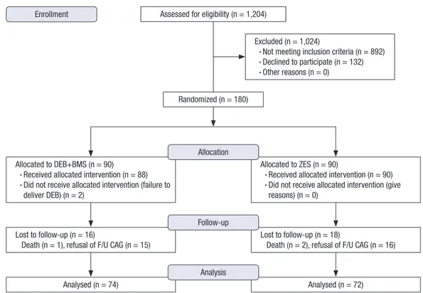
clinicaltrials.gov (NCT01539603), and a brief flowchart of the study is summarized in Fig. 1. We designed this trial in 2010. We enrolled the first patient in April, 2011 and the final patient in September, 2013.
Endpoints
The primary endpoint of the study is the in-segment late loss (LL) at 9 months measured by quantitative coronary angiogra- phy (QCA). The secondary endpoints include angiographic find- ings such as angiographic success, device success, binary angi- ographic restenosis, and clinical outcomes such as procedural success, death of all causes, myocardial infarction, target vessel revascularization, target lesion revascularization, and stent throm- bosis.
Patient population
Patients of at least 18 years of age, who had stable angina or acute coronary syndrome (unstable angina or non-ST segment eleva- tion myocardial infarction [NSTEMI]) of documented ischemia due to a significant lesion in a native coronary artery, were in- cluded in this study. Patients were eligible for inclusion if the native coronary lesion was greater than 50% diameter stenosis by visual estimation of the coronary angiogram with reference diameter between 2.5 mm and 4.0 mm and lesion length less than 28.0 mm. The following conditions were excluded from the study: ST-segment elevation myocardial infarction, unpro- tected left main lesion, ISR, intended bifurcation stenting, car- diogenic shock, chronic total occlusions, history of cerebrovas- cular accident or myocardial infarction within 1 year, and preg- nancy. If all the inclusion criteria were met and none of the ex- clusion criteria applied, the patients were asked for their written informed consent, as required by the Institutional Review Board in accordance with the Declaration of Helsinki.
Randomization and interventions
After enrollment, randomization was performed based on a single sequence of random assignments. Computer-generated random numbers were used for the sequence. The random ta- ble was concealed and independently managed at the Seoul National University Bundang Hospital Cardiovascular Research Center.
Fig. 1. The flowchart of the trial.
DEB = drug-eluting balloon, BMS = bare metal stent, ZES = zotarolimus-eluting stent, F/U CAG = follow-up coronary angiography.
Assessed for eligibility (n = 1,204) Enrollment
Excluded (n = 1,024)
• Not meeting inclusion criteria (n = 892) • Declined to participate (n = 132) • Other reasons (n = 0)
Randomized (n = 180)
Lost to follow-up (n = 16)
Death (n = 1), refusal of F/U CAG (n = 15)
Lost to follow-up (n = 18)
Death (n = 2), refusal of F/U CAG (n = 16) Follow-up
Analysed (n = 74) Analysed (n = 72)
Analysis Allocated to DEB+BMS (n = 90)
• Received allocated intervention (n = 88) • Did not receive allocated intervention (failure to
deliver DEB) (n = 2)
Allocated to ZES (n = 90)
• Received allocated intervention (n = 90) • Did not receive allocated intervention (give
reasons) (n = 0) Allocation
Index PCI
All patients received 300 mg aspirin and a loading dose of 300–
600 mg clopidogrel before the procedure, unless the patient had been taking these medications for at least 1 week prior to the procedure. Heparin was administered intravenously in boluses to maintain an activated clotting time of > 250 seconds during the procedure. Administration of glycoprotein IIb/IIIa inhibi- tors was left to the physician’s discretion. PCI was performed according to the current international guidelines (15). After ob- taining the coronary angiograms, we underwent adequate pre- dilatation of the target lesion with a plain balloon over nominal pressure. After we obtained appropriate results by the plain bal- loon angioplasty, we treated the full length of the lesion using a DEB over nominal pressure (mean ± standard deviation [SD]:
8.7 ± 3.2 atm) at least for 30 seconds (mean ± SD: 46.3 ± 14.2 seconds). Type A–D dissections occasionally occurred after the plain balloon angioplasty or DEB within the lesion but they were never extended over the DEB-treated segment. Further, we im- planted a BMS (shorter by 5 mm compared with the DEB) with- in the treated lesion in the DEB + BMS group. We implanted a ZES to encompass the full length of the lesion in the ZES group.
Therefore, the length of DEB and ZES was intended to match the lesion length, whereas the length of BMS was shorter than that of DEB to minimize geographic mismatch, and potentially optimize drug delivery to the edge of the BMS. After we implant any type of stent, we applied an adjunctive balloon at high pres- sure to minimize residual stenosis.
QCA
The coronary angiograms recorded at baseline and at the 9-month follow-up were analyzed by an independent person who was blinded to the treatment group using an automated edge detec- tion system (CASS 5.7.1; Pie Medical Imaging Systems, Maas- tricht, Netherlands). In each patient, QCA measures within the stent or the segments (including the stented region and the 5 mm edge regions) were analyzed and reported separately. LL was defined as the difference between the minimum lumen di- ameter immediately post-procedure and at 9-month follow-up, respectively. Binary restenosis was defined as > 50% diameter stenosis.
Intravascular ultrasound (IVUS)
An IVUS was recommended to all patients enrolled in the study.
We performed an IVUS before DEB or ZES deployment to as- sess the optimal size of the balloon or stent at index procedure.
IVUS imaging was performed with a 20 MHz 2.9 F, phased-ar- ray IVUS catheter (Eagle Eye; Volcano Therapeutics, Rancho Cordova, CA, USA) after administering intracoronary nitroglyc- erin (200 mg). IVUS was also performed after obtaining angio- graphically optimal results of the index procedure. If the IVUS indicated that the procedural results were not optimal, it was
left to the operator’s discretion whether to perform further post- dilatation or bailout stenting.
Post PCI medication
All the patients included in this trial were treated according to the current American College of Cardiology/American Heart Association (ACC/AHA) guidelines regarding post-stenting man- agement, which specify treatment with at least 100 mg of aspi- rin daily and 75 mg clopidogrel daily for at least 12 months after PCI (15).
Follow-up
Clinical follow-up was conducted at 1, 3, 9, and 12 months after index PCI. Routine angiographic follow-up at 9 months (per- mitted window period: ± 3 months) was performed. Copies of angiograms were submitted to the angiographic laboratory of the Cardiovascular Research Center, Seoul National University Bundang Hospital.
Sample size
We designed a trial to show that the DEB + BMS strategy would be non-inferior to the ZES one in terms of luminal LL at the 9- month follow-up. To test the hypothesis that the DEB + BMS is non-inferior to ZES and according to previous studies, we have used the SDs for the luminal LL with 0.51 mm in the DEB + BMS group and 0.26 mm in the ZES group (14,16). The non-inferiori- ty margin was defined as a luminal LL of 0.19 mm. Assuming a 2-sided alpha-level of 0.05 and a statistical power of 80%, and an estimated attrition rate of 20% (for the 9-month angiograph- ic follow-up), we would need a total of 180 patients, 90 patients in the DEB + BMS arm and 90 patients in the ZES arm. This num- ber of patients would also have 85% power to detect superiority with difference luminal LL of 0.2 mm between the groups at a 2-sided alpha-level of 0.05.
Statistical analysis
All the primary and secondary endpoints were analyzed on an intention-to-treat basis (the patients were analyzed as part of their assigned treatment group) and on per protocol basis (the patients were analyzed as part of their assigned treatment group only if they received their assigned treatment).
The baseline characteristics of the studied patients were sum- marized in terms of frequencies and percentages for categorical variables and in terms of means with SDs for continuous vari- ables. The categorical variables were compared using the Fish- er’s exact test. The continuous variables were compared using the independent 2-sample t-test. A P value of 0.05 was consid- ered as the level of statistical significance for all the tests.
Ethics statement
This study has been approved and monitored by the Institution-
al Review Board of Seoul National University Bundang Hospital (IRB No. E-1104/061-001). The written informed consent was obtained for all the subjects (Clinical trials registry, Clinical Tri- als.gov; Identifier, NCT01539603).
RESULTS Patients
Ninety patients were randomized to treatment with DEB + BMS and 90 patients to the ZES implantation alone. The baseline clin- ical characteristics of all patients were similar in the 2 groups except the presence of hypertension, which was higher in the ZES group (Table 1). There was relatively higher drop-out rate for the 9-month follow-up angiography. However, baseline char- acteristics of the patients with follow-up angiography were also similar in the 2 groups except the presence of hypertension and current smoker.
Baseline lesion and procedural characteristics
Before the index procedure, the minimal lumen diameter, ref- erence diameter, percentage diameter stenosis, and lesion length did not differ between the 2 groups (Table 2). Furthermore, fol-
lowing the index PCI, the size and length of DEB and ZES were not different between the 2 groups. In the patients randomized to treatment with DEB + BMS, the length of the BMS was 17.1 mm, on average 5.4 mm shorter than the length of deployed DEB (22.3 mm). We failed to deliver a DEB in 2 patients due to calcification and severe tortuosity of the lesion. In those cases, we inserted a ZES. Moreover, in all the cases the length of the segments treated with paclitaxel-coated balloon catheters ex- ceeded the proximal and distal end of the BMS. The results evi- denced a significant decrease in the minimal luminal diameter and less acute gain in the patients treated with DEB + BMS com- pared with those treated with ZES. The maximal pressure at stent deployment was significantly higher in the DEB + BMS group, because the nominal pressure was 10 atm for the Coroflex® Blue stent (B. Braun), compared with 9 atm for the Resolute Integri- tyTM stent (Medtronic). Further, the maximal pressure at the ad- junctive balloon was not different between the 2 groups.
QCA
Seventeen DEB + BMS and 16 ZES clinically asymptomatic pa- tients refused the angiographic follow-up. One patient in the DEB + BMS group and 2 patients in the ZES group died before Table 1. Baseline patient characteristics
Characteristics Total patients (n = 180)
DEB + BMS (n = 90)
ZES
(n = 90) P value Patients with F/U angiography
(n = 146)
DEB + BMS (n = 74)
ZES
(n = 72) P value
Age, yr 61.8 ± 11.5 61.2 ± 11.1 62.4 ± 11.9 0.457 61.4 ± 11.3 61.3 ± 10.8 61.5 ± 11.9 0.920
Man 131 (72.8) 68 (75.6) 63 (70.0) 0.503 106 (72.6) 57 (77.0) 49 (68.1) 0.267
BMI, kg/m2 25.6 ± 3.1 25.6 ± 3.1 25.7 ± 3.2 0.805 25.6 ± 3.1 25.7 ± 3.1 25.6 ± 3.2 0.773
Diabetes 54 (30.0) 28 (31.1) 26 (28.9) 0.871 40 (27.4) 21 (28.4) 19 (26.4) 0.854
Hypertension 65 (36.1) 25 (27.8) 40 (44.4) 0.029 92 (63.0) 54 (73.0) 38 (52.8) 0.016
Dyslipidemia 33 (18.3) 15 (16.7) 18 (20.0) 0.700 28 (19.2) 15 (20.3) 13 (18.1) 0.834
Current smoker 46 (25.6) 27 (30.0) 19 (21.1) 0.211 38 (26.0) 25 (33.8) 13 (18.1) 0.044
Family history of CAD 8 (5.6) 3 (3.3) 5 (5.6) 0.720 6 (4.1) 3 (4.1) 3 (4.2) 1.000
Previous MI 7 (3.9) 3 (3.3) 4 (4.4) 1.000 5 (3.4) 3 (4.1) 2 (2.8) 1.000
Previous PCI 14 (7.8) 5 (5.6) 9 (10.0) 0.405 9 (6.2) 5 (6.8) 4 (5.6) 1.000
Previous CVD 5 (2.8) 4 (4.4) 1 (1.1) 0.368 4 (2.7) 3 (4.1) 1 (1.4) 0.620
Multivessel disease 100 (55.6) 45 (50) 55 (61.1) 0.324 39 (26.7) 38 (51.4) 42 (58.3) 0.498
Clinical indication 0.216 0.362
Stable angina 85 (47.2) 42 (46.7) 43 (47.8) 66 (45.2) 32 (43.2) 34 (47.2)
Unstable angina 48 (26.7) 20 (22.2) 28 (31.1) 40 (27.4) 18 (24.3) 22 (30.6)
NSTEMI 47 (26.1) 28 (31.1) 19 (21.1) 40 (27.4) 24 (32.4) 16 (22.2)
Medication at discharge
Aspirin 179 (99.4) 89 (98.9) 90 (100.0) 1.000 146 (100.0) 74 (100.0) 72 (100.0) NA
Clopidogrel 178 (98.9) 88 (97.8) 90 (100.0) 0.497 144 (98.6) 72 (97.3) 72 (100.0) 0.497
Other antiplatelet agent 9 (5.0) 7 (7.8) 2 (2.2) 0.169 8 (5.5) 6 (8.1) 2 (2.8) 0.276
Statin 152 (84.4) 72 (80.0) 80 (88.9) 0.149 124 (84.9) 60 (81.1) 64 (88.9) 0.248
ACE inhibitor 66 (36.7) 34 (37.8) 32 (35.6) 0.877 56 (38.4) 29 (39.2) 27 (37.5) 0.866
ARB 58 (32.2) 34 (37.8) 24 (26.7) 0.151 42 (28.8) 26 (35.1) 16 (22.2) 0.101
Beta-blocker 121 (67.2) 64 (71.1) 57 (63.3) 0.341 94 (64.4) 50 (67.6) 44 (61.1) 0.490
Calcium channel blocker 55 (30.6) 26 (28.9) 29 (32.2) 0.746 43 (29.5) 23 (31.1) 20 (27.8) 0.718
Values are mean ± SD or number (%). P value was calculated using Pearson χ2 for categorical variables and Student t-test for continuous variables.
DEB = drug-eluting balloon, BMS = bare metal stent, ZES = zotarolimus-eluting stent, F/U = follow-up, BMI = body mass index, CAD = coronary artery diseases, MI = myo- car dial infarction, PCI = percutaneous coronary intervention, CVD = cerebrovascular disease, NSTEMI = non-ST segment elevation myocardial infarction, NA = not available, ACE = angiotensin-converting enzyme, ARB = angiotensin II receptor blocker, SD = standard deviation.
the angiographic follow-up. The angiographic follow-up rate was 81.1%, and was obtained at 298 days after PCI.
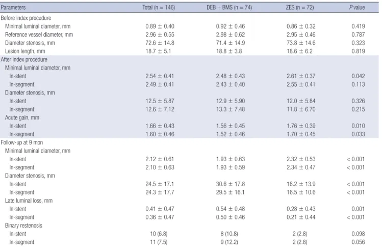
Before the index procedure, the minimal lumen diameter, reference diameter, percentage diameter stenosis, and lesion length did not differ between the 2 groups (Table 3, Fig. 2A). Af- ter the index PCI, there was significantly decreased minimal lu- minal diameter and less acute gain in the patients treated with DEB + BMS compared with those treated with ZES. The LL, thus the primary endpoint, was significantly higher in the lesions treat- ed with DEB + BMS than in those treated with ZES (0.50 ± 0.46 mm vs. 0.21 ± 0.44 mm; P < 0.001) (Fig. 2B). The mean differ- ence of the LL was 0.31 mm and 2-sided 95% confidence inter- val, 0.15–0.48 (Fig. 3), which was higher than the prespecified non-inferiority margin (P for non-inferiority = 0.138). The bina- ry restenosis rate was also higher in the DEB + BMS group alth- ough the result was not statistically significant.
Subgroup analysis
There was only one patient with chronic kidney disease (CKD) defined by serum creatinine over 1.5 mg/dL. Therefore, we did not perform any subgroup analysis of CKD. Instead, we analyzed the effect of hypertension because the proportion of hyperten-
sion was significantly different between the 2 groups. The sub- group analysis of old age, diabetes mellitus, hypertension, and multivessel stenting demonstrated that the inferiority observed when comparing the DEB + BMS groups with the ZES group was evidenced in all the subgroups as well (Fig. 3, Table 4).
Clinical follow-up
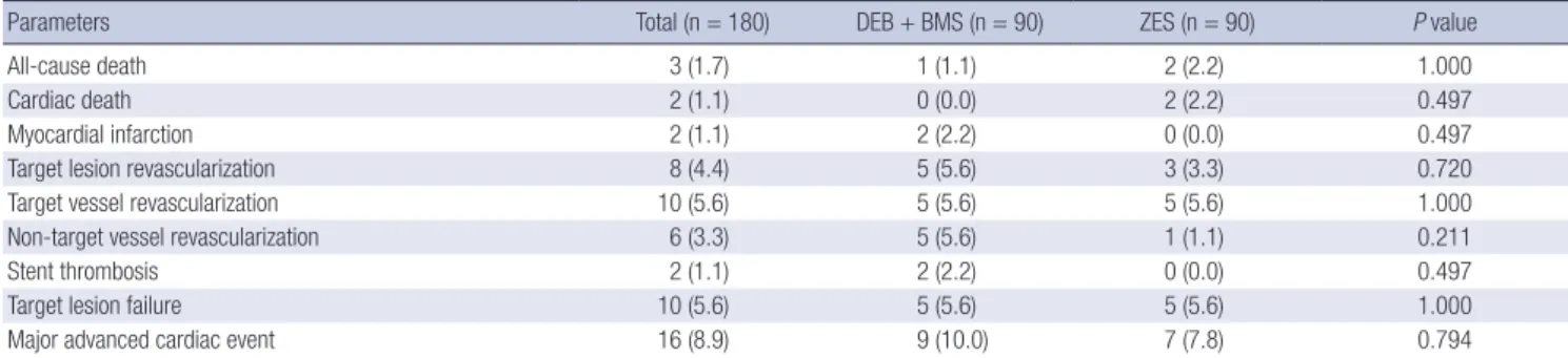
There was no clinical follow-up loss at 12 months (Table 5). Death occurred in one patient (1.1%, non-cardiac; traffic accident) from the DEB + BMS group and in 2 (2.2%, cardiac; both unexplained) from the ZES group. There were 2 myocardial infarctions in the DEB + BMS group. Both cases occurred at admission for the non- cardiac surgery department, and were associated with definite stent thrombosis during cessation of antiplatelet agents. Repeat revascularizations were performed as follows: 1) target lesion revascularization, 5/90 patients (5.6%, DEB + BMS group) and 3/90 patients (3.3%, ZES group); and 2) target vessel revascular- ization, 5/90 patients (5.6%, DEB + BMS group) and 5/90 pati- ents (5.6%, ZES group). The major adverse cardiac event was defined by the target lesion revascularization, myocardial in- farction was attributed to the target vessel, and cardiac death rate was not different when comparing the 2 groups. This study Table 2. Baseline lesion and procedural characteristics
Characteristics Total (n = 180) DEB + BMS (n = 90) ZES (n = 90) P value
Before index procedure
Lesion location 0.720
LAD 79 (43.9) 37 (41.1) 42 (46.7)
LCX 51 (28.3) 26 (28.9) 25 (27.8)
RCA 50 (27.8) 27 (30.0) 23 (25.6)
Multivessel intervention 48 (26.7) 23 (25.6) 25 (27.8) 0.866
ACC/AHA B2 or C type 142 (78.8) 69 (76.7) 73 (81.1) 0.884
Minimal luminal diameter, mm 0.89 ± 0.39 0.89 ± 0.45 0.89 ± 0.32 0.926
Reference vessel diameter, mm 2.96 ± 0.52 2.97 ± 0.59 2.96 ± 0.45 0.943
Diameter stenosis, mm 73.0 ± 14.6 72.9 ± 15.1 73.1 ± 14.1 0.951
Lesion length, mm 18.9 ± 5.5 18.9 ± 3.7 18.8 ± 6.9 0.948
After index procedure
No. of stents per patient 1.32 1.31 1.34 0.934
Stent length per lesion, mm 19.7 ± 6.0 17.1 ± 4.3 22.2 ± 6.4 < 0.001
Stent diameter, mm 3.06 ± 0.41 3.06 ± 0.43 3.06 ± 0.38 0.891
Maximal pressure at stent deployment, atm 10.4 ± 1.9 11.3 ± 1.7 9.7 ± 1.8 < 0.001
Maximal pressure at adjunctive balloon, atm 14.0 ± 4.0 13.9 ± 4.7 14.3 ± 3.3 0.600
DEB diameter, mm - 3.00 ± 0.38 - -
DEB length, mm - 22.3 ± 4.7 - -
Procedural time, min 56.9 ± 20.1 58.7 ± 23.1 55.2 ± 18.5 0.263
Contrast dye 175.0 ± 62.1 176.2 ± 65.7 173.8 ± 58.6 0.797
Minimal luminal diameter (In-stent), mm 2.55 ± 0.40 2.48 ± 0.42 2.61 ± 0.37 0.022
Minimal luminal diameter (In-segment), mm 2.51 ± 0.41 2.45 ± 0.41 2.57 ± 0.40 0.063
Diameter stenosis (In-stent), mm 12.70 ± 6.00 13.00 ± 5.99 12.40 ± 6.00 0.467
Diameter stenosis (In-segment), mm 12.8 ± 7.1 13.4 ± 7.2 12.1 ± 6.9 0.247
Lesion success 180 (100.0) 90 (100.0) 90 (100.0) -
Device success 178 (98.9) 88 (97.8) 90 (100.0) 0.497
Procedural success 180 (100.0) 89 (100.0) 90 (100.0) 1.000
Values are mean ± SD or number (%).
DEB = drug-eluting balloon, BMS = bare metal stent, ZES = zotarolimus-eluting stent, LAD = left anterior descending, LCX = left circumflex, RCA = right coronary, ACC/AHA = American College of Cardiology/American Heart Association, SD = standard deviation.
was not powered to detect differences in clinical endpoints.
DISCUSSION
The DEB + BMS strategy did not increase the procedure time or the amount of contrast dye, whereas 2/90 patients failed to ob- tain procedural success because of decreased crossability over tortuous lesions. As we intended, the DEB + BMS strategy re-
sulted in using DEB and ZES of same length and shorter BMS (BMS < ZES). Unexpectedly, the DEB + BMS strategy led to a decreased post-PCI minimal lesion diameter and less acute gain when compared with the ZES strategy, despite similar maximal pressure at adjunctive ballooning. The 9-month follow-up angi- ography revealed an inferior LL in the DEB + BMS group when compared with the ZES group. Moreover, the result of death, myocardial infarction, target lesion revascularization, target Table 3. QCA analysis (per-patient analysis, index lesion)
Parameters Total (n = 146) DEB + BMS (n = 74) ZES (n = 72) P value
Before index procedure
Minimal luminal diameter, mm 0.89 ± 0.40 0.92 ± 0.46 0.86 ± 0.32 0.419
Reference vessel diameter, mm 2.96 ± 0.55 2.98 ± 0.62 2.95 ± 0.46 0.787
Diameter stenosis, mm 72.6 ± 14.8 71.4 ± 14.9 73.8 ± 14.6 0.323
Lesion length, mm 18.7 ± 5.1 18.8 ± 3.8 18.6 ± 6.2 0.819
After index procedure Minimal luminal diameter, mm
In-stent 2.54 ± 0.41 2.48 ± 0.43 2.61 ± 0.37 0.042
In-segment 2.49 ± 0.41 2.43 ± 0.40 2.55 ± 0.41 0.113
Diameter stenosis, mm
In-stent 12.5 ± 5.87 12.9 ± 5.90 12.0 ± 5.84 0.326
In-segment 12.6 ± 7.12 13.3 ± 7.48 11.8 ± 6.70 0.215
Acute gain, mm
In-stent 1.66 ± 0.43 1.56 ± 0.45 1.76 ± 0.39 0.010
In-segment 1.60 ± 0.46 1.52 ± 0.46 1.70 ± 0.45 0.033
Follow-up at 9 mon
Minimal luminal diameter, mm
In-stent 2.12 ± 0.61 1.93 ± 0.63 2.32 ± 0.53 < 0.001
In-segment 2.10 ± 0.63 1.93 ± 0.59 2.34 ± 0.47 < 0.001
Diameter stenosis, mm
In-stent 24.5 ± 17.1 30.6 ± 17.8 18.2 ± 13.9 < 0.001
In-segment 24.3 ± 17.7 29.5 ± 16.1 16.5 ± 10.6 < 0.001
Late luminal loss, mm
In-stent 0.41 ± 0.47 0.54 ± 0.48 0.28 ± 0.43 0.001
In-segment 0.36 ± 0.47 0.50 ± 0.46 0.21 ± 0.44 < 0.001
Binary restenosis
In-stent 10 (6.8) 8 (10.8) 2 (2.8) 0.098
In-segment 11 (7.5) 9 (12.2) 2 (2.8) 0.056
Values are mean ± SD or number (%).
QCA = quantitative coronary angiography, DEB = drug-eluting balloon, BMS = bare metal stent, ZES = zotarolimus-eluting stent, SD = standard deviation.
Fig. 2. Cumulative frequency plot for minimal LL and late lumen loss. (A) Cumulative frequency plot for minimal lumen diameter before and after index procedure. (B) Cumula- tive frequency plot for late lumen loss before and after index procedure.
LL = late loss, DES = drug-eluting stent, DEB = drug-eluting balloon, BMS = bare metal stent.
(%)
Minimal lumen diameter (mm)
0.5 1.0 1.5 2.0 2.5 3.0 3.5 4.0 4.5 100
80 60 40 20 0
Baseline After treatment
DES
DEB + BMS (%)
Late loss (mm)
–1 –0.5 0 0.5 1.0 1.5 2.0 2.5 100
80 60 40 20 0
DES DEB + BMS
A B
Fig. 3. The mean difference of in-stent LL and 95% CI and its subgroup analysis. The broken line denotes the prespecified non-inferiority margin (0.19 mm).
LL = late loss, CI = confidence interval.
In-segment late loss
0 0.5 1.0
Old age ( ≥ 70) Yes No
Diabetes mellitus Yes
No Hypertension Yes No
Multivessel stenting Yes
No All patients
Table 4. Subgroup analysis of in-segment LL
Subgroups DEB + BMS ZES P value P for
interaction
Old age ( ≥ 70) 0.411
Yes 0.54 ± 0.31 0.11 ± 0.39 0.001
No 0.55 ± 0.55 0.27 ± 0.52 0.010
Diabetes mellitus 0.543
Yes 0.57 ± 0.42 0.17 ± 0.55 0.013
No 0.53 ± 0.55 0.25 ± 0.47 0.005
Hypertension 0.600
Yes 0.54 ± 0.45 0.19 ± 0.37 < 0.001
No 0.54 ± 0.66 0.28 ± 0.60 0.142
Multivessel stenting 0.077
Yes 0.74 ± 0.58 0.18 ± 0.57 0.005
No 0.47 ± 0.47 0.25 ± 0.47 0.014
Values are mean ± SD or number (%).
LL = late loss, DEB = drug-eluting balloon, BMS = bare metal stent, ZES = zotarolimus- eluting stent, SD = standard deviation.
Table 5. Cumulative incidence of clinical events up to 1 year (intention-to-treat per patient)
Parameters Total (n = 180) DEB + BMS (n = 90) ZES (n = 90) P value
All-cause death 3 (1.7) 1 (1.1) 2 (2.2) 1.000
Cardiac death 2 (1.1) 0 (0.0) 2 (2.2) 0.497
Myocardial infarction 2 (1.1) 2 (2.2) 0 (0.0) 0.497
Target lesion revascularization 8 (4.4) 5 (5.6) 3 (3.3) 0.720
Target vessel revascularization 10 (5.6) 5 (5.6) 5 (5.6) 1.000
Non-target vessel revascularization 6 (3.3) 5 (5.6) 1 (1.1) 0.211
Stent thrombosis 2 (1.1) 2 (2.2) 0 (0.0) 0.497
Target lesion failure 10 (5.6) 5 (5.6) 5 (5.6) 1.000
Major advanced cardiac event 16 (8.9) 9 (10.0) 7 (7.8) 0.794
DEB = drug-eluting balloon, BMS = bare metal stent, ZES = zotarolimus-eluting stent.
vessel revascularization, or stent thrombosis were not different between the 2 groups.
The combination of a paclitaxel-coated balloon plus BMS addresses both issues: 1) a decrease of the neointimal prolifera- tion due to homogenous administration of high concentration paclitaxel to the vessel wall, and 2) a decrease of the risk of stent thrombosis by facilitating a more rapid endothelialization due to the use of a BMS compared to the use of a DES (17). It could reduce the duration of dual antiplatelet therapy (DAPT) use in patients with high risks of bleeding. In the follow-up of the Pa- clitaxel-Eluting Percutaneous Transluminal Coronary Angio- plasty (PTCA)-Balloon Catheter to Treat Small Vessel Coronary Artery Disease (PEPCAD) I study, all the patients received at least 100 mg aspirin daily. Clopidogrel (75 mg/day) was given for one month following stand-alone DEB angioplasty, and for 3 months after additional BMS implantation. In the PEPCAD II study, all the patients received at least 100 mg of aspirin daily lifelong. Clopidogrel (75 mg/day) was given for 3 months after
DEB angioplasty and for 6 months after DES implantation. There was no late thrombosis within the 6-month follow-up. The pro- tocols of DEB studies suggest that the dual antiplatelet therapy with aspirin and clopidogrel of 4 weeks after DEB is safe and ef- fective (18).
The DEB with BMS protocol also allows for a longer paclitax- el-coated balloon to be used instead of the stented segment one.
This may be favorable since about one-third of restenosis after DES implantation occurs proximal or distal to the stent margin (17,19). To optimize the rate of drug delivery in the present study, we used a longer DEB (DEB > BMS), and estimated the diame- ter of DEB and ZES based on the IVUS findings. There were 11 binary restenoses (9 in DEB + BMS group and 2 in ZES group) as shown in Table 3. ISR pattern of DEB + BMS group included 2IB, 4IC, 1ID, 1II, and 1III (5 in-stent and 4 involving stent edge) according to Mehran ISR classification (20). ISR pattern of ZES group were 1IC and 1II (both, in-stent). Therefore, ZES showed no edge restenosis while longer DEB + short BMS did not pre- vent edge restenosis in the present study. However, the num- bers of patients and ISR occurrence were too small to conclude this issue.
Opposed to our expectation, once more the DEB + BMS strat-
egy failed to prove its non-inferiority to DES alone strategy. More- over, DES alone therapy was superior to the DEB + BMS thera- py. The design of the combination treatment (premouted vs.
sequential) or the sequence of DEB and BMS application did not seem to affect the inhibition of restenosis (21,22).
The reasons why the DEB + BMS was inferior to DES alone can be explained by the following issues. First, the efficacy of paclitaxel in inhibiting restenosis may be inherently inferior to not only sirolimus but also to zotarolimus. The paclitaxel-elut- ing stent was inferior to sirolimus-eluting stent as well as to ZES (23,24). Therefore, in the present study the paclitaxel-coated bal- loon may not inhibit neointimal hyperplasia as efficient as the sustained released of the ZES.
Second, the difference between these 2 strategies may be the drug-release kinetics. As the DEB used in the present study is known to release the coated drug within 1 month, which is mark- edly shorter than the ZES, it would influence the performance of DEB + BMS implantation strategy. Third, the DEB + BMS ther- apy resulted in a significantly decreased post-PCI minimal lu- minal diameter and less acute gain compared with the ZES ther- apy. We cannot demonstrate the mechanism of the inferior acute gain in the DEB + BMS arm. We applied the predilation with sim- ilar diameter balloons at similar pressure. The diameter of the implanted stents and the maximal pressure of the adjunctive ballooning following stent implantation were not different when comparing the 2 arms. Although the procedure methods were similar, BMS and ZES have different stent platform, which pro- vides different radial strength and conformability to the vessel wall. In the present study, the BMS had thinner strut compared with the ZES. It might result in less acute gain in DEB + BMS group.
However, it was reported that the stent with thinner struts show- ed a comparable acute gain while it elicited less angiographic and clinical restenosis compared with the thicker-strut stent (14,25). Nevertheless, in the present study, the less acute gain may partly influence the negative outcome since it is known that the late lumen diameter, late percent stenosis, and binary reste- nosis depend on the immediate lumen diameter following the procedure and the immediate residual percent stenosis, but not on the specific intervention (26).
Finally, Shin et al. (27) presented the similar efficacy and safe- ty of DEB vs. second generation DES in treatment of de novo cor- onary stenosis even without additional BMS implantation. They emphasized the perfect lesion preparation with predilation to obtain fractional flow reserve (FFR) > 0.85. In this regard, our strategy might not be sufficient to obtain a proper lesion prepa- ration for the best efficacy of the following DEB because we used gentle predilation to minimize serious dissection.
We used simple randomization based on a single sequence of random number table. Because the present study is a rela- tively small sample size clinical trial, this randomization meth- od could be problematic, resulting in an unequal number of
participants with hypertension and current smoking between the groups. The rate of drop-out was relatively high in the pres- ent study. This may be a critical methodological issue. Because we designed this study in 2010, we performed PCI to guidelines at that time. However, in more recent, e.g. focused update from 2014 or the European guidelines from 2014, the importance of physiologic assessment of coronary lesions is highlighted. There- fore, it is possible that we overestimated lesion severity on lesions in the present study. The study was not powered to detect dif- ferences in clinical endpoints.
In conclusion, the DEB + BMS strategy is inferior to the ZES one in terms of the LL at 9 months, although we do not confirm any increase in the death rate or MI when comparing the pres- ent results with those of the previous study.
DISCLOSURE
The authors have no potential conflicts of interest to disclose.
AUTHOR CONTRIBUTION
Conceptualization: Chae IH, Yoon CH. Data curation: Yoon CH.
Formal analysis: Yoon CH, Park JJ, Oh IY. Investigation: Chae IH, Yoon CH, Oh IY, Suh JW, Cho YS, Youn TJ, Choi DJ. Writing - orig- inal draft: Yoon CH. Writing - review & editing: Chae IH, Park JJ.
ORCID
In-Ho Chae http://orcid.org/0000-0003-1644-2105 Chang-Hwan Yoon http://orcid.org/0000-0001-6305-4442 Jin Joo Park http://orcid.org/0000-0001-9611-1490 Il-Young Oh http://orcid.org/0000-0002-5584-605X Jung-Won Suh http://orcid.org/0000-0002-0397-6071 Young-Seok Cho http://orcid.org/0000-0001-9944-9868 Tae-Jin Youn http://orcid.org/0000-0002-4628-9503 Dong-Ju Choi http://orcid.org/0000-0003-0146-2189
REFERENCES
1. Elezi S, Kastrati A, Neumann FJ, Hadamitzky M, Dirschinger J, Schömig A.
Vessel size and long-term outcome after coronary stent placement. Cir- culation 1998; 98: 1875-80.
2. Hoffmann R, Mintz GS, Dussaillant GR, Popma JJ, Pichard AD, Satler LF, Kent KM, Griffin J, Leon MB. Patterns and mechanisms of in-stent reste- nosis. A serial intravascular ultrasound study. Circulation 1996; 94: 1247- 54.
3. Stettler C, Wandel S, Allemann S, Kastrati A, Morice MC, Schömig A, Pfis- terer ME, Stone GW, Leon MB, de Lezo JS, et al. Outcomes associated with drug-eluting and bare-metal stents: a collaborative network meta-analy- sis. Lancet 2007; 370: 937-48.
4. Nakazawa G, Finn AV, Joner M, Ladich E, Kutys R, Mont EK, Gold HK, Burke AP, Kolodgie FD, Virmani R. Delayed arterial healing and increased late
stent thrombosis at culprit sites after drug-eluting stent placement for acute myocardial infarction patients: an autopsy study. Circulation 2008; 118:
1138-45.
5. Joner M, Finn AV, Farb A, Mont EK, Kolodgie FD, Ladich E, Kutys R, Skori- ja K, Gold HK, Virmani R. Pathology of drug-eluting stents in humans:
delayed healing and late thrombotic risk. J Am Coll Cardiol 2006; 48: 193- 202.
6. Finn AV, Nakazawa G, Joner M, Kolodgie FD, Mont EK, Gold HK, Virmani R. Vascular responses to drug eluting stents: importance of delayed heal- ing. Arterioscler Thromb Vasc Biol 2007; 27: 1500-10.
7. Kuriyama N, Kobayashi Y, Nakama T, Mine D, Nishihira K, Shimomura M, Nomura K, Ashikaga K, Matsuyama A, Shibata Y. Late restenosis follow- ing sirolimus-eluting stent implantation. JACC Cardiovasc Interv 2011; 4:
123-8.
8. Kang SJ, Mintz GS, Akasaka T, Park DW, Lee JY, Kim WJ, Lee SW, Kim YH, Whan Lee C, Park SW, et al. Optical coherence tomographic analysis of in-stent neoatherosclerosis after drug-eluting stent implantation. Circu- lation 2011; 123: 2954-63.
9. De Labriolle A, Pakala R, Bonello L, Lemesle G, Scheinowitz M, Waksman R. Paclitaxel-eluting balloon: from bench to bed. Catheter Cardiovasc In- terv 2009; 73: 643-52.
10. Scheller B, Hehrlein C, Bocksch W, Rutsch W, Haghi D, Dietz U, Böhm M, Speck U. Treatment of coronary in-stent restenosis with a paclitaxel-coat- ed balloon catheter. N Engl J Med 2006; 355: 2113-24.
11. Unverdorben M, Vallbracht C, Cremers B, Heuer H, Hengstenberg C, Mai- kowski C, Werner GS, Antoni D, Kleber FX, Bocksch W, et al. Paclitaxel- coated balloon catheter versus paclitaxel-coated stent for the treatment of coronary in-stent restenosis. Circulation 2009; 119: 2986-94.
12. Unverdorben M, Kleber FX, Heuer H, Figulla HR, Vallbracht C, Leschke M, Cremers B, Hardt S, Buerke M, Ackermann H, et al. Treatment of small coronary arteries with a paclitaxel-coated balloon catheter. Clin Res Car- diol 2010; 99: 165-74.
13. Mathey DG, Wendig I, Boxberger M, Bonaventura K, Kleber FX. Treatment of bifurcation lesions with a drug-eluting balloon: the PEPCAD V (Pacli- taxel Eluting PTCA Balloon in Coronary Artery Disease) trial. EuroInter- vention 2011; 7 Suppl K: K61-5.
14. Pöss J, Jacobshagen C, Ukena C, Böhm M. Hotlines and clinical trial up- dates presented at the German Cardiac Society Meeting 2010: FAIR-HF, CIPAMI, LIPSIA-NSTEMI, Handheld-BNP, PEPCAD III, remote ischaemic conditioning, CERTIFY, PreSCD-II, German Myocardial Infarction Regis- try, DiaRegis. Clin Res Cardiol 2010; 99: 411-7.
15. Levine GN, Bates ER, Blankenship JC, Bailey SR, Bittl JA, Cercek B, Cham- bers CE, Ellis SG, Guyton RA, Hollenberg SM, et al. 2011 ACCF/AHA/SCAI Guideline for Percutaneous Coronary Intervention. A report of the Ameri- can College of Cardiology Foundation/American Heart Association Task Force on Practice Guidelines and the Society for Cardiovascular Angiog- raphy and Interventions. J Am Coll Cardiol 2011; 58: e44-122.
16. Meredith IT, Worthley S, Whitbourn R, Walters D, Popma J, Cutlip D, Fitz- gerald P. The next-generation Endeavor Resolute stent: 4-month clinical and angiographic results from the Endeavor Resolute first-in-man trial.
EuroIntervention 2007; 3: 50-3.
17. Wöhrle J, Birkemeyer R, Markovic S, Nguyen TV, Sinha A, Miljak T, Spiess J, Rottbauer W, Rittger H. Prospective randomised trial evaluating a pacli- taxel-coated balloon in patients treated with endothelial progenitor cell capturing stents for de novo coronary artery disease. Heart 2011; 97: 1338- 42.
18. Bonaventura K, Sonntag S, Kleber FX. Antiplatelet therapy in the era of percutaneous coronary intervention with drug-eluting balloons. EuroIn- tervention 2011; 7 Suppl K: K106-11.
19. Stone GW, Ellis SG, Cannon L, Mann JT, Greenberg JD, Spriggs D, O’Shau- ghnessy CD, DeMaio S, Hall P, Popma JJ, et al. Comparison of a polymer- based paclitaxel-eluting stent with a bare metal stent in patients with com- plex coronary artery disease: a randomized controlled trial. JAMA 2005;
294: 1215-23.
20. Mehran R, Dangas G, Abizaid AS, Mintz GS, Lansky AJ, Satler LF, Pichard AD, Kent KM, Stone GW, Leon MB. Angiographic patterns of in-stent re- stenosis: classification and implications for long-term outcome. Circula- tion 1999; 100: 1872-8.
21. Gutiérrez-Chico JL, van Geuns RJ, Koch KT, Koolen JJ, Duckers H, Regar E, Serruys PW. Paclitaxel-coated balloon in combination with bare metal stent for treatment of de novo coronary lesions: an optical coherence to- mography first-in-human randomised trial, balloon first vs. stent first. Eu- roIntervention 2011; 7: 711-22.
22. Fischer D, Scheller B, Schäfer A, Klein G, Böhm M, Clever Y, Cremers B.
Paclitaxcel-coated balloon plus bare metal stent vs. sirolimus-eluting stent in de novo lesions: an IVUS study. EuroIntervention 2012; 8: 450-5.
23. Windecker S, Remondino A, Eberli FR, Jüni P, Räber L, Wenaweser P, Togni M, Billinger M, Tüller D, Seiler C, et al. Sirolimus-eluting and paclitaxel- eluting stents for coronary revascularization. N Engl J Med 2005; 353: 653- 62.
24. Xu B, Yang Y, Yuan Z, Du Z, Wong SC, Généreux P, Lu S; RESOLUTE Chi- na RCT Investigators. Zotarolimus- and paclitaxel-eluting stents in an all- comer population in China: the RESOLUTE China randomized controlled trial. JACC Cardiovasc Interv 2013; 6: 664-70.
25. Pache J, Kastrati A, Mehilli J, Schühlen H, Dotzer F, Hausleiter J, Flecken- stein M, Neumann FJ, Sattelberger U, Schmitt C, et al. Intracoronary stent- ing and angiographic results: strut thickness effect on restenosis outcome (ISAR-STEREO-2) trial. J Am Coll Cardiol 2003; 41: 1283-8.
26. Kuntz RE, Gibson CM, Nobuyoshi M, Baim DS. Generalized model of re- stenosis after conventional balloon angioplasty, stenting and directional atherectomy. J Am Coll Cardiol 1993; 21: 15-25.
27. Shin ES, Ann SH, Balbir Singh G, Lim KH, Kleber FX, Koo BK. Fractional flow reserve-guided paclitaxel-coated balloon treatment for de novo cor- onary lesions. Catheter Cardiovasc Interv 2016; 88: 193-200.