J Korean Soc Radiol 2017;76(2):96-103 https://doi.org/10.3348/jksr.2017.76.2.96
INTRODUCTION
Mammography and breast sonography are currently the pre- ferred modalities for screening and diagnosing breast cancers.
On mammography, breast composition is classified into four types as defined by the Breast Imaging Reporting and Data Sys- tem (BI-RADS) (1). Increased breast composition not only im- pairs the detection of breast lesions, but it also has been consid- ered as a risk factor of breast cancer (2). Breast sonography is useful for evaluation of masses or asymmetries. There are several
studies that investigated the sonographic findings for differentiat- ing benign breast lesions from malignant breast lesions.
With the increasing use of chest computed tomography (CT) in a variety of diagnostic pathways, breast abnormalities are be- ing identified more frequently on chest CT (3). It is important that radiologists should not overlook breast lesions on chest CT and they should be aware of CT appearances of malignant and benign breast lesions (4).
In a previous study, chest CT showed a higher interreader agree- ment for readings of breast composition as compared to mam-
Detection of Breast Abnormalities on Enhanced Chest CT:
Correlation with Breast Composition on Mammography
조영증강 흉부단층촬영검사에서 유방 병변의 발견: 유방 실질의 구성과의 연관성
Eun Mi Cho, MD, Hee Kang, MD*, Young Gyung Shin, MD, Jong Hyouk Yun, MD, Kyung Seung Oh, MD, Sekyoung Park, MD
Department of Radiology, Kosin University Gospel Hospital, Kosin University College of Medicine, Busan, Korea
Purpose: To investigate the capability of enhanced chest computed tomography (CT) for detecting breast abnormalities and to assess the influence of breast compo- sition on this detectability.
Materials and Methods: From 2000 to 2013, 75 patients who underwent mam- mography, breast sonography, and enhanced chest CT within one month and had abnormalities on sonography were included. Detection rate of breast abnormality on enhanced chest CT was compared among 4 types of breast composition by the Breast Imaging Reporting and Data System. Contribution of breast composition, size and enhancement of target lesions to detectability of enhanced chest CT was as- sessed using logistic regression and chi-square test.
Results: Of the 75 target lesions, 34 (45.3%) were detected on enhanced chest CT, corresponding with those on breast sonography; there were no significantly differ- ent detection rates among the 4 types of breast composition (p = 0.078). Breast composition [odds ratio (OR) = 1.07, p = 0.206] and enhancement (OR = 21.49, p = 0.998) had no significant effect, but size (OR = 1.23, p = 0.004) was a significant contributing factor influencing the detectability of enhanced chest CT for breast le- sions.
Conclusion: About half of the cases (45.3%) demonstrated breast lesions on chest CT corresponding with target lesions on sonography. Breast composition defined on mammography did not affect the detectability of enhanced chest CT for breast le- sions.
Index terms Breast Chest
Multidetector Computed Tomography Ultrasound
Mammography
Received May 26, 2016 Revised June 24, 2016 Accepted July 13, 2016
*Corresponding author: Hee Kang, MD Department of Radiology, Kosin University Gospel Hospital, Kosin University College of Medicine, 262 Gamcheon-ro, Seo-gu, Busan 49267, Korea.
Tel. 82-51-990-6341 Fax. 82-51-255-2764 E-mail: [email protected]
This is an Open Access article distributed under the terms of the Creative Commons Attribution Non-Commercial License (http://creativecommons.org/licenses/by-nc/3.0) which permits unrestricted non-commercial use, distri- bution, and reproduction in any medium, provided the original work is properly cited.
mography and breast composition on chest CT might provide ad- ditional information for predicting risk of breast cancers (5).
The purpose of our study is to investigate the capability of en- hanced chest CT for detecting breast abnormalities and to assess the influence of breast composition on this detectability. More- over, we tried to evaluate the CT features helpful in differentiat- ing malignant breast lesions from benign breast lesions.
MATERIALS AND METHODS
The Institutional Review Board of our institution did not re- quire any approval or informed patient consent for this type of retrospective study.
Patient Population
From January 2000 to December 2013, we retrospectively iden- tified 983 patients who underwent mammography, breast sonog- raphy, and enhanced chest CT within one month at a tertiary medical center. Patients with known breast cancer and a history of surgery for breast cancer were excluded. Finally, 75 women (age range, 35–80 years; mean age, 53.6 years), who had abnor- malities on breast sonography, were included in this study.
Clinical indications for enhanced chest CT in these 75 patients included health screening (n = 35), staging work-up for non-pul- monary malignancies (n = 18), various manifestations such as fe- ver, dyspnea, and chest pain (n = 14), abnormality on plain radi- ography (n = 6), and evaluation of pulmonary neoplasms (n = 2).
Imaging Technique
CT scans were acquired using a multi-detector CT system (So- matom Sensation 64 or dual-source Flash 128 multi-detector CT system, Siemens Medical Solutions, Erlangen, Germany). Scan- ning was performed from the lower part of the neck to the level of the middle portion of kidneys. Scanning was performed after IV administration of contrast medium (140 mL Iopamidol, Pamiray 300, Dongkook Pharm., Seoul, Korea) with a power in- jector (Mallinckrodt, Tyco and Vistron CT, Medrad, Arrendale, PA, USA) at an injection rate of 2.5 mL/sec. The scanning pa- rameters were 120 kVp; 90–150 mA; tube rotation time, 0.5 sec- onds; pitch 1.2.
Breast sonography was performed with a sonographic unit (Acuson Antares, Siemens Medical Solutions, Inc., Malvern, PA,
USA) using a standard 5–13 MHz linear array transducer by ra- diologists who had breast sonography experience ranging from 2–3 years (including reader 1–4).
All patients in this study underwent two-view mammography (both craniocaudal and mediolateral oblique views) of both breasts by a mammography unit (Affinity, Lorad/Hologic Com- pany, Danbury, CT, USA) with the film-screen system (Car- estream CR reader, Kodak DirectView, Rochester, NY, USA).
Image Review
All mammography and breast sonography examinations were retrospectively reviewed in consensus by two radiologists (reader 1, 2) who had two and three years of experience. On breast sonography, the reviewers (reader 1, 2) selected the larg- est lesion as the target lesion in each patient and described the largest diameter, location and BI-RADS assessment categories;
category 0 corresponds to an incomplete result with need for additional imaging evaluation; category 1, corresponds to a negative finding; category 2, corresponds to a benign finding;
category 3, corresponds to a probably benign finding; category 4, corresponds to a suspicious result ranging from low suspi- cion to high suspicion for malignancy; category 5, corresponds to a result that is highly suggestive of malignancy; and category 6, corresponds to the result of known biopsy-proven malignan- cy (1). The readers categorized breast composition on mam- mography. According to breast composition descriptors of the BI-RADS 5th edition, type a indicates that the breasts are almost entirely fatty. Types b and c indicate that there are scattered areas of fibroglandular density and breasts are heterogeneously dense, respectively. Type d indicates that breasts are extremely dense (1). Breast composition was also divided into dense (c and d) and non-dense (a and b) types. Moreover, they identified lesions, such as focal asymmetries or masses, and determined detection of breast lesions on mammography, according to whether there were corresponding lesions on breast sonography.
The other two radiologists (reader 3, 4) who had four and eight years of experience interpreted all enhanced chest CT examina- tions in consensus. They reviewed the unenhanced and contrast- enhanced CT scans to determine whether breast lesions, corre- sponding to the target lesions on breast sonography, could be seen. They knew about the presence of breast lesions on sonogra- phy, but they were blinded to their location and size. The readers
selected the largest lesion in each chest CT scan as the target le- sion and described its location and size; the largest diameter on an axial image, shape; oval, round, and irregular, margin; circum- scribed or not circumscribed, and presence or absence of en- hancement; enhancement indicated a more conspicuous lesion on enhanced CT images compared with unenhanced CT images.
Then, one of the reviewers (reader 3) compared the target lesions on chest CT with those on breast sonography, and decided the detection of breast lesions on chest CT according to whether tar- get lesions on enhanced chest CT corresponded with those on breast sonography. They also evaluated the presence or absence of axillary adenopathy and skin change, such as skin thickening or retraction of the involved breast (Figs. 1, 2).
Statistical Analysis
In evaluation of detectability of chest CT and mammography, differences according to the BI-RADS type of breast composition as well as the two types of breast composition were analyzed by chi-square test. Logistic regression was conducted to assess the contribution of the two types of breast composition and size and enhancement of target lesions to detectability of enhanced chest CT.
We compared the differences in CT morphology between be- nign and malignant breast lesions by the t-test for continuous variables or chi-square test for categorical variables. A p-value of less than 0.05 was considered to indicate statistical significance.
RESULTS
Detectability of Mammography and Enhanced Chest CT
The type of breast composition on mammography was as fol- lows: type a (n = 4), type b (n = 11), type c (n = 38), and type d (n
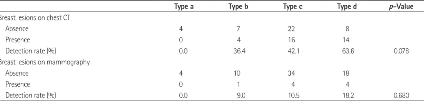
= 22). On mammography, 9 (12.0%) lesions, corresponding with the target lesions on breast sonography, were detected; breast composition type a (n = 0), type b (n = 1), type c (n = 4), and type d (n = 4). BI-RADS assessment categories for these 9 lesions iden- tified on mammography consisted of category 0 (n = 3), category 1 (n = 0), category 2 (n = 1), category 3 (n = 1), category 4 (n = 2), and category 5 (n = 2). There was no significant difference in the detection rate on mammography, according to the breast compo- sition type in chi-square test (p = 0.680) (Table 1).
A total of 75 target lesions on breast sonography were classified as BI-RADS assessment category 2 (n = 6), category 3 (n = 47), category 4 (n = 19), and category 5 (n = 3).
Of the 75 target lesions, 34 (45.3%) lesions were detected on enhanced chest CT which corresponded with the target lesions on breast sonography; breast composition type a (n = 0), type b (n = 4), type c (n = 16), and type d (n = 14). Of the 34 lesions on enhanced chest CT, 15 lesions were foci with size less than 2 mm and the remaining 19 lesions showed a mean size of 8.9 mm (range 4–54 mm). Among the 19 lesions with size more than 2 mm, the shapes were oval (n = 4), round (n = 10), and irregular (n = 5) and the margins were circumscribed (n = 12) and non- circumscribed (n = 7). In contrast-enhanced CT scans, presence or absence of enhancement in a total of 34 lesions was as fol- lows: presence (n = 15) and absence (n = 19). Associated fea- tures were axillary adenopathy (n = 5) and skin change (n = 3).
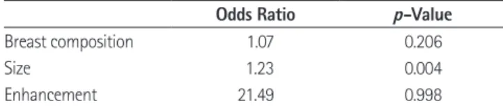
There was no significant difference in detectability of breast le- sions on chest CT, according to the four types of breast composi- tion in chi-square test (p = 0.078) (Table 1). In logistic regression analysis, two types of breast composition (dense and non-dense types) had no significant effect on detectability of breast lesions on chest CT [odds ratio (OR) = 1.07, p = 0.206]. Lesion size was a significant contributing factor for detectability of breast lesions on chest CT (OR = 1.23, p = 0.004). OR of enhancement (21.49) was very high, but there was no statistical significance (p = 0.998) (Table 2).
CT Morphologic Features of Malignant and Benign Breast Lesions
Of the 75 patients, 8 showed breast carcinoma through sonog- raphy-guided needle biopsy and 32 had benign breast diseases, which consisted of 19 pathologically proven lesions and 13 be- nign lesions categorized on breast sonography at the time of in- vestigation or follow up. In the remaining 35 patients, there were no medical records of further evaluation for breast abnormalities.
Of the 40 cases including 8 cases of malignant breast diseases and 32 cases of benign breast diseases, the morphologic features of breast lesions on enhanced chest CT were compared between benign and malignant lesions (Table 3). The mean size was sig- nificantly different between benign and malignant breast lesions (8.8 ± 4.0 and 21.5 ± 17.5, respectively; p = 0.018). The irregular shape (0% vs. 100%, p = 0.023) and non-circumscribed margin
(28.6 % vs. 71.4%, p = 0.031) were more frequently observed fea- tures in malignant lesions than in benign lesions. Axillary ade- nopathy (20% vs. 80%, p = 0.006) and skin change (0% vs. 100%,
p = 0.005) appeared more frequently in malignant breast lesions than in benign breast lesions.
Fig. 2. 42-year-old woman with fibroadenoma diagnosed through vacuum assisted biopsy.
A. Craniocaudal mammography of the right breast shows extremely dense breast and it is classified as breast composition type d. There is no ev- idence of mass nor suspicious morphology of calcification.
B. Contrast-enhanced chest CT shows a subcentimetered oval shaped enhancing mass with smooth margin in the right breast (arrow).
C. US image shows a corresponding 9 mm sized mass in the upper outer portion of right breast which is well circumscribed oval hypoechoic mass with parallel orientation and classified as BI-RADS assessment category 3.
BI-RADS = Breast Imaging Reporting and Data System, US = ultrasonography
A B C
Fig. 1. 57-year-old woman with invasive ductal carcinoma diagnosed through sonography-guided needle biopsy.
A. Craniocaudal mammography of the left breast shows heterogeneously dense breast and it is classified as breast composition type c. There is a high density mass with irregular shape, indistinct margin, fine pleomorphic calcification (arrows).
B. Contrast-enhanced chest CT shows about 53 mm sized enhancing mass in the left breast which is irregular shaped with lobulated margin. As- sociated skin thickening is also noted.
C. US image shows a corresponding about 50 mm sized mass in the lower medial portion of left breast which is hypoechoic irregular mass with a heterogeneous internal echo pattern and classified as BI-RADS assessment category 5.
BI-RADS = Breast Imaging Reporting and Data System, US = ultrasonography
A B C
Table 1. Detectability of Breast Lesions on Chest CT and Mammography, according to Types of Breast Composition
Type a Type b Type c Type d p-Value
Breast lesions on chest CT
Absence 4 7 22 8
Presence 0 4 16 14
Detection rate (%) 0.0 36.4 42.1 63.6 0.078
Breast lesions on mammography
Absence 4 10 34 18
Presence 0 1 4 4
Detection rate (%) 0.0 9.0 10.5 18.2 0.680
DISCUSSION
We investigated the capability of enhanced chest CT to detect breast lesions in patients who had abnormalities on breast sonog- raphy. The detection rate of mammography has been evaluated in previous studies and it demonstrated a wide range from 68%
to 88% (6). Kim et al. (7) reported that 86% of breast lesions were incidentally detected on chest CT and they correlated with breast sonography. On the other hand, in our study, a total of 75 pa- tients with breast abnormalities on sonography were enrolled, and of them, about half of the cases (45.3%) demonstrated breast lesions on enhanced chest CT which corresponded to the target lesions on breast sonography. Breast lesions were not incidentally detected, but the readers, who knew about the presence of breast lesions and were blinded to their location and size, selected the breast lesions on chest CT scan which corresponded with those on breast sonography.
There are several studies in which the effect of breast composi- tion on mammographic performance was evaluated (6, 8). Al- though in our study there was no significant difference in the de- tection rate among types of breast composition, which is because a very small number of lesions were detected on mammography,
in analysis of 27825 patients by Kolb et al. (6), they concluded that the sensitivity of mammography declined significantly with increasing breast composition. For enhanced chest CT scans, our results did not show two types (dense and non-dense) of breast composition or that the four types of breast composition had a significant effect on detectability of breast lesions. This might have resulted from the fact that, as a cross-sectional image, chest CT eliminates summation artifact and superimposition of glan- dular tissues.
Breast composition on mammography is a risk factor for breast cancer, as well as it affects mammographic performance (9, 10).
This has also been known that supplemental breast sonography can aid in detection of breast cancers in women with dense breasts (11). In our study, about half of the cases with breast ab- normalities were identified on enhanced chest CT. Thus for wom- en who undergo only chest CT, it may provide information on breast lesions and further evaluations including mammography or breast sonography can be suggested, especially in high risk pa- tients.
It is known that the features favoring a benign lesion on breast sonography were oval shape, parallel orientation to the skin, cir- cumscribed margin, abrupt boundary, and anechoic and hyper- echoic pattern. On the other hand, malignant features on breast sonography were irregular and round shape, non-parallel orien- tation, microlobulated, indistinct, angular, or spiculated margin, and boundary of echogenic halo (12-15). There are several stud- ies which reported the CT features of incidental breast lesions suggestive of malignancy, including irregular or spiculated mar- Table 2. Logistic Regression Analysis for Detectability of Breast Le-
sions on Enhanced Chest CT
Odds Ratio p-Value
Breast composition 1.07 0.206
Size 1.23 0.004
Enhancement 21.49 0.998
Table 3. Comparison of CT Morphology between Benign and Malignant Breast Lesions
Features Benign Lesions (n = 32) Malignant Lesions (n = 8) p-Value
Size (mm) 8.8 21.5 0.018
Shape 0.023
Round 5 (83.3) 1 (16.7)
Oval 3 (75) 1 (25)
Irregular 0 (0) 4 (100)
Margin 0.031
Circumscribed 6 (85.7) 1 (14.3)
Not circumscribed 2 (28.6) 5 (71.4)
Enhancement 0.646
Presence 4 (57.1) 3 (42.9)
Absence 1 (100) 0 (0)
Axillary lymph node 1 (20) 4 (80) 0.006
Skin thickening 0 (0) 3 (100) 0.005
Data are the numbers of patients. Numbers in parentheses are percentages.
gins, irregular shape, and rim enhancement. A washout pattern on postcontrast images and diffuse regional enhancement had high positive predictive values for malignancy (16-19). Similarly, in our study, malignant breast lesions were more irregular in shape and they demonstrated a non-circumscribed margin more frequently, compared with benign lesions (p = 0.023 and 0.031).
But the difference in enhancement between malignant and be- nign lesions was not significant (p = 0.646).
March et al. (20) studied 35 patients of breast cancer for evalu- ating detection of axillary lymph nodes on preoperative chest CT scan. They reported a positive predictive value of 89%. Another study concluded that chest CT in prone position with 5 mm sec- tions accurately predicted axillary lymph node involvement in breast cancer (21). Results of our study correlated with conclu- sions of these studies. In our study, additional findings such as axillary lymphadenopathy (p = 0.006) and skin thickening (p = 0.005) were more frequently observed in malignant breast le- sions.
There are several limitations in our study; first, the small study population and the retrospective nature of the study design may have reduced the power of this study. Contrast-enhanced chest CT for a variety of indications may be less sensitive than dedicat- ed breast CT in detecting and characterizing breast lesions (22).
Also various types of CT acquisition and injection protocols were used during the period. Therefore, delayed-phase scanning time may not have been standardized. It was limited to detect breast tumors and measure the accurate size of tumors, because breasts were compressed by gravity in the supine position. Another limi- tation was that there might be a discrepancy between the target lesions on chest CT and breast sonography, although we com- pared each of the lesions with their location and size. Because we did not consider all lesions on breast sonography, but we only considered the largest lesions, it may have influenced the detect- ability of breast lesions on chest CT.
Our study suggested that about half of the cases (45.3%) dem- onstrated breast lesions on enhanced chest CT which corre- sponded with target lesions on sonography. Also, breast compo- sition defined on mammography did not affect the detectability of enhanced chest CT for breast lesions. Therefore, cautious in- terpretation of enhanced chest CT might help detect breast ab- normalities covered with dense parenchyma.
REFERENCES
1. Sickles EA, D’Orsi CJ, Bassett LW, Appleton CM, Berg WA, Burnside ES, et al. ACR BI-RADS-Mammography 2013. In:
Reston VA, editor. ACR BI-RADS atlas breast imaging re- porting and data system. 5th ed. American College of Ra- diology, 2013:123-126
2. Tamimi RM, Byrne C, Colditz GA, Hankinson SE. Endoge- nous hormone levels, mammographic density, and subse- quent risk of breast cancer in postmenopausal women. J Natl Cancer Inst 2007;99:1178-1187
3. Meller MT, Cox JE, Callanan KW. Incidental detection of breast lesions with computed tomography. Clin Breast Cancer 2007;7:634-637
4. Yi JG, Kim SJ, Marom EM, Park JH, Jung SI, Lee MW. Chest CT of incidental breast lesions. J Thorac Imaging 2008;23:
148-155
5. Salvatore M, Margolies L, Kale M, Wisnivesky J, Kotkin S, Henschke CI, et al. Breast density: comparison of chest CT with mammography. Radiology 2014;270:67-73
6. Kolb TM, Lichy J, Newhouse JH. Comparison of the perfor- mance of screening mammography, physical examination, and breast US and evaluation of factors that influence them:
an analysis of 27,825 patient evaluations. Radiology 2002;
225:165-175
7. Kim JH, Chang YW, Hwang JH, Kim HH, Lee EH, Yang SB.
Evaluation of the significance of incidental breast lesions detected by chest CT. J Korean Soc Radiol 2013;68:229- 235
8. Boyd NF, Guo H, Martin LJ, Sun L, Stone J, Fishell E, et al.
Mammographic density and the risk and detection of breast cancer. N Engl J Med 2007;356:227-236
9. Boyd NF, Martin LJ, Rommens JM, Paterson AD, Minkin S, Yaffe MJ, et al. Mammographic density: a heritable risk fac- tor for breast cancer. Methods Mol Biol 2009;472:343-360 10. Cecchini RS, Costantino JP, Cauley JA, Cronin WM, Wick-
erham DL, Bandos H, et al. Baseline mammographic breast density and the risk of invasive breast cancer in postmeno- pausal women participating in the NSABP study of tamoxi- fen and raloxifene (STAR). Cancer Prev Res (Phila) 2012;5:
1321-1329
11. Hooley RJ, Greenberg KL, Stackhouse RM, Geisel JL, Butler
RS, Philpotts LE. Screening US in patients with mammo- graphically dense breasts: initial experience with Connecti- cut Public Act 09-41. Radiology 2012;265:59-69
12. Graf O, Helbich TH, Fuchsjaeger MH, Hopf G, Morgun M, Graf C, et al. Follow-up of palpable circumscribed noncal- cified solid breast masses at mammography and US: can biopsy be averted? Radiology 2004;233:850-856
13. Mainiero MB, Goldkamp A, Lazarus E, Livingston L, Koel- liker SL, Schepps B, et al. Characterization of breast masses with sonography: can biopsy of some solid masses be de- ferred? J Ultrasound Med 2005;24:161-167
14. Stavros AT, Thickman D, Rapp CL, Dennis MA, Parker SH, Sisney GA. Solid breast nodules: use of sonography to dis- tinguish between benign and malignant lesions. Radiology 1995;196:123-134
15. Hong AS, Rosen EL, Soo MS, Baker JA. BI-RADS for sonog- raphy: positive and negative predictive values of sonograph- ic features. AJR Am J Roentgenol 2005;184:1260-1265 16. Porter G, Steel J, Paisley K, Watkins R, Holgate C. Inciden-
tal breast masses detected by computed tomography: are any imaging features predictive of malignancy? Clin Radi-
ol 2009;64:529-533
17. Moyle P, Sonoda L, Britton P, Sinnatamby R. Incidental breast lesions detected on CT: what is their significance? Br J Radiol 2010;83:233-240
18. Surov A, Fiedler E, Wienke A, Holzhausen HJ, Spielmann RP, Behrmann C. Intramammary incidental findings on staging computer tomography. Eur J Radiol 2012;81:2174-2178 19. Lin WC, Hsu HH, Li CS, Yu JC, Hsu GC, Yu CP, et al. Inciden-
tally detected enhancing breast lesions on chest computed tomography. Korean J Radiol 2011;12:44-51
20. March DE, Wechsler RJ, Kurtz AB, Rosenberg AL, Needleman L. CT-pathologic correlation of axillary lymph nodes in breast carcinoma. J Comput Assist Tomogr 1991;15:440-444 21. Hata Y, Ogawa Y, Nishioka A, Inomata T, Yoshida S, Toki T.
[Evaluation of thin section CT scanning in the prone posi- tion of metastatic axillary lymph nodes for breast cancer].
Nihon Igaku Hoshasen Gakkai Zasshi 1996;56:1027-1031 22. Prionas ND, Lindfors KK, Ray S, Huang SY, Beckett LA,
Monsky WL, et al. Contrast-enhanced dedicated breast CT:
initial clinical experience. Radiology 2010;256:714-723
조영증강 흉부단층촬영검사에서 유방 병변의 발견:
유방 실질의 구성과의 연관성
조은미 · 강 희* · 신영경 · 윤종혁 · 오경승 · 박세경
목적: 조영증강 흉부 전산화단층촬영에서 유방 병변의 발견율과 이에 대해 유방 실질의 밀도가 영향을 미치는지 알아보 고자 하였다.
대상과 방법: 한 달 이내에 유방 초음파와 단순유방촬영, 조영증강 흉부 전산화단층촬영을 모두 시행 받은 환자 중 유방 초음파에서 병변이 발견된 환자를 대상으로 하였다. 단순유방촬영에서 Breast Imaging Reporting and Data System에 의 해 정의된 4 유형의 유방 실질의 구성에서 흉부단층촬영상 유방 병변의 발견 빈도를 비교하였다. 유방 실질의 밀도와 유방 초음파상 병변의 크기, 조영증강 여부가 유방 병변의 발견율에 어떤 영향을 미치는지 카이 제곱 검정과 로지스틱 회귀분석 을 통해 알아보았다.
결과: 총 75명의 환자 중 34명(45.3%)에서 유방초음파에서 확인된 병변과 일치하는 병변이 흉부단층촬영에서 발견되었 으며 4 유형의 유방 실질의 밀도에 따라 발견율의 유의한 차이는 없었다(p = 0.078). 로지스틱 회귀분석 결과 유방 실질 의 밀도[odds ratio (이하 OR) = 1.07, p = 0.206]와 조영증강 여부(OR = 21.49, p = 0.998)는 흉부단층촬영에서 유방 병변의 발견율에 유의한 영향을 미치지 않았으며 병변의 크기(OR = 1.23, p = 0.004)는 통계적으로 유의한 기여 인자로 나 타났다.
결론: 조영증강 흉부 전산화단층촬영상 45.3%에서 초음파에 상응하는 유방 병변을 관찰할 수 있었으며 유방 실질의 밀 도는 발견율에 유의한 영향을 미치지 않았다.
고신대학교 의과대학 고신대학교복음병원 영상의학과