Human Papillomavirus Prevalence and Genotype Distribution among HIV-Infected Women in Korea
The epidemiology on human papillomavirus (HPV) among human immunodeficiency virus (HIV)-infected women in Korea is not well established. A retrospective study was conducted to determine the prevalence and genotype distribution of HPV infection among HIV- infected women in Korea. HPV DNA genotype and cervical cytology were examined in 60 HIV-positive women and 1,938 HIV-negative women. HPV genotypes were analyzed by using a HPV DNA chip. HIV-infected women had higher prevalence of high-risk HPV (hr- HPV) infection (30% vs 4.9%, adjusted odds ratio [AOR], 6.96; 95% confidence interval [CI], 3.63-13.34, P < 0.001) and abnormal cervical cytology (18.3% vs 1.8%, AOR, 10.94; 95% CI, 5.18-23.1, P < 0.001) compared with controls. The most common hr-HPV genotype detected in HIV-infected women was HPV 16 (10%), followed by 18 (6.7%) and 52 (5%). Prevalence of quadrivalent vaccine-preventable types (HPV 6, 11, 16, and 18) was 21.7% and 2.3% in HIV-positive women and HIV-negative women, respectively. Age was a significant risk factor for hr-HPV infection in HIV-infected women (P = 0.039). The presence of hr-HPV was significantly associated with abnormal cervical cytology (P < 0.001).
These findings suggest that HPV testing for cervical cancer screening in HIV-infected women would be necessary, particularly among young age group.
Keywords: HIV; Women; Human Papillomavirus; Prevalence; Genotype Eun Kyoung Park,1 Heerim Cho,1
Sun Hee Lee,1 Seung Geun Lee,1 Sang Yeup Lee,2 Ki Hyung Kim,3 Chang Hun Lee,4 Joo Seop Chung,1 and Ihm Soo Kwak1
1Deparment of Internal Medicine, Pusan National University School of Medicine, Medical Research Institute, Pusan National University Hospital, Busan;
2Deparment of Family Medicine, Yangsan Pusan National University Hospital, Yangsan; 3Deparment of Obstetrics & Gynecology, Pusan National University Hospital, Busan; 4Deparment of Pathology, Pusan National University Hospital, Busan, Korea Received: 22 August 2013
Accepted: 8 November 2013 Address for Correspondence:
Sun Hee Lee, MD
Division of Infectious Diseases, Department of Internal Medicine, Pusan National University Hospital, 179 Gudeok-ro, Seo-gu, Busan 602-739, Korea
Tel: +82.51-240-7673, Fax: +82.51-247-3213 E-mail: [email protected]
This study was supported by Medical Research Institute Grant (2005-5), Pusan National University.
http://dx.doi.org/10.3346/jkms.2014.29.1.32 • J Korean Med Sci 2014; 29: 32-37
INTRODUCTION
Human immunodeficiency virus (HIV)-infected women have higher rates of human papillomavirus (HPV) infection than HIV-uninfected women, and are also at higher risk for persis- tent infection and progression to malignancy (1-4). In addition, HPV may be a cofactor in HIV acquisition (5, 6). Persistent in- fections with high risk HPV (hr-HPV) are known to be a neces- sary first step in the development of cervical cancer. The preva- lence of hr-HPV infection and genotype distribution varied in different geographic areas (7). With the introduction of HPV vaccine, knowledge regarding hr-HPV genotype distribution has become increasingly important.
As of December 2010, a total of 623 women were diagnosed with HIV infection in Korea (8). Following the first case of HIV infection was reported in a foreign worker in 1985, active screen- ing test for HIV infection was performed for commercial sex workers (CSWs) who might be contacted with the foreigners (9, 10). Eleven HIV-infected female CSWs were detected between 1985 and 1988. Since AIDS Prevention Law was enacted in 1987, mandatory screening for HIV infection was extended to all CSWs,
overseas sailors, and people working in “hygiene related jobs”
requiring health certificates, such as restaurants, coffee shops, barbershops, bars, and hotels. As a result, a sharp increase in the number of cases was detected among overseas sailors, who were responsible for 34% of people diagnosed with HIV infec- tion until the mandatory screening test for this group ended in 1993 (9-11). HIV infection was also detected among their wives and other female sex partners. The proportion of women with HIV infection has remained stable and the man-to-woman ra- tio of patients infected with HIV was 11:1. A previous study show- ed that 67.6% of HIV-infected women in Korea had regular sex- ual partners; 26.1% were tested for HIV diagnosis because of their HIV-positive sex partners. These findings suggested that the majority of HIV-infected women were infected from their regular male sex partners, such as husband or person living to- gether without being legally married (12).
There are limited data on the prevalence of HPV infection and hr-HPV genotypes among HIV-infected women in Korea, and little is known about their relationship with cervical cytolo- gy in this population. The objective of this study was to examine the prevalence and genotype distribution of HPV infection among Infectious Diseases, Microbiology & Parasitology
HIV-infected women in Korea.
MATERIALS AND METHODS Study population
We retrospectively reviewed the medical records for HIV-infected women attending Pusan National University Hospital between January 2009 and December 2012. The Hospital is a 1,220-bed university-affiliated teaching hospital and provides HIV care for HIV-infected patients in southeastern region of Korea. The study included HIV-infected ethnically Korean women who were aged 18 yr and older, and tested cervical cytology and HPV genotype in the study hospital. HIV-infected women who had current pregnancy or history of prior hysterectomy or coniza- tion were excluded. During that period, 78 HIV-infected wom- en attended to the study hospital, and 60 (76.9%) were included in the study. For comparison, we used the data for women who visited Health Promotion Centers of Pusan National University Hospital and Yangsan Pusan National University Hospital for a regular medical check-up during the same period. Patients aged 18 yr and older who tested cervical cytology with HPV genotype were included, and HIV seropositive patients were excluded.
Information obtained from database of the Health Promotion Centers included age, marital status, smoking history, parity, comorbidity, cervical cytology, HPV genotype, and serology for hepatitis B virus (HBV), hepatitis C virus (HCV) and syphilis.
When multiple tests had been conducted from the same patient at different times, only the first test was used for assessing the prevalence and genotype distribution of HPV infection. Clinical categories were defined by the 1993 Centers for Disease Con- trol and Prevention (CDC) classification criteria (13).
Cervical cytology and human papillomavirus genotyping Cervical samples for cytology and HPV genotyping were obtain- ed by gynecologists as a routine care or a part of regular medi- cal check-up. Pap smear results were categorized according to the 2001 Bethesda classification system terminology (14). HPV genotype was determined using an PCR based DNA microar- ray system, the HPV DNA chip (MyHPV Chip Kit®, BioMedLab, Seoul, Korea). This contains 24 type-specific probes detecting 13 types of high risk HPV (16, 18, 31, 33, 35, 39, 45, 51, 52, 56, 58, 59, and 68) and 11 types of low risk HPV (6, 11, 34, 40, 42, 43, 44, 53, 54, 66, and 70).
Statistical analysis
Categorical variables were compared using Pearson’s chi-square test or Fisher’s exact test, whereas non-categorical variables were tested with the Mann-Whitney U-test. Comparison be- tween HIV-positive women and HIV-negative women was con- ducted by using multiple logistic regression techniques while controlling for potential confounding variables. In multivariate
analysis, variables were entered into the model simultaneously, not using an automatic model selection. All tests were consid- ered statistically significant at P < 0.05. The statistical analyses were conducted using PASW Statistics 18 (SPSS Inc., Chicago, IL, USA).
Ethics statement
This study protocol was approved by the institutional review board (IRB) of Pusan National University Hospital (IRB No. E- 2013013) and Yangsan Pusan National University Hospital (IRB No. 05-2013-044), respectively. Informed consent was waived by the boards.
RESULTS
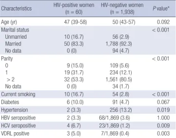
A total of 60 HIV-positive women and 1,938 HIV-negative wom- en were included in this study. Their demographic characteris- tics are described in Table 1. Median age was 47 yr old (inter- quartile range 39-58) for HIV-positive women and 50 yr old (in- terquartile range 43-57) for HIV-negative women (P = 0.092).
The marital status, parity, smoking, comorbidity, HCV and syph- ilis serology showed significant difference between two groups.
By using the multiple logistic regression model, we compared the prevalence of HPV infection and the results of cervical cy- tology between two groups (Table 2). Overall, HPV infection rate was significantly higher in HIV-positive women than HIV- negative women (46.7% vs 14.1%; adjusted odds ratio [AOR], 4.48; 95% confidence interval [CI], 2.56-7.83, P < 0.001). Multi- ple HPV types were detected in 18.3% of HIV-positive women and 1.5% of HIV-negative women (AOR, 12.87; 95% CI, 5.43- 30.54, P < 0.001). HIV-positive women had a significantly great-
Table 1. Baseline characteristics of HIV-positive women and HIV-negative controls Characteristics HIV-positive women
(n = 60) HIV-negative women
(n = 1,938) P value*
Age (yr) 47 (39-58) 50 (43-57) 0.092
Marital status Unmarried Married No data
10 (16.7) 50 (83.3)
0 (0)
56 (2.9) 1,788 (92.3)
94 (4.7)
< 0.001
Parity 0 1
> 2 No data
9 (15.0) 19 (31.7) 32 (53.3)
0 (0)
109 (5.6) 234 (12.1) 1,561 (80.5) 34 (1.7)
< 0.001
Current smoking 10 (16.7) 54 (2.8) < 0.001
Diabetes 6 (10.0) 91 (4.7) 0.067
Hypertension 2 (3.3) 256 (13.2) 0.019
HBV seropositive 2 (3.3) 68/1,869 (3.6) 1.000
HCV seropositive 4 (6.7) 23/1,869 (1.2) 0.009
VDRL positive 3 (5.0) 7/1,869 (0.4) 0.003
Data are number (%) of patients, unless otherwise indicated. *Calculated using chi- square test, Fisher’s exact test, or the Mann-Whitney U-test. HIV, human immunode- ficiency virus; HBV, hepatitis B virus; HCV, hepatitis C virus; VDRL, venereal disease research laboratory.
Table 2. Prevalence of human papillomavirus infection and abnormal cervical cytology in 60 HIV-positive women and 1,938 HIV-negative women, and adjusted odds ratios and 95% confidence intervals determined by multiple logistic regression model using HIV status as an independent variable
Variables HIV-positive women (n = 60) HIV-negative women (n = 1,938) Adjusted odds ratio 95% Confidence interval P value*
Any HPV positive 28 (46.7) 274 (14.1) 4.48 2.56-7.83 < 0.001
Multiple types of any HPV 11 (18.3) 29 (1.5) 12.87 5.43-30.54 < 0.001
Hr-HPV positive 18 (30.0) 95 (4.9) 6.96 3.63-13.34 < 0.001
Multiple types of hr-HPV 6 (10.0) 12 (0.6) 12.63 3.75-42.56 < 0.001
Abnormal cervical cytology ASCUS
LSIL HSIL AGC
11 (18.3) 4 (6.7) 3 (5.0) 4 (6.7) 0 (0)
35 (1.8) 14 (0.7) 11 (0.6) 8 (0.4) 2 (0.1)
10.94 5.18-23.10 < 0.001
Data are number (%) of patients, unless otherwise indicated. *Calculated using multivariate logistic regression analysis. HIV, human immunodeficiency virus; HPV, humanpapil- loma virus; Hr, high risk; ASCUS, atypical squamous cells of undetermined significance; LSIL, low-grade squamous intraepithelial lesions; HSIL, high-grade squamous intraepi- thelial lesions; AGC, atypical glandular cells.
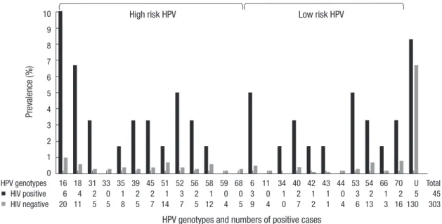
Fig. 1. Human papillomavirus (HPV) genotypic distribution in 60 human immunodeficiency virus (HIV)-positive and 1,938 HIV-negative women. U, un-typeable HPV.
High risk HPV Low risk HPV
Prevalence (%)
HPV genotypes and numbers of positive cases
HPV genotypes 16 18 31 33 35 39 45 51 52 56 58 59 68 6 11 34 40 42 43 44 53 54 66 70 U Total
HIV positive 6 4 2 0 1 2 2 1 3 2 1 0 0 3 0 1 2 1 1 0 3 2 1 2 5 45
HIV negative 20 11 5 5 8 5 7 14 7 5 12 4 5 9 4 0 7 2 1 4 6 13 3 16 130 303
10 9 8 7 6 5 4 3 2 1 0
er hr-HPV infection rate than women without HIV infection (30% vs 4.9%; AOR, 6.96; 95% CI, 3.63-13.34, P < 0.001). Multi- ple hr-HPV types were detected in 10% of HIV-positive women and 0.6% of HIV-negative women (AOR, 12.63; 95% CI, 3.75- 42.56, P < 0.001).
Fig. 1 shows the type-specific HPV prevalence of both HIV- positive and HIV-negative women. The most common hr-HPV genotype detected in HIV-positive women was HPV 16 (10%), followed by 18 (6.7%) and 52 (5%). Other common hr-HPV types detected in HIV-positive women were HPV 31 (3.3%), 39 (3.3%), 45 (3.3%), and 56 (3.3%). In HIV-negative women, the most pre- valent hr-HPV was HPV 16 (1.0%), followed by 51 (0.7%), 58 (0.6%), and 18 (0.6%). Other commonly detected hr-HPV types in HIV-negative women were HPV 35 (0.4%), 45 (0.4%), and 52 (0.4%). Commonly encountered non-oncogenic viruses were HPV 70 (0.8%), 54 (0.7%), 6 (0.5%), 40 (0.4%), and 53 (0.3%) in HIV-negative women, and HPV 6 (5%), 53 (5%), 40 (3.3%), 54 (3.3%), and 70 (3.3%) in HIV-positive women. Five samples
(8.3%) in HIV-positive women and 130 (6.7%) in HIV-negative women were unable to type. Prevalence of quadrivalent vac- cine-preventable types (HPV 6, 11, 16, and 18) was 21.7% (13/60) and 2.3% (44/1,938) in HIV-positive women and HIV-negative women, respectively.
A total of 11 HIV-positive women (18.3%) and 35 HIV-nega- tive women (1.8%) had abnormal cytology (AOR, 10.94; 95% CI, 5.18-23.1, P < 0.001) (Table 2). HIV-infected women with hr- HPV were more likely to have abnormal cytology than those without hr-HPV (P < 0.001) (Table 3). All 7 HIV-positive women with high-grade squamous intraepithelial lesion (HSIL) or low- grade squamous intraepithelial lesion (LSIL) tested positive for hr-HPV and had multiple HPV types, five of which also had multiple hr-HPV. A total of 12 hr-HPV were detected in 7 HIV- positive women with HSIL or LSIL, and the most frequently de- tected hr-HPV was HPV 16 (3/7). Other detected hr-HPV were 18, 31, 35, 39, 45, 51, 52, 56, and 58 (by 1 occurrence for each).
When we compared the predictors for hr-HPV infection be-
tween hr-HPV positive and hr-HPV negative HIV-infected wom- en, age was a significant risk factor for hr-HPV infection (P = 0.039). Other factors were not associated with hr-HPV detection (Table 3).
DISCUSSION
Several studies have consistently shown that HIV-infected wo- men are at increased risk for HPV infection, although the prev- alence and genotype distribution varied in a different geographic area (15-21). This is the first report of prevalence of HPV among HIV-infected women in Korea to our knowledge. In this study, we found significantly higher prevalence of HPV infection in HIV-infected women, 46.7% for any HPV and 30% for hr-HPV,
compared to HIV-negative controls, 14.1 and 4.9%, respectively.
Hr-HPV was about 7 times more commonly detected among HIV-infected patients than among HIV-negative controls when adjusted with other demographic factors. The frequency of ab- normal cervical cytology was about 11 times higher in HIV-in- fected women than in HIV-negative women. Our findings are in line with the results from other studies in other countries that reported hr-HPV prevalence rates ranging from 35.3% to 56% in HIV-infected women (15-21).
The prevalence of hr-HPV in HIV-negative women in our study is slightly lower than those reported in previous popula- tion based studies conducted in Busan, where it has been re- ported between 6% and 9%, 6.3% in the general population, 8.1% in the women attending cervical cancer screening pro- Table 3. Comparison of demographic characteristics and cervical cytology results between 18 high risk HPV positive HIV-infected women and 42 high risk HPV negative coun- terparts
Characteristics Hr-HPV positive HIV-infected women
(n = 18)
Hr-HPV negative HIV-infected women
(n = 42) P value*
Age, median (IQR), yr 40.5 (33-53.8) 52.5 (40-58) 0.039
Marital status Unmarried Maintained marriage Divorced
Separated by death
4 (22.2) 10 (55.6) 2 (11.1) 2 (11.1)
6 (14.3) 17 (40.5) 10 (23.8) 9 (21.4)
0.482
Route of transmission Heterosexual
Unknown 18 (100)
0 (0) 41 (97.6)
1 (2.4)
1.000
HIV positive sexual partners Husband
Other known HIV positive partners Unknown
7 (38.9) 6 (33.3) 5 (27.8)
18 (42.9) 6 (14.3) 18 (42.9)
0.268
Ever been a sex worker 4 (22.2) 6 (14.3) 0.468
Duration from HIV diagnosis to HPV test, median (IQR), yr 3.2 (0.3-8.5) 3.85 (1.6-8.7) 0.367
CDC Clinical category at the time of HPV test A
B C
6 (33.3) 10 (55.6) 2 (11.1)
24 (57.1) 11 (26.2) 7 (16.7)
0.114
ART at the time of HPV test None
< 1 yr
> 1 yr
6 (33.3) 1 (5.6) 11 (61.1)
13 (31.0) 8 (19.0) 21 (50.0)
0.504
CD4 cell counts at the time of HPV test, median (IQR), cells/μL
> 350 201-350
≤ 200
307 (181-480.5) 7 (38.9) 6 (33.3) 4 (22.2)
351 (252-460) 21 (50.0) 13 (31.0) 8 (19.0)
0.669
Viral suppression at the time of HPV test Undetectable
Detectable ( > 50 copies/mL)
8 (44.4) 10 (55.6)
17 (40.5) 25 (59.5)
0.783
HBV seropositive 1 (5.6) 1 (2.4) 0.514
HCV seropositive 1 (5.6) 3 (7.1) 1.000
Syphilis seropositive 1 (5.6) 2 (4.8) 1.000
Cervical cytology Negative ASCUS LSIL HSIL
10 (55.6) 1 (5.6) 3 (16.7) 4 (22.2)
39 (92.9) 3 (7.1) 0 (0) 0 (0)
< 0.001
Data are number (%) of patients, unless otherwise indicated. *Calculated using chi-square test, Fisher’s exact test, or the Mann-Whitney U-test. HIV, human immunodeficiency virus; Hr, high risk; HPV, humanpapilloma virus; IQR, interquartile range; CDC, Centers for Disease Control and Prevention; ART, anti-retroviral therapy; HBV, hepatitis B virus;
HCV, hepatitis C virus; ASCUS, atypical squamous cells of undetermined significance; LSIL, low-grade squamous intraepithelial lesions; HSIL, high-grade squamous intraepithe- lial lesions.
gram, and 8.6% in female university students (22-25). The dif- ferences between the studies are probably due to differences in the study population, detection method, and the definition of hr-HPV type.
In this study, HPV 16 was the most prevalent hr-HPV type detected in both HIV-positive and HIV-negative women. HPV 18, 52, 31, 39, 45, and 56 were also common among HIV-infect- ed women with prevalence ranging from 3.3% to 6.7%. Other commonly detected hr-HPV types in HIV-negative women were 51, 58, 18, 35, 45, and 52 with prevalence ranging from 0.4% to 0.7%. There were some differences in the distribution of HPV genotypes between HIV-positive and HIV-negative women ex- cept for HPV 16. In a previous study evaluating the concordance of HPV infection among heterosexually active couples and the impact of HIV coinfection on the prevalence of HPV, HIV coin- fection in one partner had a significant impact on the preva- lence of HPV infection in the other partner (26). Concordance of the same HPV genotypes was more commonly found among couples where one or both partners were HIV-infected, com- pared with HIV-uninfected couples (26). The majority of HIV- infected women were infected by their male sexual partners in Korea (12), and our HIV-positive women had regular sexual partners in 61.7%. Therefore, HPV type may be diverse depend- ing on types of their regular sexual partners. Although there is no study regarding HPV types of HIV-positive regular sexual partners, this may explain some differences in diverse type dis- tribution of HPV infection between HIV-positive and HIV-neg- ative women. In the previous studies conducted with CSWs in Korea, HPV 16, 18, and 58 were the most frequently detected hr- HPV types (27, 28). In our study, about 17% of HIV-infected wo- men had ever been a CSW, and this also may contribute some differences in the distribution of HPV genotypes between HIV- positive and HIV-negative women.
Quadrivalent HPV vaccine was immunogenic and well toler- ated in HIV-infected young women in a recent study (29). HPV vaccine is recommended by the Advisory Committee on Im- munization Practices (ACIP) for HIV-infected individuals through age 26 yr for those who did not get any or all doses when they were younger (30). Prevalence of vaccine-preventable types (HPV 6, 11, 16, and 18) in our HIV-infected women was 21.7%, suggesting that HPV DNA or serologic screening would be need- ed before immunization in HIV-infected women in Korea, al- though the ACIP does not recommend HPV DNA or serologic screening before immunization (30).
We also investigated risk factors for carrying hr-HPV in HIV- infected women. Age was a significantly important risk factor of hr-HPV infection in HIV-infected women. Sexually active young HIV-infected women tend to have hr-HPV more frequently.
However, other socio-demographic factors including marital status, route of transmission, presence of HIV- positive regular sex partners, history of female CSW, duration from HIV diagno-
sis to test, CDC clinical category, CD4 cell counts, ART, viral suppression, syphilis, and HBV/HCV coinfection were not sig- nificantly associated with the presence of hr-HPV in HIV-infect- ed women. The presence of hr-HPV was significantly associat- ed with abnormal findings on Pap smear. All HIV-infected wo- men with LSIL or worse on Pap smear reacted positive for hr- HPV in our study.
This study has some limitations. First, this study was a retro- spective cross-sectional study. The longitudinal follow-up data were not available, and the behavioral risk factors, such as the number of lifetime sex partners, were not considered in more detail. Second, our study was conducted in the southeastern region of Korea, and the number of HIV-infected women was relatively small, therefore our findings may not be generalized to other region of the country. Third, we did not test all geno- types of HPV; the proportion of persons with un-typeable HPV was relatively high, particularly in HIV-negative controls (31).
Even with these limitations, our study could reflect the overall situation of HPV infection among HIV-infected women in Korea.
In conclusion, this study demonstrated that hr-HPV infec- tion was common among HIV-infected women in Korea. The most frequent hr-HPV was HPV 16, followed by HPV 18 and 52.
Age was a significantly important risk factor of hr-HPV infec- tion in HIV-infected women. The presence of hr-HPV was sig- nificantly associated with abnormal cervical cytology. These findings suggest that HPV testing for cervical cancer screening in HIV-infected women would be necessary in Korea, particu- larly among young age group.
DISCLOSURE
The authors have no conflicts of interest to disclose.
REFERENCES
1. Ellerbrock TV, Chiasson MA, Bush TJ, Sun XW, Sawo D, Brudney K, Wright TC Jr. Incidence of cervical squamous intraepithelial lesions in HIV-infected women. JAMA 2000; 283: 1031-7.
2. Hawes SE, Critchlow CW, Faye Niang MA, Diouf MB, Diop A, Touré P, Aziz Kasse A, Dembele B, Salif Sow P, Coll-Seck AM, et al. Increased risk of high-grade cervical squamous intraepithelial lesions and invasive cervical cancer among African women with human immunodeficiency virus type 1 and 2 infections. J Infect Dis 2003; 188: 555-63.
3. Guiguet M, Boué F, Cadranel J, Lang JM, Rosenthal E, Costagliola D;
Clinical Epidemiology Group of the FHDH-ANRS CO4 Cohort. Effect of immunodeficiency, HIV viral load, and antiretroviral therapy on the risk of individual malignancies (FHDH-ANRS CO4): a prospective co- hort study. Lancet Oncol 2009; 10: 1152-9.
4. Abraham AG, D’Souza G, Jing Y, Gange SJ, Sterling TR, Silverberg MJ, Saag MS, Rourke SB, Rachlis A, Napravnik S, et al. Invasive cervical can- cer risk among HIV-infected women: a North American multicohort col- laboration prospective study. J Acquir Immune Defic Syndr 2013; 62:
405-13.
5. Lissouba P, Van de Perre P, Auvert B. Association of genital human pap- illomavirus infection with HIV acquisition: a systematic review and me- ta-analysis. Sex Transm Infect 2013; 89: 350-6.
6. Houlihan CF, Larke NL, Watson-Jones D, Smith-McCune KK, Shiboski S, Gravitt PE, Smith JS, Kuhn L, Wang C, Hayes R. Human papillomavi- rus infection and increased risk of HIV acquisition: a systematic review and meta-analysis. AIDS 2012; 26: 2211-22.
7. Guan P, Howell-Jones R, Li N, Bruni L, de Sanjosé S, Franceschi S, Clif- ford GM. Human papillomavirus types in 115,789 HPV-positive women:
a meta-analysis from cervical infection to cancer. Int J Cancer 2012; 131:
2349-59.
8. Korea Centers for Disease Control and Prevention. Annual report on the notified HIV/AIDS in Korea. Available at http://stat.mw.go.kr [ac- cessed on 1 July 2013].
9. Shin Y, Kee M. Health care systems in transition II. Korea, part II. the current status of HIV-AIDS in Korea. J Public Health Med 1998; 20: 47- 51.
10. Oh MD, Choe K. Epidemiology of HIV infection in the Republic of Korea.
J Korean Med Sci 1999; 14: 469-74.
11. Choe PG, Park WB, Song JS, Kim NH, Park JY, Song KH, Park SW, Kim HB, Kim NJ, Oh MD. Late presentation of HIV disease and its associated factors among newly diagnosed patients before and after abolition of a government policy of mass mandatory screening. J Infect 2011; 63: 60-5.
12. Lee JH, Lee EJ, Kim SS, Nam JG, Whang J, Kee MK. Epidemiological characteristics of HIV-infected women in the Republic of Korea: a low HIV prevalence country. J Public Health Policy 2009; 30: 342-55.
13. 1993 revised classification system for HIV infection and expanded sur- veillance case definition for AIDS among adolescents and adults. MM- WR Recomm Rep 1992; 41: 1-19.
14. Solomon D, Davey D, Kurman R, Moriarty A, O’Connor D, Prey M, Raab S, Sherman M, Wilbur D, Wright T Jr, et al. The 2001 Bethesda system:
terminology for reporting results of cervical cytology. JAMA 2002; 287:
2114-9.
15. Veldhuijzen NJ, Braunstein SL, Vyankandondera J, Ingabire C, Ntirush- wa J, Kestelyn E, Tuijn C, Wit FW, Umutoni A, Uwineza M, et al. The ep- idemiology of human papillomavirus infection in HIV-positive and HIV- negative high-risk women in Kigali, Rwanda. BMC Infect Dis 2011; 11:
333.
16. Dartell M, Rasch V, Kahesa C, Mwaiselage J, Ngoma T, Junge J, Gernow A, Ejlersen SF, Munk C, Iftner T, et al. Human papillomavirus prevalence and type distribution in 3603 HIV-positive and HIV-negative women in the general population of Tanzania: the PROTECT study. Sex Transm Dis 2012; 39: 201-8.
17. McKenzie ND, Kobetz EN, Hnatyszyn J, Twiggs LB, Lucci JA 3rd. Wom- en with HIV are more commonly infected with non-16 and -18 high-risk HPV types. Gynecol Oncol 2010; 116: 572-7.
18. Luque AE, Hitti J, Mwachari C, Lane C, Messing S, Cohn SE, Adler D, Rose R, Coombs R. Prevalence of human papillomavirus genotypes in HIV-1-infected women in Seattle, USA and Nairobi, Kenya: results from the Women’s HIV Interdisciplinary Network (WHIN). Int J Infect Dis 2010;
14: e810-4.
19. Sarkar K, Pal R, Bal B, Saha B, Bhattacharya S, Sengupta S, Mazumdar
PP, Chakraborti S. Oncogenic HPV among HIV infected female popula- tion in West Bengal, India. BMC Infect Dis 2011; 11: 72.
20. Dols JA, Reid G, Brown JM, Tempelman H, Bontekoe TR, Quint WG, Boon ME. HPV type distribution and cervical cytology among HIV-posi- tive Tanzanian and South African women. ISRN Obstet Gynecol 2012;
2012: 514146.
21. Mane A, Nirmalkar A, Risbud AR, Vermund SH, Mehendale SM, Sa- hasrabuddhe VV. HPV genotype distribution in cervical intraepithelial neoplasia among HIV-infected women in Pune, India. PLoS One 2012; 7:
e38731.
22. Clifford GM, Shin HR, Oh JK, Waterboer T, Ju YH, Vaccarella S, Quint W, Pawlita M, Franceschi S. Serologic response to oncogenic human papil- lomavirus types in male and female university students in Busan, South Korea. Cancer Epidemiol Biomarkers Prev 2007; 16: 1874-9.
23. Oh JK, Franceschi S, Kim BK, Kim JY, Ju YH, Hong EK, Chang YC, Rha SH, Kim HH, Kim JH, et al. Prevalence of human papillomavirus and Chlamydia trachomatis infection among women attending cervical cancer screening in the Republic of Korea. Eur J Cancer Prev 2009; 18:
56-61.
24. Shin HR, Lee DH, Herrero R, Smith JS, Vaccarella S, Hong SH, Jung KY, Kim HH, Park UD, Cha HS, et al. Prevalence of human papillomavirus infection in women in Busan, South Korea. Int J Cancer 2003; 103: 413- 21.
25. Shin HR, Franceschi S, Vaccarella S, Roh JW, Ju YH, Oh JK, Kong HJ, Rha SH, Jung SI, Kim JI, et al. Prevalence and determinants of genital in- fection with papillomavirus, in female and male university students in Busan, South Korea. J Infect Dis 2004; 190: 468-76.
26. Mbulawa ZZ, Coetzee D, Marais DJ, Kamupira M, Zwane E, Allan B, Constant D, Moodley JR, Hoffman M, Williamson AL. Genital human papillomavirus prevalence and human papillomavirus concordance in heterosexual couples are positively associated with human immunodefi- ciency virus coinfection. J Infect Dis 2009; 199: 1514-24.
27. Yun H, Park J, Choi I, Kee M, Choi B, Kim S. Prevalence of human papil- lomavirus and herpes simplex virus type 2 infection in Korean commer- cial sex workers. J Microbiol Biotechnol 2008; 18: 350-4.
28. Rhee JE, Shin MY, Kim CM, Kee HY, Chung JK, Min SK, Kim SJ, Jang DH, Kim SS, Choi BS. Prevalence of human papillomavirus infection and genotype distribution among high-risk Korean women for prospect- ing the strategy of vaccine development. Virol J 2010; 7: 201.
29. Kahn JA, Xu J, Kapogiannis BG, Rudy B, Gonin R, Liu N, Wilson CM, Worrell C, Squires KE. Immunogenicity and safety of the human papil- lomavirus 6, 11, 16, 18 vaccine in HIV-infected young women. Clin Infect Dis 2013; 57: 735-44.
30. Bridges CB, Woods L, Coyne-Beasley T; Centers for Disease Control and Prevention (CDC), ACIP Adult Immunization Work Group. Advi- sory Committee on Immunization Practices (ACIP) recommended im- munization schedule for adults aged 19 years and older: United States, 2013. MMWR Surveill Summ 2013; 62: 9-19.
31. Lee EH, Um TH, Chi HS, Hong YJ, Cha YJ. Prevalence and distribution of human papillomavirus infection in Korean women as determined by restriction fragment mass polymorphism assay. J Korean Med Sci 2012;
27: 1091-7.