Changes in Bone Mineral Density of Both Proximal Femurs after Total Knee Arthroplasty
Kwang Kyoun Kim, MD, Ye Yeon Won, MD*, Youn Moo Heo, MD, Dae Hee Lee, MD, Jeong Yong Yoon, MD, Won Sub Sung, MD
Department of Orthopedic Surgery, Konyang University College of Medicine, Daejeon,
*Department of Orthopedic Surgery, Ajou University School of Medicine, Suwon, Korea
Received November 25, 2012; Accepted May 8, 2013 Correspondence to: Ye Yeon Won, MD
Department of Orthopedic Surgery, Ajou University School of Medicine, 206 World cup-ro, Yeongtong-gu, Suwon 443-721, Korea
Tel: +82-31-219-5220, Fax: +82-31-219-5229 E-mail: jyyoon17@naver.com
Due to a growing aging population and prevalence of os- teoarthritis, rates of total knee arthroplasty (TKA) have increased. TKA offers relief from pain, and improvement in knee function and quality of life. However, TKA may result in postoperative complications, such as infection, thromboembolism, and nerve injury.
The authors experienced one patient who com-
Background: This study investigated the effects of total knee arthroplasty (TKA) on bone mineral density (BMD) of the proximal femur in patients who underwent the procedure.
Methods: Forty-eight patients scheduled to undergo unilateral TKA because of primary knee osteoarthritis were included in this study, which was conducted at a medical center between October 2006 and October 2009. In these 48 patients, 96 hips were evaluated. Measurement of BMD was performed preoperatively and one month, three months, six months, and one year after unilateral TKA. Repeated measured analysis of variance and paired t-tests for comparison of two repeated samples were used to compare differences between time points (preoperation, one, three, six, and 12 months) and between the operative and nonopera- tive sides.
Results: Preoperatively, BMD of the femoral neck, trochanter, and total hip on the operative side were lower than on the nonop- erative side; however, there was no statistical difference. BMD of both femoral neck areas was significantly lower than preopera- tive BMD at one month and three months after TKA. BMD of both trochanter areas was significantly lower than preoperative BMD at one month and three months after TKA. BMD of both total hips was significantly lower than preoperative BMD at three months after TKA. However, no statistical differences of changes in BMD were observed between the operative and nonoperative sides at each measurement time.
Conclusions: According to our results, TKA was found to affect both proximal femurs during the acute period. However, TKA did not affect a change in BMD of the proximal femur during one year postoperative.
Keywords: Proximal hip, Bone mineral density, Total knee arthroplasty
plained of groin pain of the ipsilateral side without a his- tory of trauma at three months after TKA, and we found a femoral neck fracture. We searched reports and journals on femoral neck stress fracture following TKA. Some studies have reported ipsilateral femoral neck fracture as a rare complication of TKA. Morbidity associated with hip fractures may include serious complications, such as deep vein thrombosis, postoperative infection, pain, and loss of mobility. Therefore, we thought that femoral neck fracture following TKA was a very serious problem and that is why we embarked on this study.
With increased demand of daily activity, microfrac- tures during surgery due to hammer blows, the design of prosthesis, and decreased bone mineral density (BMD)
Copyright © 2014 by The Korean Orthopaedic Association
This is an Open Access article distributed under the terms of the Creative Commons Attribution Non-Commercial License (http://creativecommons.org/licenses/by-nc/3.0) which permits unrestricted non-commercial use, distribution, and reproduction in any medium, provided the original work is properly cited.
Clinics in Orthopedic Surgery • pISSN 2005-291X eISSN 2005-4408
after TKA, a few hypotheses regarding the etiologies of femoral neck stress fractures have been proposed.1-5) How- ever, reports regarding changes of BMD in the proximal hip after TKA have been rare.6,7) Therefore, we first in- vestigated whether TKA can affect changes in proximal hip BMD. Second, if so, does TKA have different effects on BMD of the operative and nonoperative sides? Some cases have reported the occurrence of femoral neck stress fracture within one year after an operation. Therefore, we studied the change in BMD within one year after TKA.
METHODS
Patients
Among patients who underwent TKA because of pri- mary knee osteoarthritis from October 2006 to October 2009, we included only patients who had BMD measured with unilateral TKA at preoperation, and one, three, six, and 12 months after the operation. TKA was performed with patients who had a radiographic Kellgren-Lawrence grade III8) or greater and wanted the operation due to severe knee pain. We excluded patients who had under- gone a contralateral TKA (seven cases) within one year after TKA or who had any neurological issues (cerebral infarction, 1 case; Parkinson disease, 3 cases; peroneal nerve injury, 1 case) or took medicine (steroid, 32 cases;
osteoporosis drug, 41 cases). A total of 48 consecutive patients (11 males and 37 females) were included in this study. The mean age of the patients was 63 years (range, 53 to 76 years). The 40 cases of the nonoperative side were Kellgren-Lawrence grade II or less. Eight cases had Kell- gren Lawrence grade III or more, but did not receive TKA.
They did not feel the need to undergo surgery of the con-
tralateral knee.
Measurement of BMD
BMD of both hips was measured at baseline and at one, three, six, and 12 months follow-up. The femoral neck, trochanter, and total area of the hip were measured in the proximal femur. BMD was measured using dual energy X-ray absorptiometry (DEXA; lunar PPX-L, Medison, WI, USA). The in vivo coefficients of variation for BMD were 1.4% for the lumbar spine, 2.1% for the femoral neck, 2.1%
for ward’s triangle, 1.1% for the greater trochanter, and 1.0% for the total proximal femur. We used a leg holding device provided by the manufacturer to gain high-quality anteroposterior scans of the hips. The least significant change (LSC) was calculated for each skeletal variable studied using the formula CV% × 2 × 1.41, which would represent a significant statistical difference at the 95% con- fidence level.9)
Statistics
SPSS ver. 18.0 (SPSS Inc., Chicago, IL, USA) was used for statistical analysis. Repeated measured analysis of variance (ANOVA) was performed for testing changes of BMD at each measurement time on the operative side and nonop- erative side. A paired t-test was performed for testing the difference in changes of BMD between the operative side and nonoperative side at each time point (baseline, one, three, six, and 12 months). Statistical significance was de- fined as p < 0.05.
RESULTS
In this study, the authors classified two categories from
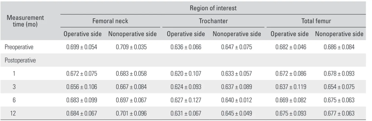
Table 1. Preoperative and Postoperative BMDs of Both Sides of the Hip
Measurement time (mo)
Region of interest
Femoral neck Trochanter Total femur
Operative side Nonoperative side Operative side Nonoperative side Operative side Nonoperative side Preoperative 0.699 ± 0.054 0.709 ± 0.035 0.636 ± 0.066 0.647 ± 0.075 0.682 ± 0.046 0.686 ± 0.084 Postoperative
1 0.672 ± 0.075 0.683 ± 0.058 0.620 ± 0.107 0.633 ± 0.057 0.672 ± 0.086 0.678 ± 0.093 3 0.656 ± 0.106 0.667 ± 0.084 0.624 ± 0.093 0.637 ± 0.089 0.637 ± 0.119 0.654 ± 0.075 6 0.683 ± 0.099 0.697 ± 0.067 0.627 ± 0.127 0.640 ± 0.012 0.669 ± 0.082 0.675 ± 0.063 12 0.684 ± 0.067 0.701 ± 0.096 0.631 ± 0.067 0.645 ± 0.049 0.675 ± 0.093 0.677 ± 0.063 BMD: bone mineral density, TKA: total knee arthroplasty.
each subject, one was bone density of the proximal femur that underwent TKA (ipsilateral side), and the other was bone density of the proximal femur that did not undergo TKA (contralateral side). There were no statistically sig- nificant differences in mean baseline BMD of the femur neck, trochanter, or total area between the ipsilateral and the contralateral side. (femur neck, p = 0.153; trochanter, p
= 0.184; total area, p = 0.169).
On the ipsilateral side, BMD in the femur neck was measured as 0.672 ± 0.075 g/cm2 after postoperative one month, and 0.656 ± 0.106 g/cm2 after postoperative three months, and these values were significantly lower than baseline BMD. BMD in the trochanter was measured as 0.620 ± 0.107 g/cm2 after postoperative one month and 0.624 ± 0.093 g/cm2 after postoperative three months, and these values were significantly lower than baseline BMD.
BMD in the total area was measured as 0.637 ± 0.119 g/
cm2 after postoperative three months, and these values were significantly lower than baseline BMD.
On the contralateral side, BMD in the femur neck was measured as 0.683 ± 0.058 g/cm2 after postoperative one month, and 0.667 ± 0.084 g/cm2 after postoperative three months, and these values were significantly lower than baseline BMD. BMD in the trochanter was measured as 0.633 ± 0.057 g/cm2 after postoperative one month, and 0.637 ± 0.089 g/cm2 after postoperative three months, and these values were significantly lower than baseline BMD.
BMD in the total area was measured as 0.654 ± 0.075 g/
cm2, and these values were significantly lower than base- line BMD (Table 1). However, when the LSC was not included in the calculation, only BMDs in the femoral neck and total area after postoperative three months were significantly lower than baseline BMD.
No statistical differences in changes of BMD were observed between the two groups at each measurement time (Table 2). The changes (%) of BMD after postopera- tive one year are shown in Table 3.
DISCUSSION
Severe complications in elderly patients with a hip frac- ture included pneumonia, thromboembolism, and death.
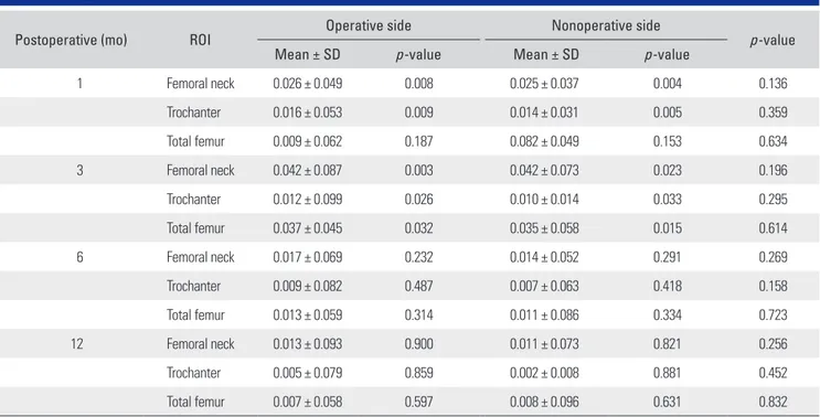
Known risk factors for hip fracture include falls, proximal femoral geometry, vitamin D level, muscle strength, and Table 2. Changes in BMD from the Preoperative Baseline after TKA
Postoperative (mo) ROI Operative side Nonoperative side
p-value
Mean ± SD p-value Mean ± SD p-value
1 Femoral neck 0.026 ± 0.049 0.008 0.025 ± 0.037 0.004 0.136
Trochanter 0.016 ± 0.053 0.009 0.014 ± 0.031 0.005 0.359
Total femur 0.009 ± 0.062 0.187 0.082 ± 0.049 0.153 0.634
3 Femoral neck 0.042 ± 0.087 0.003 0.042 ± 0.073 0.023 0.196
Trochanter 0.012 ± 0.099 0.026 0.010 ± 0.014 0.033 0.295
Total femur 0.037 ± 0.045 0.032 0.035 ± 0.058 0.015 0.614
6 Femoral neck 0.017 ± 0.069 0.232 0.014 ± 0.052 0.291 0.269
Trochanter 0.009 ± 0.082 0.487 0.007 ± 0.063 0.418 0.158
Total femur 0.013 ± 0.059 0.314 0.011 ± 0.086 0.334 0.723
12 Femoral neck 0.013 ± 0.093 0.900 0.011 ± 0.073 0.821 0.256
Trochanter 0.005 ± 0.079 0.859 0.002 ± 0.008 0.881 0.452
Total femur 0.007 ± 0.058 0.597 0.008 ± 0.096 0.631 0.832
BMD: bone mineral density, TKA: total knee arthroplasty, ROI: region of interest, SD: standard deviation.
Table 3. Changes (%) in BMD at Postoperative One Year
Variable Operative side (%) Nonoperative side (%)
Femur neck 2.14 1.12
Trochanter 0.78 0.03
Total femur 1.01 1.31
BMD: bone mineral density.
BMD.10,11) Among these, decrease in BMD is an important factor. Melton et al.12) reported that the more BMD de- creased, the greater the risk of femur neck fracture as the square of BMD change, and greater the risk of trochanteric fracture as the cube of BMD change.
If a certain area of an object under loading shows a change in material property or mechanical property or mi- crostructure, the transmission of stress is changed. Chang- es of the knee, such as osteoarthritis or TKA would have an effect on the mechanical properties of surrounding regions.13,14) Many studies have reported that knee osteo- arthritis influences bone strength of the proximal femur and patients with osteoarthritis usually have increased BMD in the femur neck.15-18) In our study, preoperatively, BMD of the hip on the operative side was lower than on the nonoperative side in accordance with the study re- ported by Ishii et al.7) However, no statistical differences were observed. Soininvaara et al.6) reported that preopera- tive BMD of the proximal femur on the operative side was significantly lower than that of the contralateral side in all region of interests. Considering the results, degree of exercise restriction due to pain and limited range of mo- tion of the joint, degree of osteoarthritis, pain scale before operation, dominant-leg, individual demands in daily life, and discordance between BMD of the left- and right-hip were considered compositively.19-22) Also, according to the results of our analysis, the fact that 83% of the nonopera- tive side showed Kellgren-Lawrence stage II or less (83%) should be considered, and there was a limit to the applica- tion of the result to severe osteoarthritis of both knees.
Regarding discordance between BMD of the left- and right-hip, many studies have reported that the differ- ence was not statistically significant, and BMD measure- ments of both hips were meaningless.22) However, some studies have reported that measurement of BMD of both hips has an effect on diagnosis according to WHO osteo- porosis diagnostic criteria, asserting the need for measure- ment of BMD in both hips.22) However, in our study, we analyzed changes of BMD on each side and differences of the change in BMD between the operative side and non- operative side. Therefore, discordance between BMD of the left- and right-hip was not our concern.
BMD in the femoral neck and total area showed a significant decrease after postoperative three months on both the nonoperative side and the operative side.
However, BMD at twelve months after operation showed similar values compared to preoperative levels, but lower than baseline, and followed predicted rates of age related loss.17,21,23,24) We believe that an early decrease of BMD occurred with decreased activity and weight load due to
postoperative pain, and less recovery of general condition and nutritional state before postoperative three months and recovery to baseline BMD after six months were due to their restoration.
Our results followed predicted rates of age related loss at postoperative one year. Age-related loss of BMD after postoperative one year differs according to ethnic group. Hannan et al.21) reported BMD losses of 0.04%–
2.85% per year. Dennison et al.23) reported that women aged 60–75 years showed a BMD loss of 1.43% naturally.
Young et al.24) reported that changes of BMD in white people was 0.35%–0.96% in the femur neck, 0.32%–0.95%
in total, and 0.36%–1.14% in the trochanter. In our study, for change in BMD after TKA at postoperative one year, recovery of BMD after the operation at one year was not sufficient to reach a baseline state and followed age related loss. This differed from the report by Ishii et al.,7) which indicated that early intervention by TKA might also be protective against later hip fracture.
We checked the difference in BMD of both hips in order to determine whether TKA can affect proximal BMD differently between the operative side and nonop- erative side. According to our results, there was no signifi- cant difference in change of BMD between the operative side and nonoperative side. Therefore, we suggest that changes in general condition, nutritional state, and degree of weight bearing did not influence BMD of either hip dif- ferently. In addition, factors directly and indirectly related to TKA, such as less recovery of range of motion and de- crease of periprosthetic BMD, did not influence either hip differently. In considering these results, a decrease in post- operative BMD is not a direct cause of ipsilateral femoral neck stress fracture after TKA.1-5) We deem there could be another cause, such as the occurrence of femoral neck microfracture intraoperatively because of hammering or forceful manipulation in osteoporotic patients. A con- strained type design would be a cause that would demand increased range of motion and result in increased bending and shearing force on the hip joint.
There were some limitations of our study. First, the number of patients was only 49 of 871 cases who under- went an operation during the research period. Second, our study may include more ambulating patients and patients who showed good results, who visited the hospital regular- ly and accepted measurements of BMD five times for a set period. Third, we evaluated bone strength with only BMD.
We suggest that further study is needed, with inclusion of a larger number of cases, and additional analysis together with bone quality, such as the microstructure.
Although TKA has an effect on BMD in both the
femur neck and trochanter after postoperative one month and three months, it does not affect the ipsilateral side and contralateral side differently. Therefore, we thought that a temporary decrease in BMD after TKA was not the direct cause of ipsilateral femoral stress fracture, but the indirect cause of ipsilateral femur neck stress fractures after TKA.
Therefore, there is a need to conduct a variable study on
the causes of ipsilateral femoral neck stress fracture after TKA.
CONFLICT OF INTEREST
No potential conflict of interest relevant to this article was reported.
REFERENCES
1. Joshi N, Pidemunt G, Carrera L, Navarro-Quilis A. Stress fracture of the femoral neck as a complication of total knee arthroplasty. J Arthroplasty. 2005;20(3):392-5.
2. Hendel D, Beloosesky Y, Weisbort M. Fracture of the hip af- ter knee arthroplasty: an unusual case with pain in the knee.
Acta Orthop Scand. 2001;72(2):194-5.
3. Pankaj A, Malhotra R, Logani V, Bhan S. Bilateral femoral neck stress fractures following total knee arthroplasty: a case report and review of literature. Arch Orthop Trauma Surg.
2007;127(7):549-52.
4. Atalar H, Aytekin MN, Gunay C, Yavuz OY. Stress fracture of the femoral neck as a complication of revision arthroplas- ty of the knee: a case report. Acta Orthop Belg. 2008;74(3):
418-20.
5. Lesniewski PJ, Testa NN. Stress fracture of the hip as a com- plication of total knee replacement: case report. J Bone Joint Surg Am. 1982;64(2):304-6.
6. Soininvaara TA, Miettinen HJ, Jurvelin JS, Alhava EM, Kroger HP. Bone mineral density in the proximal femur and contralateral knee after total knee arthroplasty. J Clin Densi- tom. 2004;7(4):424-31.
7. Ishii Y, Yagisawa K, Ikezawa Y. Changes in bone mineral density of the proximal femur after total knee arthroplasty. J Arthroplasty. 2000;15(4):519-22.
8. Kellgren JH, Lawrence JS. Radiological assessment of osteo- arthrosis. Ann Rheum Dis. 1957;16(4):494-502.
9. Gluer CC. Monitoring skeletal changes by radiological tech- niques. J Bone Miner Res. 1999;14(11):1952-62.
10. Im GI, Lim MJ. Proximal hip geometry and hip fracture risk assessment in a Korean population. Osteoporos Int.
2011;22(3):803-7.
11. Nguyen TV, Center JR, Eisman JA. Femoral neck bone loss predicts fracture risk independent of baseline BMD. J Bone Miner Res. 2005;20(7):1195-201.
12. Melton LJ 3rd, Wahner HW, Richelson LS, O'Fallon WM, Riggs BL. Osteoporosis and the risk of hip fracture. Am J
Epidemiol. 1986;124(2):254-61.
13. Soininvaara T, Nikola T, Vanninen E, Miettinen H, Kroger H.
Bone mineral density and single photon emission computed tomography changes after total knee arthroplasty: a 2-year follow-up study. Clin Physiol Funct Imaging. 2008;28(2):
101-6.
14. Liu TK, Yang RS, Chieng PU, Shee BW. Periprosthetic bone mineral density of the distal femur after total knee arthro- plasty. Int Orthop. 1995;19(6):346-51.
15. Naitou K, Kushida K, Takahashi M, Ohishi T, Inoue T. Bone mineral density and bone turnover in patients with knee os- teoarthritis compared with generalized osteoarthritis. Calcif Tissue Int. 2000;66(5):325-9.
16. Burger H, van Daele PL, Odding E, et al. Association of ra- diographically evident osteoarthritis with higher bone min- eral density and increased bone loss with age: the Rotterdam Study. Arthritis Rheum. 1996;39(1):81-6.
17. Hart DJ, Cronin C, Daniels M, Worthy T, Doyle DV, Spector TD. The relationship of bone density and fracture to inci- dent and progressive radiographic osteoarthritis of the knee:
the Chingford Study. Arthritis Rheum. 2002;46(1):92-9.
18. Leblanc AD, Schneider VS, Evans HJ, Engelbretson DA, Krebs JM. Bone mineral loss and recovery after 17 weeks of bed rest. J Bone Miner Res. 1990;5(8):843-50.
19. van der Poest Clement E, van der Wiel H, Patka P, Roos JC, Lips P. Long-term consequences of fracture of the lower leg:
cross-sectional study and long-term longitudinal follow-up of bone mineral density in the hip after fracture of lower leg.
Bone. 1999;24(2):131-4.
20. Weinreb M, Rodan GA, Thompson DD. Osteopenia in the immobilized rat hind limb is associated with increased bone resorption and decreased bone formation. Bone.
1989;10(3):187-94.
21. Hannan MT, Felson DT, Dawson-Hughes B, et al. Risk fac- tors for longitudinal bone loss in elderly men and women:
the Framingham Osteoporosis Study. J Bone Miner Res.
2000;15(4):710-20.
22. Petley GW, Taylor PA, Murrills AJ, Dennison E, Pearson G, Cooper C. An investigation of the diagnostic value of bilateral femoral neck bone mineral density measurements.
Osteoporos Int. 2000;11(8):675-9.
23. Dennison E, Eastell R, Fall CH, Kellingray S, Wood PJ, Cooper C. Determinants of bone loss in elderly men and
women: a prospective population-based study. Osteoporos Int. 1999;10(5):384-91.
24. Young R, May H, Murphy S, Grey C, Compston JE. Rates of bone loss in peri- and postmenopausal women: a 4 year, prospective, population-based study. Clin Sci (Lond).
1996;91(3):307-12.