A cross-sectional retrospective study to analyze the underlying causes and clinical characteristics of children with reactive
thrombocytosis at a Korean tertiary medical center
Juhee Shin, Dong Hyun Lee, Nani Jung, Hee Joung Choi, Ye Jee Shim
Department of Pediatrics, Keimyung University School of Medicine and Dongsan Medical Center, Daegu, Korea
p-ISSN 2287-979X / e-ISSN 2288-0011 https://doi.org/10.5045/br.2018.53.3.233 Blood Res 2018;53:233-239.
Received on April 2, 2018 Revised on June 4, 2018 Accepted on June 26, 2018
Background
Reactive thrombocytosis (RT) is a common condition among children, although no stud- ies have examined the etiology or clinical characteristics of RT among Korean children.
Methods
This retrospective study evaluated children with RT at a single Korean tertiary center dur- ing a 10-year period.
Results
RT accounted for 13.5% of children who were admitted to the pediatric ward (4,113/30,355): mild RT, 82.7%; moderate RT, 14.1%; severe RT, 1.1%; and extreme RT, 2.1%. There was a negative correlation between platelet count and Hb level (P=0.008).
There were positive correlations between platelet count and WBC (P=0.001), eryth- rocyte sedimentation rate (ESR) (P=0.007), and admission duration (P=0.006). The most common cause of RT was infection and the second most common was Kawasaki disease (KD). The highest proportion of lower respiratory tract infection was observed in extreme RT (P<0.001). The proportion of KD was highest in extreme RT (P<0.001) and in children aged 1‒7.9 years (P<0.001). The proportion of refractory KD was highest in extreme RT (P=0.005). In cases of KD, there was a positive correlation between platelet count and fever duration (P=0.006). Non-KD autoimmune inflammation was only observed in mild/moderate RT, and its proportion was highest in children aged 8‒18 years (P<0.001).
Conclusion
In children, more severe RT was associated with lower Hb, increased WBC, ESR, and pro- longed admission. With respiratory infection or KD, extreme RT was associated with more severe disease course.
Key Words Reactive thrombocytosis, Extreme thrombocytosis, Children, Etiology, Kawasaki disease
Correspondence to Ye Jee Shim, M.D., Ph.D.
Department of Pediatrics, Keimyung University School of Medicine, Dongsan Medical Center, Dalseong-ro 56, Jung-gu, Daegu 41931, Korea
E-mail: [email protected]
Ⓒ 2018 Korean Society of Hematology
INTRODUCTION
Thrombocytosis is a common condition among children, and it is a frequent cause of referral to a pediatric hema- to-oncologist or medical institution for further testing, ac- counting for 6–15% of hospitalizations and/or visits to an ambulatory/emergency clinic among children [1-3]. Thro- mbocytosis can be classified as primary or reactive thrombo- cytosis (RT), with primary thrombocytosis involving a pri- mary bone marrow disorder, such as essential thrombocy-
tosis, polycythemia vera, chronic myeloid leukemia, or mye- lodysplastic syndrome [4]. In contrast, RT is much more frequent than primary thrombocytosis and is caused by non-bone marrow related medical or surgical conditions, which are most often viral or bacterial infections [1, 5].
There are national reports regarding the etiology and clinical characteristics of RT among children from several countries [2, 6-11], although there are no data regarding RT among children in Korea. The present retrospective study aimed to determine the etiology and clinical characteristics of RT among children who were admitted to a single Korean center
Table 1. Baseline characteristics and underlying causes reactive thrombocytosis in 4,113 children.
Characteristics N
Age in years, median (range) 0.3 (0–16.9)
<1 yr, N (%) 2,922 (71.0)
1–7.9 yr, N (%) 1,066 (25.9)
8–18 yr, N (%) 125 (3.1)
Gender, male:female 1,780:2,333
Platelet count ×103/L, median (range) 579 (501–1,732) Severity classification, N (%)
Mild (grade 1) 3,400 (82.7)
Moderate (grade 2) 580 (14.1)
Severe (grade 3) 46 (1.1)
Extreme (grade 4) 87 (2.1)
Underlying disease, N (%)
Infection 3,216 (78.2)
Lower respiratory tract infection 1,145
Upper respiratory tract infection 714
Gastrointestinal infection 471
Urinary tract infection 416
Meningitis 169
Bacteremia or septicemia 138
Other infections 163
Kawasaki diseasea) 247 (6.0) Atypical Kawasaki disease 101
Refractory Kawasaki disease 90
Tissue damage 149 (3.6) Congenital malformationb) 45
Gastroesophageal reflux disease 32
Hemorrhage 19
Intussusception 8
Appendicitis 7
Colon polyp 5
Gastric ulcer 7
Foreign body 5
Fracture or trauma 5
Burn 2
Others 14
Neurologic disease 115 (2.8) Seizure 76
Other neurologic problems 39
Allergy 76 (1.9) Allergic colitis 52
Atopic dermatitis & contact dermatitis 9
Urticaria 8
Anaphylaxis 2
Other allergy 5
Perinatal disease 74 (1.8) Neonatal jaundice 50
Feeding difficulty or vomiting in newborns 16
Neonatal pneumonia 5
Others 3
Hemato-oncologic disease 61 (1.5) Malignant neoplasm 21
Benign neoplasm 16
Anemia 15
Others 10
Autoimmune inflammationa) 54 (1.3) Henoch-Schönlein purpura 24
Inflammatory bowel disease 23
Juvenile idiopathic arthritis 7
Others 121 (2.9)
a)Autoimmune inflammation was classified separately from Kawasaki disease. b)Congenital malformation includes hypertrophic pyloric stenosis, Hirschsprung disease, biliary atresia, choledochal cyst, congenital hydronephrosis, imperforate anus, and tracheoeso- phageal fistula.
during a 10-year period, and to compare the results to those from other countries.
MATERIALS AND METHODS
Participants and ethics statement
In this retrospective study, we collected data for children
<18 years old with a platelet count of >500×109/L, who were admitted to the pediatric ward in Keimyung University Dongsan Medical Center between January 2006 to December 2016. We excluded healthy newborns, premature infants who were admitted to the neonatal intensive care unit, and patients with primary thrombocytosis. The study protocol was approved by the institutional review board of Keimyung University Dongsan Medical Center (2017-11-038).
Definitions
Cases of RT were classified based on previous criteria [9, 12] as mild (grade 1, platelet count of 501–700, ×109/L), moderate (grade 2, platelet count of 701–900, ×109/L), severe (grade 3, platelet count of 901–1,000, ×109/L), and extreme (grade 4, platelet count of >1,000, ×109/L).
Kawasaki disease (KD) is diagnosed by prolonged fever (≥5 days) and at least four of five clinical features: changes in the extremities, polymorphous exanthema, bilateral bulbar conjunctival injection without exudate, changes in the lips and oral cavity, and cervical lymphadenopathy (>1.5 cm diameter) [13]. According to the American Heart Association, atypical KD is possible in children (≥6 mo) with prolonged fever and two or three principle features or in infants with fever (≥7 days) without other explanation [13]. Refractory KD is defined as persistent fever that lasts for more than 24–36 hours after the end of initial KD treatment [14].
Statistical analysis
Patients’ baseline characteristics were reported as median and range, although intragroup comparisons were performed using mean and 95% confidence interval (CI) values. Analysis of variance with Bonferroni adjustment was used to compare mean values between the groups, and intragroup comparisons of proportions were performed using the linear by linear chi-square statistic. Pearson’s correlation analysis was per- formed to define correlations between the variables. All stat- istical analyses were performed using IBM SPSS version 23.0 (IBM Corp., Armonk, NY, USA), and P-values <0.05 were considered statistically significant.
RESULTS
Baseline characteristics and underlying causes of RT Patients’ baseline characteristics and underlying causes of RT are described in Table 1. During the study period, RT accounted for 13.5% of children who were admitted to the pediatric ward (4,113/30,355). The median age of children with RT was 0.3 years, and the proportion aged
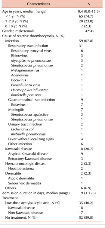
Table 2. Baseline characteristics of 87 children with extreme reactive thrombocytosis.
Characteristics N
Age in years, median (range) 0.4 (0.0–15.8)
<1 yr, N (%) 65 (74.7)
1–7.9 yr, N (%) 20 (23.0)
8–18 yr, N (%) 2 (2.3)
Gender, male:female 42:45
Cause of reactive thrombocytosis, N (%)
Infection 59 (67.8)
Respiratory tract infection 31 Respiratory syncytial virus 6
Rhinovirus 3
Mycoplasma pneumoniae 3
Streptococcus pneumoniae 2
Metapneumovirus 1
Adenovirus 1
Bocavirus 1
Parainfluenza virus 1
Haemophilus influenzae 1
Bordetella pertussis 1
Gastrointestinal tract infection 9
Rotavirus 7
Meningitis 7
Streptococcus agalactiae 3 Streptococcus pneumoniae 1
Urinary tract infection 2
Escherichia coli 1
Klebsiella pneumoniae 1 Fever without localizing signs 4
Other infection 6
Kawasaki disease 18 (20.7)
Atypical Kawasaki disease 9 Refractory Kawasaki disease 3
Hemato-oncologic disease 2 (2.3)
Hepatoblastoma 2
Dermatitis 2 (2.3)
Atopic dermatitis 1
Seborrheic dermatitis 1
Others 6 (6.9)
Admission duration in days, median (range) 9 (3–123) Treatment
Low-dose acetylsalicylic acid, N (%) 35 (40.2)
Kawasaki disease 18
Non-Kawasaki disease 17
No treatment, N (%) 52 (59.8)
<1 year was 71.0%. Among a total 4,113 children, 82.7%
of cases involved mild RT, 14.1% involved moderate RT, 1.1% involved severe RT, and 2.1% of cases involved extreme RT. The most common cause of RT was infection (78.2%) and the second most common cause was KD (6.0%). The characteristics and underlying clinical conditions of the 87 children with extreme RT are described in Table 2. The median age at diagnosis of extreme RT was 0.4 years, and the proportion of children who were <1 year old was 74.7%.
The most common cause of extreme RT was infection (67.8%), which most frequently involved respiratory tract
infection; the second most common cause of extreme RT was KD (20.7%).
Comparisons according to RT severity
Table 3 shows the variables according to RT severity.
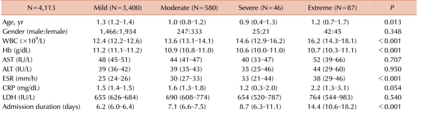
Significant differences according to RT severity were de- tected for hemoglobin (Hb) levels, white blood cell (WBC) count, erythrocyte sedimentation rate (ESR), and admission duration. No significant differences were detected in terms of age, gender ratio, liver function results, C-reactive protein levels, and lactate dehydrogenase levels. Correlation analysis revealed a negative correlation between platelet count and Hb level (r=-0.083, P<0.001), as well as positive correlations between platelet count and WBC count (r=0.145, P<0.001), between platelet count and ESR (r=0.158, P<0.001), and between platelet count and admission duration (r=0.188, P< 0.001) (Fig. 1). After controlling for patient age, disease cat- egory, liver function, C-reactive protein levels, and lactate dehydrogenase levels, the negative correlation between plate- let count and Hb level persisted (r=-0.251, P=0.008). In addi- tion, independent positive correlations persisted between pla- telet count and WBC count (r=0.305, P=0.001), between pla- telet count and ESR (r=0.252, P=0.007), and between platelet count and admission duration (r=0.257, P=0.006).
Comparing underlying medical conditions according to RT severity
In cases of respiratory tract infection, the proportions of lower and upper respiratory tract infections were sig- nificantly different according to the severity of RT (Table 4). The highest proportion of lower respiratory tract infection was observed in the extreme RT group (P<0.001). In cases of KD, the proportions of RT cases that involved KD were 5.2% (178/3,400) for mild RT, 7.9% (46/580) for moderate RT, 10.9% (5/46) for severe RT, and 20.7% (18/87) for ex- treme RT (Fig. 2). The differences according to RT severity were significant, and the highest proportion was observed in the extreme RT group (P<0.001). The proportions of refractory KD were significantly different according to RT severity (P=0.005) (Table 4). The proportions of atypical KD were not significantly different according to the RT severity (P=0.319). In cases of children with RT who had KD, correlation analysis revealed a positive correlation be- tween platelet count and fever duration (r=0.175, P=0.006).
Non-KD autoimmune inflammation (Henoch-Schönlein purpura, juvenile idiopathic arthritis, Crohn disease, or ulcer- ative colitis) was not involved in cases of severe or extreme RT, and was limited to mild RT cases (1.4%, 46/3,400) and moderate RT cases (1.7%, 10/580) (Fig. 2).
Age-related distributions of underlying diseases
Children were classified by age into the following groups:
children <1 year old, those 1–7.9 years old, and those 8–18 years old. The proportions of KD were 2.6% (77/2,922) in the <1-year-old group, 15.6% (166/1,066) in the 1–
7.9-year-old group, and 3.2% (4/124) in the 8–18-year-old group. The proportions of non-KD autoimmune in-
Table 3. Comparison of variables according to the severity of reactive thrombocytosis.
N=4,113 Mild (N=3,400) Moderate (N=580) Severe (N=46) Extreme (N=87) P
Age, yr 1.3 (1.2–1.4) 1.0 (0.8–1.2) 0.9 (0.4–1.3) 1.2 (0.7–1.7) 0.013
Gender (male:female) 1,466:1,934 247:333 25:21 42:45 0.348
WBC (×109/L) 12.4 (12.2–12.6) 13.6 (13.1–14.1) 14.6 (12.9–16.2) 16.2 (14.3–18.1) <0.001
Hb (g/dL) 11.2 (11.1–11.2) 10.9 (10.8–11.0) 10.6 (10.0–11.0) 10.7 (10.3–11.1) <0.001
AST (IU/L) 48 (45–51) 44 (41–47) 40 (33–47) 52 (39–66) 0.707
ALT (IU/L) 39 (36–42) 39 (35–43) 35 (25–46) 44 (29–60) 0.950
ESR (mm/h) 25 (24–26) 30 (27–33) 33 (21–44) 38 (29–46) <0.001
CRP (mg/dL) 1.5 (1.4–1.5) 1.6 (1.3–1.8) 1.2 (0.3–2.0) 2.2 (1.3–3.1) 0.054
LDH (IU/L) 655 (626–684) 690 (608–774) 654 (520–787) 764 (544–983) 0.540
Admission duration (days) 6.2 (6.0–6.4) 7.1 (6.6–7.5) 8.7 (6.3–11.1) 14.4 (10.6–18.2) <0.001 Values are presented as mean and 95% confidence intervals.
Abbreviations: ALT, alanine transaminase; AST, aspartate transaminase; CRP, C-reactive protein; ESR, erythrocyte sedimentation rate; LDH, lactate dehydrogenase.
Fig. 1. Correlation analysis revealed a negative correlation between platelet count and hemoglobin level (A), as well as positive correlations between platelet count and white blood cell count (B), between platelet count and erythrocyte sedimentation rate (C), and between platelet count and admission duration (D).
flammation (Henoch-Schönlein purpura, juvenile idiopathic arthritis, Crohn disease, or ulcerative colitis) were 0%
(0/2,922) in the <1-year-old group, 2.4% (26/1,066) in the
1–7.9-year-old group, and 21.8% (27/124) in the 8–
18-year-old group. The proportion of KD was significantly higher in the 1–7.9-year-old group and the proportion of
Table 4. Comparison of the proportions of lower and upper respiratory infections, and refractory and atypical Kawasaki disease, according to the severity of reactive thrombocytosis.
(N=1,546)Mild Moderate
(N=258) Severe
(N=21) Extreme
(N=34) P
Respiratory tract infection (N, %)
Lower respiratory tract 928 (60.0) 173 (67.1) 16 (76.2) 28 (82.4) <0.001
Upper respiratory tract 618 (40.0) 85 (32.9) 5 (23.8) 6 (17.6)
(N=178)Mild Moderate
(N=46) Severe
(N=5) Extreme
(N=18) P
Typical and atypical Kawasaki disease (N, %)
Typical 108 (60.7) 25 (54.3) 3 (60.0) 9 (50.0) 0.319
Atypical 70 (39.3) 21 (45.7) 2 (40.0) 9 (50.0)
Refractory and non-refractory Kawasaki disease (N, %)
Refractory 56 (31.5) 21 (45.7) 2 (40.0) 11 (61.1) 0.005
Non-refractory 122 (68.5) 25 (54.3) 3 (60.0) 7 (38.9)
Fig. 2. The proportion of Kawasaki disease (navy arrow) according to the severity of reactive thrombocytosis (RT) among children who were admitted to a single Korean tertiary center. The proportion of Kawasaki disease was significantly different between the groups by reactive thrombocytosis severity, with the highest proportion in the extreme RT group (P<0.001).
Fig. 3. The proportions of Kawasaki disease (navy arrow) and autoimmune inflammation (white arrow with black outline) according to patient age. The proportion of Kawasaki disease was highest in the 1–7.9-year-old group, and the proportion of inflammation was highest in the 8–18-year-old group (P<0.001). Autoimmune inflammation includes Henoch-Schönlein purpura, juvenile idiopathic arthritis, Crohn disease, and ulcerative colitis.
autoimmune inflammation was significantly higher in the 8–18-year-old group (both P<0.001) (Fig. 3).
Treatment and clinical outcome of children with RT Treatment of underlying disorders was performed in par- ticipants, and platelet counts gradually decreased after the underlying cause was resolved. Among the 4,113 children, 2.1% of cases had extreme RT. Approximately 40% of chil- dren with extreme RT received low-dose acetylsalicylic acid;
of these children, over 50% had KD and the remainder did not (Table 2). None of the patients experienced clinical complications (such as thromboembolic events or bleeding)
that were associated with extreme RT.
DISCUSSION
Among the children who were admitted to our center, the prevalence of RT was 13.5% (4,113/30,355), which is similar to the prevalence in other countries [1-3]. The in- cidence of RT is age-dependent, with much higher rates among young children, especially those who are <1–2 years old [2, 3, 6, 7, 9-11] and much lower rates among children
aged >11–15 years [2, 3]. In the present study, infants who were <1 year old accounted for >70% of all RT cases.
This phenomenon is related to the correlation between serum thrombopoietin (TPO) levels and age-dependent platelet changes [15], with serum TPO levels being highest during the neonatal period and gradually decreasing with age [15].
Despite the prevalence of RT among children, extreme RT is very rare and accounts for approximately 2% of RT cases [3, 6], which is similar to the rate in the present study (2.1%, 87/4,331). As previously reported [3], extreme RT was associated with low Hb levels, high WBC counts, and a prolonged admission period. Platelet counts were not corre- lated with C-reactive protein levels, as has been previously reported [3], although extreme RT was associated with high ESR, and platelet count was positively correlated with ESR in the present study.
Respiratory tract infections are the most common causes of RT and extreme RT [8, 10, 16], with upper respiratory tract infections being common in cases of mild thrombocy- tosis and lower respiratory tract infections being common in cases of severe thrombocytosis [8]. In the present study, the highest proportion of lower respiratory tract infection was observed in the extreme RT group. In other words, more severe RT was associated with more severe respiratory tract infection. In the present study, respiratory syncytial virus (RSV) and rhinovirus were the most common pathogens in cases of respiratory tract infection with extreme RT (Table 2), and similar results have been previously reported [17].
Levels of interleukin (IL)-6 or IL-8 can increase during RSV infection [18], which is related to the activation of bronchial epithelial cell secretion and stimulation of platelet production [18].
In this study, KD was the second most common cause of all cases of RT (6.0%, 247/4,113) and extreme RT (20.7%, 18/87). Similarly, KD causes RT in approximately 9.4% of Japanese pediatric cases [2]; these results are markedly higher than those in other countries [6-11], which is likely related to the high prevalence of KD in Asian countries, including Korea and Japan [19, 20]. Interestingly, the proportions of both KD and refractory KD were highest in the extreme RT subgroup in this study. Further, correlation analysis re- vealed a positive correlation between platelet count and fever duration in children with KD and RT. In other words, more severe RT was associated with more severe KD.
Thrombocytosis in cases of KD is an important condition, as it increases the risk of coronary aneurysm thrombosis, myocardial infarction, and death [21]. Among patients with KD, RT can be affected by the levels of various cytokines, such as IL-1, IL-6, TPO, and tumor necrosis factor α[21], with TPO being the main cytokine that affects platelet pro- duction and megakaryocyte development [21]. The liver and kidneys produce TPO, and serum concentrations are regu- lated through binding to c-Mpl on platelets and mega- karyocytes [22]. Interestingly, TPO levels are usually ele- vated during the first week of the acute infection phase, with subsequent decreases that correspond to improvement in the symptoms of infection [23, 24]. However, platelet
counts increase during the second and third weeks of in- fection [23, 24], which may reflect a gradual effect of TPO levels on platelet production through the c-Mpl ligand mech- anism [23, 24].
In a Japanese study, autoimmune diseases (juvenile idio- pathic arthritis, inflammatory bowel diseases, and KD) ac- counted for 4–11% of pediatric RT cases [2], with KD being the principal cause of RT among children <7 years old and other diseases being the main causes among those aged
>11 years [2]. In the present study, the cause of RT was classified as KD or non-KD autoimmune inflammation, which included Henoch-Schönlein purpura, juvenile idio- pathic arthritis, and inflammatory bowel diseases. Similar to the findings of the Japanese study [2], the present study revealed that the proportion of KD was highest in the 1–
7.9-year-old group and the proportion of non-KD auto- immune inflammation was highest in the 8–18-year-old group (P<0.001).
In most cases, RT is benign and the platelet count is nor- malized after treatment of the primary disease, in the absence of complications [10, 25]. Furthermore, most patients with RT do not require prophylactic treatment using anti- coagulants or platelet aggregation inhibitors [12]. Even in extreme cases of RT, there is no established prophylactic treatment or evidence to support the use of prophylactic drugs for asymptomatic children [12]. Thus, if thrombocy- tosis persists after treatment of RT that is based on the under- lying etiology of RT, treatment can be considered for throm- bocytosis [25].
There are some limitations in our study. Most patients with RT did not undergo blood testing until platelet counts had dropped to completely normal levels. Therefore, the exact duration of the platelet increase could not be calculated and was insufficient to be included as statistical data.
In conclusion, infection, especially respiratory tract in- fection, was the most common cause of RT among Korean children in this study, and KD was the second most common cause. Extreme RT was associated with leukocytosis, anemia, increased ESR, prolonged admission duration, more severe respiratory tract infection, and more severe KD. Therefore, RT could be a factor in evaluating the severity of disease and predicting hospitalization duration. In addition, the pres- ent study revealed that KD was an important cause of extreme RT and all RT among 1–7.9-year-old patients, whereas non-KD autoimmune inflammation was more likely to cause mild-to-moderate RT, especially among children ≥8 years old. However, the present study is limited by its single-center design, and further evaluation is needed to better understand the status of RT in Korea.
AuthorsÊ Disclosures of Potential Conflicts of Interest No potential conflicts of interest relevant to this article were reported.
REFERENCES
1. Mantadakis E, Tsalkidis A, Chatzimichael A. Thrombocytosis in childhood. Indian Pediatr 2008;45:669-77.
2. Matsubara K, Fukaya T, Nigami H, et al. Age-dependent changes in the incidence and etiology of childhood thrombocytosis. Acta Haematol 2004;111:132-7.
3. Wang JL, Huang LT, Wu KH, Lin HW, Ho MY, Liu HE.
Associations of reactive thrombocytosis with clinical characteristics in pediatric diseases. Pediatr Neonatol 2011;52:261-6.
4. Sanchez S, Ewton A. Essential thrombocythemia: a review of diagnostic and pathologic features. Arch Pathol Lab Med 2006;130:1144-50.
5. Kucine N, Chastain KM, Mahler MB, Bussel JB. Primary thrombocytosis in children. Haematologica 2014;99:620-8.
6. Demidowicz E, Moppert J, Nowacka AZ, Styczynski J, Wysocki M. Essential thrombocythemia and reactive thrombocytosis in children. EC Paediatrics 2016;2:107-15.
7. Heng JT, Tan AM. Thrombocytosis in childhood. Singapore Med J 1998;39:485-7.
8. Özcan C, Şaylı TR, Koşan-Çulha V. Reactive thrombocytosis in children. Turk J Pediatr 2013;55:411-6.
9. Sutor AH. Thrombocytosis in childhood. Semin Thromb Hemost 1995;21:330-9.
10. Vora AJ, Lilleyman JS. Secondary thrombocytosis. Arch Dis Child 1993;68:88-90.
11. Yohannan MD, Higgy KE, al-Mashhadani SA, Santhosh-Kumar CR. Thrombocytosis. Etiologic analysis of 663 patients. Clin Pediatr (Phila) 1994;33:340-3.
12. Dame C, Sutor AH. Primary and secondary thrombocytosis in childhood. Br J Haematol 2005;129:165-77.
13. McCrindle BW, Rowley AH, Newburger JW, et al. Diagnosis, treatment, and long-term management of Kawasaki disease: a scientific statement for health professionals from the American Heart Association. Circulation 2017;135:e927-99.
14. Kim HJ, Lee HE, Yu JW, Kil HR. Clinical outcome of patients with refractory Kawasaki disease based on treatment modalities.
Korean J Pediatr 2016;59:328-34.
15. Ishiguro A, Nakahata T, Matsubara K, et al. Age-related changes in thrombopoietin in children: reference interval for serum thrombopoietin levels. Br J Haematol 1999;106:884-8.
16. O'Shea J, Sherlock M, Philip R. Thrombocytosis in childhood.
Acta Haematol 2005;113:212.
17. Zheng SY, Xiao QY, Xie XH, et al. Association between secondary thrombocytosis and viral respiratory tract infections in children.
Sci Rep 2016;6:22964.
18. Lee FE, Walsh EE, Falsey AR, et al. Human infant respiratory syncytial virus (RSV)-specific type 1 and 2 cytokine responses ex vivo during primary RSV infection. J Infect Dis 2007;195:1779-88.
19. Park YW, Han JW, Park IS, et al. Kawasaki disease in Korea, 2003-2005. Pediatr Infect Dis J 2007;26:821-3.
20. Newburger JW, Taubert KA, Shulman ST, et al. Summary and abstracts of the Seventh International Kawasaki Disease Symposium: December 4-7, 2001, Hakone, Japan. Pediatr Res 2003;53:153-7.
21. Emmons RV, Reid DM, Cohen RL, et al. Human thrombopoietin levels are high when thrombocytopenia is due to megakaryocyte deficiency and low when due to increased platelet destruction.
Blood 1996;87:4068-71.
22. Kaushansky K. Thrombopoietin: the primary regulator of platelet production. Blood 1995;86:419-31.
23. Ishiguro A, Ishikita T, Shimbo T, et al. Elevation of serum thrombopoietin precedes thrombocytosis in Kawasaki disease.
Thromb Haemost 1998;79:1096-100.
24. Ishiguro A, Suzuki Y, Mito M, et al. Elevation of serum throm- bopoietin precedes thrombocytosis in acute infections. Br J Haematol 2002;116:612-8.
25. Sutor AH. Screening children with thrombosis for thrombophilic proteins. Cui bono? J Thromb Haemost 2003;1:886-8.