Highlights
• There are many unresolved clinical problems about mild traumatic brain injury (mTBI) such as a low sensitivity of standard neuroimaging studies, and absence of reliable predicting models.
• It is very difficult to diagnose mTBI in symptomatic patients in the absence of witnesses, clear signs of head trauma, and abnormalities on neuroimaging.
• Blood proteins have great potential as diagnostic and prognostic biomarkers of mTBI.
• Technological advances in the targeted proteomics are expected to realize the clinical potential of blood-based protein biomarkers.
Review
Received: Mar 12, 2017 Revised: Mar 20, 2017 Accepted: Mar 29, 2017 Correspondence to Byung-Mo Oh
Department of Rehabilitation Medicine, Seoul National University College of Medicine, Seoul National University Hospital, 101 Daehak-ro, Jongno-gu, Seoul 03080, Korea.
Tel: +82-2-2072-2619 Fax: +82-2-743-7473 E-mail: keepwiz@gmail.com
Current State and Prospects of Development of Blood-based
Biomarkers for Mild Traumatic Brain Injury
Hyun Haeng Lee, Woo Hyung Lee, Han Gil Seo, Dohyun Han, Youngsoo Kim, Byung-Mo Oh
ABSTRACT
The current understanding of the pathophysiology of mild traumatic brain injury (mTBI) is, without doubt, incomplete. Nevertheless, we tried to summarize the state-of-the-art explanation of how the brain is continuously injured even after a single impact. We also reviewed the real struggle of diagnosing mTBI, which culminated in showing the potential of blood-based biomarkers as an alternative or complementary way to overcome this difficulty.
Pathophysiology of mTBI is subdivided into primary and secondary injuries. Primary injury is caused by a direct impact on the head and brain. Secondary injury refers to the changes in energy metabolism and protein synthesis/degradation resulting from the biochemical cascades as follows; calcium influx, mitochondrial dysfunction, fractured microtubules, and Wallerian degeneration, neuroinflammation, and toxic proteinopathy. Since the diagnosis of mTBI is made through the initial clinical information, it is difficult and inaccurate to diagnose mTBI without the absence of a witness or sign of head trauma. Blood-based biomarkers are expected to play an important role in diagnosing mTBI and predicting functional outcomes, due to their feasibility and the recent progress of targeted proteomics techniques (i.e., liquid chromatography tandem mass spectrometry [LC-MS/MS]).
Keywords: Biomarkers; Brain Concussion; Blood; Proteomics
INTRODUCTION
Traumatic brain injury (TBI), which is known as a “silent epidemic,” is no longer silent. The report to Congress on TBI in the United States estimated that > 2 million people with TBI in the United States visit the emergency department each year [1]. Although there is no corresponding national statistic yet, according to the “2009 Trauma Statistics” published by the Centers for Disease Control and Prevention (CDC) of Korea, approximately 160,000 people experience TBI in South Korea annually. Mild TBI (mTBI), which accounts for approximately 80%–90% of all the traumatic brain damage, is also called concussion and is reported to occur in approximately 100–300 per million population worldwide [2]. However, considering that many patients with mTBI do not seek medical attention [3], the actual incidence of mTBI should be much more
Review
Received: Mar 12, 2017 Revised: Mar 20, 2017 Accepted: Mar 29, 2017 Correspondence to Byung-Mo Oh
Department of Rehabilitation Medicine, Seoul National University College of Medicine, Seoul National University Hospital, 101 Daehak-ro, Jongno-gu, Seoul 03080, Korea.
Tel: +82-2-2072-2619 Fax: +82-2-743-7473 E-mail: keepwiz@gmail.com Copyright © 2017. Korea Society for Neurorehabilitation
This is an Open Access article distributed under the terms of the Creative Commons Attribution Non-Commercial License (https://
creativecommons.org/licenses/by-nc/4.0) which permits unrestricted non-commercial use, distribution, and reproduction in any medium, provided the original work is properly cited.
Conflict of Interest
The author has no potential conflicts of interest to disclose.
Current State and Prospects of Development of Blood-based
Biomarkers for Mild Traumatic Brain Injury
Hyun Haeng Lee,1 Woo Hyung Lee,1 Han Gil Seo,1 Dohyun Han,2 Youngsoo Kim,3 Byung-Mo Oh1
1 Department of Rehabilitation Medicine, Seoul National University College of Medicine, Seoul National University Hospital, Seoul, Korea
2Proteomics Core Facility, Biomedical Research Institute, Seoul National University Hospital, Seoul, Korea
3Department of Biomedical Engineering, Seoul National University College of Medicine, Seoul, Korea
than 300 per million each year [2], even greater than 3 times the incidence of breast cancer [4]. Although the after-effects of mTBI are often mild, it has tremendous significance in public health. Some researchers report that approximately 44% of the medical expenditures associated with TBI are due to mTBI. The incidence of TBI, especially mTBI, is on the rise owing to an increase in fall cases among the elderly, increased accidents secondary to an increase in leisure sports, and an increase in regional tension and terrorism worldwide [5].
However, studies on the pathophysiology and long-term complications of mTBI are still at the infancy stage. The reasons are varied, but above all, most patients with mTBI appear almost completely recovered within a few months. A considerable number of persons with mTBI experience persistent symptoms after trauma for months, or even years. Patients with sequelae lasting > 1 year account for approximately 15% of all mTBI cases [6]; however, there is no reliable model to predict which patients will continue to have symptoms. Moreover, recent studies have shown that mTBI could be a risk factor for neurodegenerative diseases in the long term; hence, there is an increasing need for a prediction model [7-9]. The fact that it is difficult to assess functional decline and recovery in patients with mTBI also hampers research [10]. There is also a lack of sensitive and feasible tools for the measurements of mild cognitive impairment, decrease in thought speed, and change in behavior, which are the frequent complaints of patients with mTBI. Repeated measurements with typical neurocognitive tests provide the most sensitive results; however, these tests are expensive and require much effort.
In this article, we reviewed the pathophysiology of adult mTBI and the unresolved issues that arise from the clinical course of this condition, and subsequently explored the potentials of blood-based biomarkers in contributing to coping with these challenges. In children, mTBI is very common and involves unique clinical problems that differ from those in adults; however, they are beyond the scope of this review.
DEFINITION AND DIAGNOSTIC CRITERIA FOR MILD TBI
Clinical criteria should be used to diagnose mTBI because objective tests such as computed tomography (CT) and blood tests usually do not show any abnormal findings. Although the definition of mTBI is still controversial, the most widely accepted diagnostic criteria are those proposed by the American Congress of Rehabilitation Medicine (ACRM), which are used by the Centers for Disease Control and Prevention and the World Health Organization (WHO) [11] (Table 1). In short, mTBI is defined in terms of the duration of the initial Glasgow coma
Table 1. The mTBI diagnostic criteria proposed by ACRM [11]
Diagnostic criteria A patient with mTBI is a person who has had a traumatically induced physiological disruption of the brain function, as manifested by one or more of the following conditions:
- Any period of loss of consciousness for up to 30 min
- Any loss of memory for events immediately before or after the accident for up to 24 hr
- Any alteration of the mental state at the time of the accident (e.g., feeling dazed, disoriented, or confused) - Focal neurological deficit(s) that may or may not be transient
Exclusion criteria However, the severity of the injury should not exceed the following conditions:
- Loss of consciousness exceeding 30 min - PTA longer than 24 hr
- A GCS score that falls below 13 after 30 min
• Such anomalies should not be due to alcohol, recreational drugs, medications, systemic diseases, or extracranial damage • There should be no abnormality on imaging modalities such as CT or MRI (DVA and DoD guidelines)
mTBI, mild traumatic brain injury; ACRM, American Congress of Rehabilitation Medicine; PTA, post-traumatic amnesia; GCS, Glasgow coma scale; CT, computed tomography; MRI, magnetic resonance imaging; DVA, Department of Veterans Affairs; DoD, Department of Defense.
scale (GCS) score, duration of loss of consciousness, and post-traumatic amnesia (PTA). The WHO task force team added the exclusion criteria that these abnormalities should not be due to alcohol or other recreational drugs, medications, systemic diseases, or extracranial injuries [12]. According to the US Department of Veterans Affairs (DVA) and the Department of Defense (DoD) guidelines, lesions identified by using structural imaging modalities such as magnetic resonance imaging (MRI) or CT suggest that the condition is more severe than mild [13].
PATHOPHYSIOLOGY
In mTBI, neurological abnormalities, such as changes in consciousness or memory loss, appear immediately after the injury and improve within a relatively short period. Symptoms such as dizziness and impaired concentration mostly disappear within 1–2 weeks [14]. Clinically, the symptoms improve within 1–2 weeks, but 30–45 days are required for a complete recovery based on neuropsychological tests [15]. Nevertheless, it does not necessarily mean that the brain physiology recovers to normal. Many studies on the pathophysiology of brain damage have been conducted in animal models; however, they considered moderate to severe brain injury, and hence, not much is known about the pathophysiology of mTBI.
In general, the pathophysiology of brain damage is usually distinguished based on whether the injury is primary or secondary. A primary injury results from a direct impact to the head. Primary injuries consist of contact loading and inertial loading, characterized by the acceleration and rotation of the brain parenchyma in the cranium. Occasionally, an inertial load alone can cause brain damage without a contact load. A typical example is that of a passenger wearing a seatbelt, who sustains brain damage during an automobile rollover without direct impact to the head. Secondary injury is a result of the biochemical cascade following the primary injury in TBI, due to changes in the energy metabolism and protein synthesis/degradation [16].
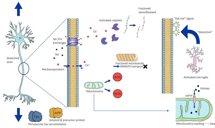
From a neurophysiological point of view, mechanical load can induce a stretching load on the axons that constitute the white matter, leading to diffuse axonal injury. The pathophysiology of secondary injury to the axons caused by an excessive stretching load has been described in many studies (Fig. 1).
Calcium influx
A physical load on the axons is known to cause the elevation of intracellular Ca2+ ions via multiple pathways. One such pathway is through the inflow of Na+ via voltage-gated channels and the subsequent reversal of the Na+/Ca2+ exchanger that results in an increase in the intracellular Ca2+ concentration [17]. Another pathway of calcium influx is via
“mechanoporation.” Mechanoporation is the term used to describe an opening in the axolemma caused by a stretch load on the axon [16]. The increase in intracellular calcium concentration is known to activate calpain, a Ca2+-activated protease, leading to proteolysis of structural proteins such as neurofilaments [18]. Calpain is also known to be involved in the later stages of Wallerian degeneration in the peripheral and central nervous systems [18].
Mitochondrial dysfunction
To compensate for the disturbance in intracellular ionic currents, various membrane ion pumps are recruited. During this process, glucose metabolism increases, resulting in the depletion of intracellular adenosine triphosphate (ATP) and the introduction of calcium into
the mitochondria, leading to increased oxidative stress [19,20]. In addition, the influx of calcium triggers the formation of mitochondrial permeability pores on the inner mitochondrial membranes, promoting the migration of molecules (< 1.5 kDa) into the mitochondria. This process ultimately leads to the swelling and death of mitochondria [21,22].
Fractured microtubules and Wallerian degeneration
Increased intracellular oxidative stress increases intracellular lactate levels and causes cell edema [23]. Axon swelling is known to cause neurofilament accumulation and microtubule dysfunction and fracture [24]. This results in impaired axonal transport leading to the accumulation of synthesized proteins. Consequently, axonal bulbs are formed as they accumulate at specific sites, and in severe cases, the axons are cut at these sites [25]. This is mainly observed at the cortical-subcortical boundary. Fractured microtubules will be unable to carry nicotinamide mononucleotide adenylyltransferase-2 (NMNAT2), resulting in Wallerian degeneration, mediated by sterile alpha and toll/interleukin-1 receptor (TIR) motif- containing protein 1 (SARM1) proteins via the intracellular signaling pathways [26,27].
Phagocytosis and neuroinflammation
Following axonal damage, phosphatidylserine residues on the cell membrane translocate out of the cell. Activated microglia recognize the corresponding residue, the so-called “eat- me” signal, and proceed with phagocytosis [28]. Activated microglia were found to become activated even after the acute phase [29,30]. Ongoing activation of microglia has also been reported post-mortem in patients with TBI [31]. Activated microglia promote inflammation by releasing cytokines and chemokines. Proinflammatory cytokines are known to cause caspase-mediated proteolysis and microglia recruitment [16].
Stretched axon
Fractured neurofilament
Perivascular tau accumulation
Mitochondria
Activated calplain
Activated microglia
Mitochondria swelling ---> Death Solutes mPTP
“Eat-me” signal
“Detection”
Fractured microtubule NMNST2 transport
Tau APP
ROS ROS
Amyloid β precursor protein Mechanoporation
Na⁺ Na⁺/Ca⁺⁺
Exchanger
Ca⁺⁺
Ca⁺⁺
Fig. 1. Pathophysiology occurring in the stretched axon.
APP, amyloid-β precursor protein; NMNAT2, nicotinamide mononucleotide adenylyltransferase-2; ROS, reactive oxygen species.
Toxic proteinopathies and neurodegeneration
Dysfunction of axonal transport, caused by microtubule disruption, results in the accumulation of amyloid-β precursor protein (APP) [24]. This APP is cleaved into amyloid-β peptides, which aggregate into amyloid plaques, the pathological hallmark finding in Alzheimer's disease (AD).
Tau, a microtubule-stabilizing protein, has been shown to accumulate perivascularly during the disruption of microtubules [32], suggesting that TBI may be associated with neurodegenerative diseases such as AD [16].
The pathophysiological changes in the axons as described above may persist for years after the injury. It has been reported that the neurofibrillary tangles seen in AD are also seen in the brains of boxers and football players who have had repeated concussions [33]. Recent studies have shown that cis-phosphorylated tau protein levels increase throughout the mouse brain after moderate to severe TBI or mTBI. Monoclonal antibodies to cis-phosphorylated tau protein appeared to improve the pathophysiology and behavioral outcome, thus displaying therapeutic potential [34].
UNRESOLVED CHALLENGES OF MTBI: THE NEED FOR BIOMARKERS
Diagnosis
As mentioned earlier, patients with mTBI do not have any abnormality on standard
neuroimaging; hence, the diagnosis is based mainly on the patient's symptoms and signs and the testimony of witnesses. This reiterates the essential limitation that it is very difficult to diagnose mTBI in patients who present to the emergency department without any objective evidence, such as recorded image data, and in those with confusion or disorientation without any trace of head trauma. The problem becomes more complicated, especially when issues of litigation or secondary gain are involved.
In addition, the diagnostic criteria, including imaging techniques, need more clarification.
The criterion of “no abnormality on CT or MR imaging” recommended by the US DVA/DoD guidelines is too ambiguous to be applied in clinical practice because MRI sequences are very diverse and not specified. Microbleeding, which is not clearly detected with traditional methods, may be detected by using modern sequences such as susceptibility-weighted images. It is necessary to modify the diagnostic criteria in light of recent advances in modern medicine with new equipment and imaging techniques being developed rapidly.
There are several other problems associated with the ACRM diagnostic criteria for mTBI. The upper limits of the duration of loss of consciousness or memory loss after trauma are specified as 30 minutes and 24 hours, respectively, with no mention of the lower limits. Consequently, patients who had loss of consciousness for 20 minutes and 10 seconds are both classified into one category. There is also a claim that patients with GCS scores of 13 have a different clinical course from those with scores of 14 and 15 [35]. Further studies are needed to overcome these limitations. Thus, it is difficult to diagnose mTBI in a symptomatic patient in the absence of witnesses, signs of head trauma, and abnormalities on neuroimaging. Despite these difficulties, efforts have been made to accurately diagnose mTBI. Magnetoencephalography (MEG) and electroencephalography (EEG) have been explored as part of such efforts. In a recently published case-control study, it has been found that the control and mTBI groups could be distinguished with 100% accuracy by using MEG [36]; therefore, the application of these
modalities is being actively researched. Additionally, EEG is also being studied as a diagnostic tool for mTBI. Recent studies have shown that the EEG slow wave quantity is a sensitive indicator in the diagnosis and prognosis of mTBI [37]. Recently, blood-based biomarkers that can be measured easily and quantitatively have been recognized as useful in the diagnosis of mTBI. Recent studies have shown glial fibrillary acidic protein (GFAP) and ubiquitin carboxy- terminal hydrolase-L1 (UCH-L1) as emerging biomarker candidates for diagnosing TBI [38].
Prognosis
A typical mTBI is known to resolve within 24 hours of visiting the emergency department, and symptoms such as headaches, dizziness, loss of concentration, and body and cognitive symptoms disappear within 12 weeks. However, studies have shown that 50% of patients remain symptomatic even after 3 months [39]. In addition, approximately 15% of patients continue to have neurological disorders or symptoms even after 1 year, which is sometimes referred to as post-concussion syndrome (PCS) [40]. Although various studies have been performed to detect PCS subgroups early, there is no reliable clinical model predicting progression to PCS [41]. In addition, a prognostic model with clinical variables for moderate to severe brain injury has been proposed; however, but its prediction accuracy is low [42]. Therefore, if a body fluid- based biomarker is developed to predict the prognosis of mTBI, it is expected to help in clinical decision-making. In addition, there are a few predictive models currently available for long- term complications such as chronic traumatic encephalopathy (CTE); however, in the future, biomarkers in blood are expected to be used in making such predictive models.
BODY FLUID CANDIDATES FOR BIOMARKER RESEARCH IN MTBI
Cerebrospinal fluid (CSF)
Theoretically, CSF is the closest body fluid to the target organ, the brain, and is most likely to reflect the pathophysiology of TBI [14]. However, in practice, there are various associated problems. When the blood-brain barrier (BBB) is damaged along with brain damage, various substances are introduced into the CSF from the blood as well as the brain. Comparing this with signal processing, the substance introduced from the brain to the CSF would be a signal, and the substance introduced from the blood to the CSF would be noise. Hence, the noise in CSF can be increased after TBI.
Another limitation is that CSF is difficult to obtain. In patients with moderate to severe TBI, who are hospitalized and undergo extraventricular drainage (EVD), it is relatively easier to obtain CSF; however, obtaining CSF from mTBI patients is impractical. Patients with mTBI are more likely to refuse to undergo the relatively more invasive lumbar puncture, which can also aggravate headache. In addition, as there are differences in the components of CSF obtained through lumbar puncture and EVD, the interpretation of test results and their application to clinical decision making can be complex.
Blood
As blood is the most common body fluid specimen used for various tests in hospitals, it is easy to obtain patient's consent. Compared with CSF, it is one step more distal to the target organ; however, it has the advantage that the measurement results do not change significantly even with BBB compromise. The fact that reference values for various blood markers are well known is also an advantage [10].
A disadvantage of using blood specimens is that biomarkers released from the brain into the bloodstream are likely to have low levels in blood. The levels of most candidate protein marker levels are in the picogram range (10−12 g/mL); hence, the measurement itself is technically challenging. Moreover, to date, the time to detection in blood after the injury is different for each biomarker. For example, GFAP levels in the plasma were reported to increase significantly 8 hours after the injury, whereas markers such as UCH-L1 increase significantly at baseline and then return to normal levels after 12 hours [38]. When the biomarkers are not specific to the brain, blood concentration is vulnerable to “noise” from other organs. For example, s100B levels increase in blood after a brain injury; however, as it is also produced in tissues other than the brain, its blood levels increase even after trauma to other parts of the body without any brain damage [43].
Urine
Urine is a body fluid that can ideally be used when the target organ is the kidney. In TBI, as the target organ is the brain, the concentrations of protein markers in urine are likely to show smaller changes than in blood. Changes in the femtogram range (10−15 g/mL) are very difficult to measure with traditional immunological methods. In addition, in the research stage, it is necessary to collect a 24-hour urine sample instead of a spot urine sample at a specific time point, and the hassle of obtaining 24-hour urine samples is also a big obstacle.
BLOOD-BASED BIOMARKERS UNDER RESEARCH
The advantages and disadvantages of individual blood-based biomarkers were summarized (Table 2). Details of the blood-based biomarkers are shown below.
s100B
s100B is a calcium-binding protein that exists in astrocytes and Schwann cells. It is known to affect intracellular calcium homeostasis, and is responsible for intracellular signal transduction [44]. Blood and CSF concentrations of s100B increase after trauma, Table 2. Potential blood-based biomarkers
Biomarker Related structure Advantages Disadvantages
s100B Astroglial cells - Hemolyzed samples do not show false positives - Increase in concentration even in adipocytes, cartilage cells, and melanoma
- Specific time window for rise (12–48 hr after injury) - Short half-life (< 2 hr) - A significant difference in concentration according to the
severity of TBI
- Useful for determining the need for brain CT imaging in mTBI patients (Scandinavian CT guidelines)
UCH-L1 Neuronal cytoplasm - High correlation with initial GCS score - Lower accuracy compared to GFAP in determining the need for neurosurgical intervention
- Good correlation with abnormalities on CT (better than GFAP and s100B)
SBDPs Neuronal cytoplasm - Increases within 15 min of TBI, and continues to increase
until 3–24 hr after TBI - Not specific to brain damage
- Good correlation with mortality after TBI
NSE Neuronal cytoplasm - Indicator of post-TBI functional level and mortality - False positives in hemolyzed specimens GFAP Astroglial cells - Specific for brain damage - Delayed rise (> 8 hr after TBI)
- Useful for determining the need for brain CT imaging in
mTBI patients (better than s100B) - “Return to work” ratio at 6 mon after TBI cannot be predicted
miRNA Unknown - After brain injury, the miRNA expression levels are
significantly changed - Technically challenging
- Downregulation of miR-23a and miR-27a can be confirmed within 4 hr of brain injury
TBI, traumatic brain injury; CT, computed tomography; mTBI, mild TBI; UCH-L1, ubiquitin carboxy-terminal hydrolase-L1; GCS, Glasgow coma scale; GFAP, glial fibrillary acidic protein; SBDPs, spectrin breakdown products; NSE, neuron-specific enolase; miRNA, micro RNA.
and unlike neuron-specific enolase (NSE), its concentration does not change much in hemolyzed specimens. Therefore, it has been highlighted as a biomarker of brain damage and many studies have been conducted to date [45]. Unfortunately, s100B is also produced by oligodendrocytes, neural progenitor cells, adipocytes, cartilage cells, and tumor tissues such as melanoma, and its levels may also increase after BBB dysfunction [46-49]. Therefore, its concentration increases even in polytrauma without brain damage and may be increased by the exacerbation of systemic conditions such as hemorrhagic shock. Furthermore, it is known that the darker the skin color, the higher the levels of s100B [50]. Therefore, skin color must also be considered when interpreting s100B levels. As s100B is excreted 100%
through the kidneys, care must be taken while interpreting s100B levels in patients with renal insufficiency. However, it is known that there are no significant differences in the blood concentrations of s100B in patients with mild to moderate renal dysfunction [51,52].
Several studies have shown the biological half-life of s100B to be 25 minutes to 2 hours, and it is difficult to specify a detection time window due to the relatively short half-life. However, based on the recently published dynamic model of s100B concentration, it was found that the increase in s100B concentration due to brain damage could be specifically confirmed 12–48 hours after the TBI [53]. In addition, the secondary increase of s100B, confirmed via serial sampling, has been shown to reflect secondary brain damage [54-56], increasing the expectations of the clinical use of s100B.
s100B showed a significant difference in concentration according to the degree of brain damage (mild, moderate, and severe TBI) [57,58], and 2 reviews showed a correlation with the GCS scores at admission [59,60]. In addition, a newer version of the Scandinavian CT guidelines published in 2013 may help determine whether brain CT scans are needed for patients with mTBI visiting the emergency department. If the s100B concentration is < 0.10 µg/L, measured within 6 hours from the time of injury, brain CT may not be needed [61]. On the contrary, the predictive power was not sufficiently high for PCS in children [62].
UCH-L1
UCH-L1 is a cysteine protease present in neurons and accounts for approximately 2% of the total soluble protein in the brain [63]. Recent studies have shown that UCH-L1 levels rapidly increase in the plasma after brain injury and rapidly decrease subsequently within 36 hours [38]. In a recent study involving mild and moderate TBI, UCH-L1 levels were highly correlated with the GCS scores and the presence of lesions on brain CT [64]. In another prospective study, the UCH-L1 concentration in blood was found to be superior to that of GFAP or s100B in predicting the presence of CT lesions within 6 hours after mild and moderate brain injury [65]. However, a recently published prospective cohort study confirmed that the accuracy of UCH-L1 was lower than that of GFAP in determining the presence of brain damage, presence of lesions on CT, and necessity of neurosurgical intervention [38].
Spectrin breakdown products (SBDPs)
Spectrin is a major component of the cytoskeleton, broken down by calpain after brain damage, resulting in 2 αII-spectrin fragments: 150 kDa (SBDP150) and 145 kDa (SBDP145).
The αII-spectrin fragments are present in the axons and pre-synaptic terminals of neurons, and caspase-3 again degrades it into 120 kDa (SBDP120) [66]. Although SBDPs reflect the necrosis and apoptosis processes in the brain, they are not specific for brain damage. They also occur in cerebral ischemia, neurodegenerative disease (i.e., AD), and normal aging [67,68]. Increased concentrations of SBDPs occurred within 15 minutes of brain damage and
were found to increase significantly from 3 to 24 hours after brain injury [69,70]. The SBDP concentrations in the CSF are associated with mortality after brain injury [67]. Although techniques to reduce spectrin breakdown by using calpain inhibitors have been introduced, their therapeutic effect on axon survival is yet to be determined in brain injury [71,72].
NSE
An enzyme involved in glycolysis, NSE is a marker that reflects apoptosis and is present in the neuron cell body [73]. Unfortunately, hemolyzed samples show false positives [74]. Recent meta-analyses have shown that NSE can be used as an indicator of functional levels and mortality rates after TBI [75].
GFAP
GFAP is a protein found only in the central nervous system and constitutes the cytoskeleton of astrocytes. It is known that when astrocytes are damaged, the production of GFAP is greatly increased [76]. It is a marker specific to brain damage because it does not increase after trauma to other parts of the body; however, after TBI, it takes 8 hours for its blood levels to increase [38]. The GFAP blood concentrations in patients with mTBI in the emergency department is better than the s100B blood concentrations in predicting the need for brain CT. Especially, in cases of extracranial injuries, GFAP blood concentrations were also found to have a higher specificity for detecting lesions that could be confirmed with brain CT [77].
In addition, in 2 reviews, the concentrations of GFAP correlated with the initial GCS scores, as with s100B [59,60]. However, the outcome of patients at 6 months after TBI could not be predicted precisely based on the GFAP blood concentrations [45].
Micro RNA (miRNA)
miRNA binds to the 3' untranslated region of messenger RNA (mRNA) and causes degradation of mRNA or inhibits mRNA translation. Even a single miRNA is known to regulate the entire mRNA network [78], and is expected to be used in therapeutics by blocking the pathophysiological cascade after brain injury. In particular, miRNAs are abundantly expressed in the brain tissue, and some are known to have brain-specific
functions [79-81]. After brain injury, miRNA expression levels are known to vary significantly [82,83], with the downregulation of miR-23a and miR-27a being confirmed within 4 hours of the brain injury. These changes are believed to be related to the increased expression of proapoptotic Bcl-2 family members (NoxA, Puma, and Bax) [83].
Other biomarkers
In addition, several studies have shown that other biomarker candidates, including inflammatory cytokines, amyloid-related markers, phosphorylated tau, TAR DNA-binding protein 43, hormones (steroid and pituitary hormones), neurofilament light chain, and myelin basic protein have the potential for diagnosing mTBI.
PROSPECTS AND FUTURE CHALLENGES
Recently, a quantum jump in the technology of protein biomarker development has occurred, which involved the mass spectrometry (MS)-based proteomics workflow based on high-throughput proteomics by using high-performance MS including Orbitrap® liquid chromatography-tandem mass spectrometry (LC-MS/MS; Thermo Fisher Scientific, Waltham, MA, USA) and triple quadrupole LC-MS/MS. Those high-performance technologies
generated huge amount of proteome information. Subsequently, a bioinformatics algorithm was developed for processing large data, which can expedite both the identification and quantitation of proteome data sets. In addition, the tools of systems biology guided the functional annotation among all bits and pieces of proteome data.
However, the importance of clinical knowledge and experience is more emphasized than ever even with the advances in technology because well-designed biorepository and comprehensive functional evaluation is the pivotal requirement for biomarker discovery and validation.
REFERENCES
1. Centers for Disease Control and Prevention (US). Report to congress on traumatic brain injury in the United States: epidemiology and rehabilitation. Atlanta (GA): Centers for Disease Control and Prevention; 2015.
2. Cassidy JD, Carroll LJ, Peloso PM, Borg J, von Holst H, Holm L, et al. Incidence, risk factors and prevention of mild traumatic brain injury: results of the WHO Collaborating Centre Task Force on Mild Traumatic Brain Injury. J Rehabil Med 2004:28-60.
PUBMED | CROSSREF
3. Setnik L, Bazarian JJ. The characteristics of patients who do not seek medical treatment for traumatic brain injury. Brain Inj 2007;21:1-9.
PUBMED | CROSSREF
4. Li C. Breast cancer epidemiology. New York (NY): Springer-Verlag New York; 2010.
5. Levin HS, Diaz-Arrastia RR. Diagnosis, prognosis, and clinical management of mild traumatic brain injury. Lancet Neurol 2015;14:506-517.
PUBMED | CROSSREF
6. Ponsford J, Willmott C, Rothwell A, Cameron P, Kelly AM, Nelms R, et al. Impact of early intervention on outcome following mild head injury in adults. J Neurol Neurosurg Psychiatry 2002;73:330-332.
PUBMED | CROSSREF
7. Fleminger S, Oliver DL, Lovestone S, Rabe-Hesketh S, Giora A. Head injury as a risk factor for Alzheimer's disease: the evidence 10 years on; a partial replication. J Neurol Neurosurg Psychiatry 2003;74:857-862.
PUBMED | CROSSREF
8. Jafari S, Etminan M, Aminzadeh F, Samii A. Head injury and risk of Parkinson disease: a systematic review and meta-analysis. Mov Disord 2013;28:1222-1229.
PUBMED | CROSSREF
9. Chen H, Richard M, Sandler DP, Umbach DM, Kamel F. Head injury and amyotrophic lateral sclerosis.
Am J Epidemiol 2007;166:810-816.
PUBMED | CROSSREF
10. Di Battista AP, Rhind SG, Baker AJ. Application of blood-based biomarkers in human mild traumatic brain injury. Front Neurol 2013;4:44.
PUBMED | CROSSREF
11. Greenwald BD, Ambrose AF, Armstrong GP. Mild brain injury. Rehabil Res Pract 2012;2012:469475.
12. Saatman KE, Duhaime AC, Bullock R, Maas AI, Valadka A, Manley GTWorkshop Scientific Team and Advisory Panel Members. Classification of traumatic brain injury for targeted therapies. J Neurotrauma 2008;25:719-738.
PUBMED | CROSSREF
13. Cifu D, Hurley R, Peterson M, Cornis-Pop M, Rikli PA, Ruff RL, et al. VA/DoD clinical practice guideline for management of concussion/mild traumatic brain injury. J Rehabil Res Dev 2009;46:CP1-CP68.
PUBMED | CROSSREF
14. Zetterberg H, Smith DH, Blennow K. Biomarkers of mild traumatic brain injury in cerebrospinal fluid and blood. Nat Rev Neurol 2013;9:201-210.
PUBMED | CROSSREF
15. Iverson GL, Gaetz M, Lovell MR, Collins MW. Cumulative effects of concussion in amateur athletes. Brain Inj 2004;18:433-443.
PUBMED | CROSSREF
16. Hill CS, Coleman MP, Menon DK. Traumatic axonal injury: mechanisms and translational opportunities.
Trends Neurosci 2016;39:311-324.
PUBMED | CROSSREF
17. Iwata A, Stys PK, Wolf JA, Chen XH, Taylor AG, Meaney DF, et al. Traumatic axonal injury induces proteolytic cleavage of the voltage-gated sodium channels modulated by tetrodotoxin and protease inhibitors. J Neurosci 2004;24:4605-4613.
PUBMED | CROSSREF
18. Ma M, Ferguson TA, Schoch KM, Li J, Qian Y, Shofer FS, et al. Calpains mediate axonal cytoskeleton disintegration during Wallerian degeneration. Neurobiol Dis 2013;56:34-46.
PUBMED | CROSSREF
19. Cheng G, Kong RH, Zhang LM, Zhang JN. Mitochondria in traumatic brain injury and mitochondrial- targeted multipotential therapeutic strategies. Br J Pharmacol 2012;167:699-719.
PUBMED | CROSSREF
20. Siedler DG, Chuah MI, Kirkcaldie MT, Vickers JC, King AE. Diffuse axonal injury in brain trauma: insights from alterations in neurofilaments. Front Cell Neurosci 2014;8:429.
PUBMED | CROSSREF
21. Halestrap AP. What is the mitochondrial permeability transition pore? J Mol Cell Cardiol 2009;46:821-831.
PUBMED | CROSSREF
22. Mazzeo AT, Beat A, Singh A, Bullock MR. The role of mitochondrial transition pore, and its modulation, in traumatic brain injury and delayed neurodegeneration after TBI. Exp Neurol 2009;218:363-370.
PUBMED | CROSSREF
23. Giza CC, Hovda DA. The neurometabolic cascade of concussion. J Athl Train 2001;36:228-235.
PUBMED
24. Tang-Schomer MD, Johnson VE, Baas PW, Stewart W, Smith DH. Partial interruption of axonal transport due to microtubule breakage accounts for the formation of periodic varicosities after traumatic axonal injury. Exp Neurol 2012;233:364-372.
PUBMED | CROSSREF
25. Barkhoudarian G, Hovda DA, Giza CC. The molecular pathophysiology of concussive brain injury. Clin Sports Med 2011;30:33-48, vii-iii.
PUBMED | CROSSREF
26. Yang J, Wu Z, Renier N, Simon DJ, Uryu K, Park DS, et al. Pathological axonal death through a MAPK cascade that triggers a local energy deficit. Cell 2015;160:161-176.
PUBMED | CROSSREF
27. Osterloh JM, Yang J, Rooney TM, Fox AN, Adalbert R, Powell EH, et al. dSarm/Sarm1 is required for activation of an injury-induced axon death pathway. Science 2012;337:481-484.
PUBMED | CROSSREF
28. Brown GC, Neher JJ. Microglial phagocytosis of live neurons. Nat Rev Neurosci 2014;15:209-216.
PUBMED | CROSSREF
29. Ramlackhansingh AF, Brooks DJ, Greenwood RJ, Bose SK, Turkheimer FE, Kinnunen KM, et al.
Inflammation after trauma: microglial activation and traumatic brain injury. Ann Neurol 2011;70:374-383.
PUBMED | CROSSREF
30. Loane DJ, Kumar A, Stoica BA, Cabatbat R, Faden AI. Progressive neurodegeneration after experimental brain trauma: association with chronic microglial activation. J Neuropathol Exp Neurol 2014;73:14-29.
PUBMED | CROSSREF
31. Johnson VE, Stewart JE, Begbie FD, Trojanowski JQ, Smith DH, Stewart W. Inflammation and white matter degeneration persist for years after a single traumatic brain injury. Brain 2013;136:28-42.
PUBMED | CROSSREF
32. Hay J, Johnson VE, Smith DH, Stewart W. Chronic traumatic encephalopathy: the neuropathological legacy of traumatic brain injury. Annu Rev Pathol 2016;11:21-45.
PUBMED | CROSSREF
33. Tokuda T, Ikeda S, Yanagisawa N, Ihara Y, Glenner GG. Re-examination of ex-boxers' brains using immunohistochemistry with antibodies to amyloid beta-protein and tau protein. Acta Neuropathol 1991;82:280-285.
PUBMED | CROSSREF
34. Kondo A, Shahpasand K, Mannix R, Qiu J, Moncaster J, Chen CH, et al. Antibody against early driver of neurodegeneration cis P-tau blocks brain injury and tauopathy. Nature 2015;523:431-436.
PUBMED | CROSSREF
35. Mena JH, Sanchez AI, Rubiano AM, Peitzman AB, Sperry JL, Gutierrez MI, et al. Effect of the modified Glasgow Coma Scale score criteria for mild traumatic brain injury on mortality prediction: comparing classic and modified Glasgow Coma Scale score model scores of 13. J Trauma 2011;71:1185-1192.
PUBMED | CROSSREF
36. Dimitriadis SI, Zouridakis G, Rezaie R, Babajani-Feremi A, Papanicolaou AC. Functional connectivity changes detected with magnetoencephalography after mild traumatic brain injury. Neuroimage Clin 2015;9:519-531.
PUBMED | CROSSREF
37. Modarres M, Kuzma NN, Kretzmer T, Pack AI, Lim MM. EEG slow waves in traumatic brain injury:
convergent findings in mouse and man. Neurobiol Sleep Circadian Rhythms 2016;1:S2451994416300025.
PUBMED | CROSSREF
38. Papa L, Brophy GM, Welch RD, Lewis LM, Braga CF, Tan CN, et al. Time course and diagnostic accuracy of glial and neuronal blood biomarkers GFAP and UCH-L1 in a large cohort of trauma patients with and without mild traumatic brain injury. JAMA Neurol 2016;73:551-560.
PUBMED | CROSSREF
39. Lundin A, de Boussard C, Edman G, Borg J. Symptoms and disability until 3 months after mild TBI. Brain Inj 2006;20:799-806.
PUBMED | CROSSREF
40. Burke MJ, Fralick M, Nejatbakhsh N, Tartaglia MC, Tator CH. In search of evidence-based treatment for concussion: characteristics of current clinical trials. Brain Inj 2015;29:300-305.
PUBMED | CROSSREF
41. McCauley SR, Boake C, Levin HS, Contant CF, Song JX. Postconcussional disorder following mild to moderate traumatic brain injury: anxiety, depression, and social support as risk factors and comorbidities. J Clin Exp Neuropsychol 2001;23:792-808.
PUBMED | CROSSREF
42. MRC CRASH Trial CollaboratorsPerel P, Arango M, Clayton T, Edwards P, Komolafe E, et al. Predicting outcome after traumatic brain injury: practical prognostic models based on large cohort of international patients. BMJ 2008;336:425-429.
PUBMED | CROSSREF
43. Olsson B, Zetterberg H, Hampel H, Blennow K. Biomarker-based dissection of neurodegenerative diseases. Prog Neurobiol 2011;95:520-534.
PUBMED | CROSSREF
44. Heizmann CW, Fritz G, Schäfer BW. S100 proteins: structure, functions and pathology. Front Biosci 2002;7:d1356-d1368.
PUBMED
45. Metting Z, Wilczak N, Rodiger LA, Schaaf JM, van der Naalt J. GFAP and S100B in the acute phase of mild traumatic brain injury. Neurology 2012;78:1428-1433.
PUBMED | CROSSREF
46. Donato R, Sorci G, Riuzzi F, Arcuri C, Bianchi R, Brozzi F, et al. S100B's double life: intracellular regulator and extracellular signal. Biochim Biophys Acta 2009;1793:1008-1022.
PUBMED | CROSSREF
47. Steiner J, Bernstein HG, Bielau H, Berndt A, Brisch R, Mawrin C, et al. Evidence for a wide extra-astrocytic distribution of S100B in human brain. BMC Neurosci 2007;8:2.
PUBMED | CROSSREF
48. Blyth BJ, Farahvar A, He H, Nayak A, Yang C, Shaw G, et al. Elevated serum ubiquitin carboxy-terminal hydrolase L1 is associated with abnormal blood-brain barrier function after traumatic brain injury. J Neurotrauma 2011;28:2453-2462.
PUBMED | CROSSREF
49. Lopez NE, Krzyzaniak MJ, Blow C, Putnam J, Ortiz-Pomales Y, Hageny AM, et al. Ghrelin prevents disruption of the blood-brain barrier after traumatic brain injury. J Neurotrauma 2012;29:385-393.
PUBMED | CROSSREF
50. Ben Abdesselam O, Vally J, Adem C, Foglietti MJ, Beaudeux JL. Reference values for serum S-100B protein depend on the race of individuals. Clin Chem 2003;49:836-837.
PUBMED | CROSSREF
51. Jönsson H, Johnsson P, Höglund P, Alling C, Blomquist S. Elimination of S100B and renal function after cardiac surgery. J Cardiothorac Vasc Anesth 2000;14:698-701.
PUBMED | CROSSREF
52. Usui A, Kato K, Abe T, Murase M, Tanaka M, Takeuchi E. S-100ao protein in blood and urine during open- heart surgery. Clin Chem 1989;35:1942-1944.
PUBMED
53. Ercole A, Thelin EP, Holst A, Bellander BM, Nelson DW. Kinetics of serum S100b after traumatic brain injury [Internet]. Available at https://www.repository.cam.ac.uk/handle/1810/254934 [accessed on 8 February 2017].
54. Raabe A, Kopetsch O, Woszczyk A, Lang J, Gerlach R, Zimmermann M, et al. S-100B protein as a serum marker of secondary neurological complications in neurocritical care patients. Neurol Res 2004;26:440-445.
PUBMED | CROSSREF
55. Thelin EP, Nelson DW, Bellander BM. Secondary peaks of S100B in serum relate to subsequent radiological pathology in traumatic brain injury. Neurocrit Care 2014;20:217-229.
PUBMED | CROSSREF
56. Undén J, Astrand R, Waterloo K, Ingebrigtsen T, Bellner J, Reinstrup P, et al. Clinical significance of serum S100B levels in neurointensive care. Neurocrit Care 2007;6:94-99.
PUBMED | CROSSREF
57. Herrmann M, Jost S, Kutz S, Ebert AD, Kratz T, Wunderlich MT, et al. Temporal profile of release of neurobiochemical markers of brain damage after traumatic brain injury is associated with intracranial pathology as demonstrated in cranial computerized tomography. J Neurotrauma 2000;17:113-122.
PUBMED | CROSSREF
58. Romner B, Ingebrigtsen T, Kongstad P, Børgesen SE. Traumatic brain damage: serum S-100 protein measurements related to neuroradiological findings. J Neurotrauma 2000;17:641-647.
PUBMED | CROSSREF
59. Mondello S, Muller U, Jeromin A, Streeter J, Hayes RL, Wang KK. Blood-based diagnostics of traumatic brain injuries. Expert Rev Mol Diagn 2011;11:65-78.
PUBMED | CROSSREF
60. Kövesdi E, Lückl J, Bukovics P, Farkas O, Pál J, Czeiter E, et al. Update on protein biomarkers in traumatic brain injury with emphasis on clinical use in adults and pediatrics. Acta Neurochir (Wien) 2010;152:1-17.
PUBMED | CROSSREF
61. Undén J, Ingebrigtsen T, Romner BScandinavian Neurotrauma Committee (SNC). Scandinavian guidelines for initial management of minimal, mild and moderate head injuries in adults: an evidence and consensus-based update. BMC Med 2013;11:50.
PUBMED | CROSSREF
62. Babcock L, Byczkowski T, Wade SL, Ho M, Bazarian JJ. Inability of S100B to predict postconcussion syndrome in children who present to the emergency department with mild traumatic brain injury: a brief report. Pediatr Emerg Care 2013;29:458-461.
PUBMED | CROSSREF
63. Boutté AM, Yao C, Kobeissy F, May Lu XC, Zhang Z, Wang KK, et al. Proteomic analysis and brain- specific systems biology in a rodent model of penetrating ballistic-like brain injury. Electrophoresis 2012;33:3693-3704.
PUBMED | CROSSREF
64. Papa L, Lewis LM, Silvestri S, Falk JL, Giordano P, Brophy GM, et al. Serum levels of ubiquitin C-terminal hydrolase distinguish mild traumatic brain injury from trauma controls and are elevated in mild and moderate traumatic brain injury patients with intracranial lesions and neurosurgical intervention. J Trauma Acute Care Surg 2012;72:1335-1344.
PUBMED | CROSSREF
65. Welch RD, Ayaz SI, Lewis LM, Unden J, Chen JY, Mika VH, et al. Ability of serum glial fibrillary acidic protein, ubiquitin C-terminal hydrolase-L1, and S100B to differentiate normal and abnormal head computed tomography findings in patients with suspected mild or moderate traumatic brain injury. J Neurotrauma 2016;33:203-214.
PUBMED | CROSSREF
66. Berger RP, Hayes RL, Richichi R, Beers SR, Wang KK. Serum concentrations of ubiquitin C-terminal hydrolase-L1 and αII-spectrin breakdown product 145 kDa correlate with outcome after pediatric TBI. J Neurotrauma 2012;29:162-167.
PUBMED | CROSSREF
67. Mondello S, Robicsek SA, Gabrielli A, Brophy GM, Papa L, Tepas J 3rd, et al. αII-spectrin breakdown products (SBDPs): diagnosis and outcome in severe traumatic brain injury patients. J Neurotrauma 2010;27:1203-1213.
PUBMED | CROSSREF
68. Kobeissy FH, Liu MC, Yang Z, Zhang Z, Zheng W, Glushakova O, et al. Degradation of βII-spectrin protein by calpain-2 and caspase-3 under neurotoxic and traumatic brain injury conditions. Mol Neurobiol 2015;52:696-709.
PUBMED | CROSSREF
69. Aikman J, O'Steen B, Silver X, Torres R, Boslaugh S, Blackband S, et al. Alpha-II-spectrin after controlled cortical impact in the immature rat brain. Dev Neurosci 2006;28:457-465.
PUBMED | CROSSREF
70. McGinn MJ, Kelley BJ, Akinyi L, Oli MW, Liu MC, Hayes RL, et al. Biochemical, structural, and biomarker evidence for calpain-mediated cytoskeletal change after diffuse brain injury uncomplicated by contusion.
J Neuropathol Exp Neurol 2009;68:241-249.
PUBMED | CROSSREF
71. Posmantur R, Kampfl A, Siman R, Liu J, Zhao X, Clifton GL, et al. A calpain inhibitor attenuates cortical cytoskeletal protein loss after experimental traumatic brain injury in the rat. Neuroscience 1997;77:875-888.
PUBMED | CROSSREF
72. Saatman KE, Creed J, Raghupathi R. Calpain as a therapeutic target in traumatic brain injury.
Neurotherapeutics 2010;7:31-42.
PUBMED | CROSSREF
73. Selakovic V, Raicevic R, Radenovic L. The increase of neuron-specific enolase in cerebrospinal fluid and plasma as a marker of neuronal damage in patients with acute brain infarction. J Clin Neurosci 2005;12:542-547.
PUBMED | CROSSREF
74. Ramont L, Thoannes H, Volondat A, Chastang F, Millet MC, Maquart FX. Effects of hemolysis and storage condition on neuron-specific enolase (NSE) in cerebrospinal fluid and serum: implications in clinical practice. Clin Chem Lab Med 2005;43:1215-1217.
PUBMED | CROSSREF
75. Cheng F, Yuan Q, Yang J, Wang W, Liu H. The prognostic value of serum neuron-specific enolase in traumatic brain injury: systematic review and meta-analysis. PLoS One 2014;9:e106680.
PUBMED | CROSSREF
76. Nylén K, Ost M, Csajbok LZ, Nilsson I, Blennow K, Nellgård B, et al. Increased serum-GFAP in patients with severe traumatic brain injury is related to outcome. J Neurol Sci 2006;240:85-91.
PUBMED | CROSSREF
77. Papa L, Silvestri S, Brophy GM, Giordano P, Falk JL, Braga CF, et al. GFAP out-performs S100β in detecting traumatic intracranial lesions on computed tomography in trauma patients with mild traumatic brain injury and those with extracranial lesions. J Neurotrauma 2014;31:1815-1822.
PUBMED | CROSSREF
78. Lim LP, Lau NC, Garrett-Engele P, Grimson A, Schelter JM, Castle J, et al. Microarray analysis shows that some microRNAs downregulate large numbers of target mRNAs. Nature 2005;433:769-773.
PUBMED | CROSSREF
79. Bak M, Silahtaroglu A, Møller M, Christensen M, Rath MF, Skryabin B, et al. MicroRNA expression in the adult mouse central nervous system. RNA 2008;14:432-444.
PUBMED | CROSSREF
80. Bhalala OG, Srikanth M, Kessler JA. The emerging roles of microRNAs in CNS injuries. Nat Rev Neurol 2013;9:328-339.
PUBMED | CROSSREF
81. Coolen M, Bally-Cuif L. MicroRNAs in brain development and physiology. Curr Opin Neurobiol 2009;19:461-470.
PUBMED | CROSSREF
82. Balakathiresan N, Bhomia M, Chandran R, Chavko M, McCarron RM, Maheshwari RK. MicroRNA let-7i is a promising serum biomarker for blast-induced traumatic brain injury. J Neurotrauma 2012;29:1379-1387.
PUBMED | CROSSREF
83. Hu Z, Yu D, Almeida-Suhett C, Tu K, Marini AM, Eiden L, et al. Expression of miRNAs and their cooperative regulation of the pathophysiology in traumatic brain injury. PLoS One 2012;7:e39357.
PUBMED | CROSSREF
![Table 1. The mTBI diagnostic criteria proposed by ACRM [11]](https://thumb-ap.123doks.com/thumbv2/123dokinfo/5211999.120998/3.892.63.834.869.1059/table-mtbi-diagnostic-criteria-proposed-acrm.webp)