• 검색 결과가 없습니다.

Development of Neuropathic Pain Behavior and Expression of CCL2/CCR2 and CX3CL1/CX3CR1 after Spinal Cord Hemisection

N/A
N/A
Protected

Academic year: 2021

Share "Development of Neuropathic Pain Behavior and Expression of CCL2/CCR2 and CX3CL1/CX3CR1 after Spinal Cord Hemisection"

Copied!
7
0
0

로드 중.... (전체 텍스트 보기)

전체 글

(1)

Hea-Woon Park, MD, PhD; Jo-Young Son, MS; Yun-Woo Cho, MD, PhD

1

; Se-Jin Hwang, MD, PhD

2

; Su-Jeong Kim, PhD

3

; Sang-Ho Ahn, MD, PhD

1

; Sung-Ho Jang, MD, PhD

1

; Yong-Jae Jung, MD

1

Department of Rehabilitation Medicine, School of Medicine, Catholic University of Deagu;

1

Department of Rehabilita- tion Medicine, College of Medicine, Yeungnam University;

2

Department of Anatomy and Cell Biology, College of Medicine, Hanyang University;

3

Institute of Medical Science, Yeungnam University

Purpose: The purpose of this study was to evaluate the development of pain behavior and the expression of CCL2/CCR2 and CX3CL1/CX3CR1 at above and below the level of hemisection of the spinal cord in a rat model.

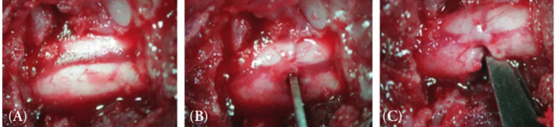
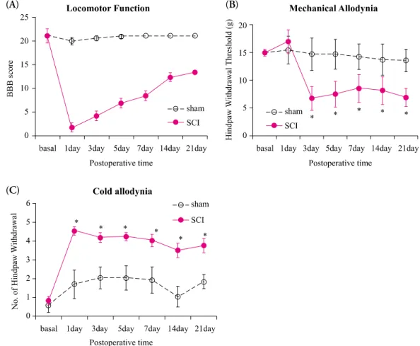
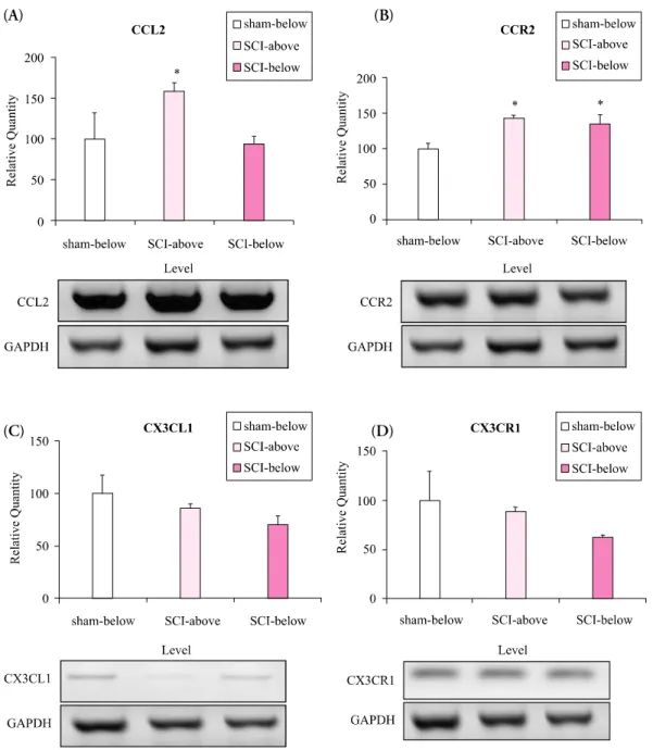
Methods: Spinal cords of adult female Sprague-Dawley rats (n= 16, 200~250 g, 6~8 weeks old) were hemisected at T13 on the right side to develop the spinal hemisection injury model. We compared behavioral responses of the hemisection and of a sham surgery group. Behavioral tests for motor function (by the BBB locomotor scale), and for pain response for mechanical and cold allodynia were assessed postoperatively (PO) for 21 days. Expression of mRNA for chemokines and their receptors (CCL2/CCR2 and CX3CL1/CX3CR1) below and above the level of the spinal cord dissection were examined by RT-PCR.

Results: We observed gradual motor improvement and the development of mechanical and cold allodynia on the ipsilateral hindpaw after spinal hemisection injury. We also found upregulation of mRNA expression of CCL2/CCR2 both above and below the level of spinal cord dissection but CX3CL1/CX3CR1 mRNA expression.

Conclusion: Upregulation of CCL2/CCR2 is associated with neuropathic pain after spinal hemisection injury. CCL2/CCR2 may play an important role in the development of neuropathic pain after SCI as well as of peripheral neuropathic pain. These findings may improve understanding of the pathophysiological mechanism of neuropathic pain after SCI.

Keywords: Spinal cord injury, Neuropathic pain, Chemokine, CCL2/CCR2, CX3CL1/CX3CR1 Received: May 19, 2010

Revised: June 9, 2010 Accepted: June 15, 2010

Corresponding author: Se-Jin Hwang, hwangsj@hanyang.ac.kr; Sang-Ho Ahn, spineahn@ynu.ac.kr

CCL2/CCR2 and CX3CL1/CX3CR1 after Spinal Cord Hemisection

The Journal Korean Society of Physical Therapy

I. Introduction

Many patients with spinal cord injury (SCI) suffer from various sequalae which include paralysis, sensory loss, intractable pain, pressure sores, neurogenic bladder and bowel, and so on. Among them, chronic pain is one of important factors diminishing their quality of life. Pain after SCI is characterized as nociceptive and neuropathic pain, and it occurs above- , at- , and below-level of lesion.

1

Patients with SCI usually present the pain sensation of lower extremities below the level of injury.

2

The neuropathic pain in SCI patients has been usually difficult to manage by conventional treatment and the mechanism of neuropathic pain has not been understood well. Further studies for pathophysiologic

mechanism of neuropathic pain after SCI and new therapeutic challenge targeting to neuropathic pain secondary to SCI may be important.

Recently, it has been known that chemokines are associated

with the development and maintenance of neuropathic pain

after peripheral nerve injury.

3-6

Chemokines modulates the elec-

trical activity of neurons including increased release of neurotrans-

mitters through Ca-dependent mechanisms and transactivation

of transient receptor channels.

5,6

They contribute to chronic

neuropathic pain by mediation of neuronal signaling from

neurons to glial cells in spinal dorsal horn. Two chemokines known

to be related to neuropathic pain are monocyte chemoattractant

protein-1 (MCP-1/CCL2) and its receptor (CCR2), and frac-

(2)

(A) (B) (C)

Figure 1. Operative procedure for spinal cord hemisection injury model. After laminec- tomy of T11-12 vertebra segments (A), T13 spinal cord was hemisected with #11 blade (B, C).

talkine (CX3CL1) and its receptor, CX3CR1.

4,7

In the condi- tion of neuropathic pain after peripheral nerve injury, activation of CCR2 by CCL2 induces membrane depolarization and sen- sitizes nociceptors, and then triggers spinal neuroimmune re- action.

3

Neuronal fractalkine is cleaved by cathepsin S released at activated microglia.

3

Liberated fractalkine activates CX3CR1 receptors on microglia leading to release of nociceptive media- tors.

3

It has been also supposed that microglial activation and various chemokines may be involved in the development of below-level neuropathic pain after SCI.

8,9

However, litte has been reported on the development of neuropathic pain in asso- ciation with the expression of various chemokines after SCI.

The purpose of this study was to evaluate the development of pain behavior after SCI and the expression of CCL2/CCR2 and CX3CL1/CX3CR1 at above and below level of spinal cord in a rat model of spinal cord hemisection.

II. Materials and Methods

1. Spinal cord hemisection injury

All experimental procedures were carried out according to the Institutional Animal Care and Use Committee guidelines at the Yeungnam University, South Korea. Adult female Sprague- Dawley rats (n=16, 200∼250 g, 6∼8 weeks old) were used for this study. Animals were acclimatized to controlled laboratory environments (12 hr light/dark cycle) with free access to food and water. For spinal cord injury rat model, animals were anes- thetized by injection of Zoletil (50 mg/kg, Intraperitoneal (IP), Virbac Laboratories, France). Under anesthesia, laminectomy of T11-T12 vertebral segments was performed and exposed the T13 spinal cord segment. Andthen, the spinal cord was hemi- sected at T13 on right side with #11 blade without damage of major vessels (Figure 1).

10

The musculature, fascia and skin were

sutured in layers after surgery and animals were warmed with heating pad to restore the temperature. For sham surgery, all procedures were done identical without hemisection of spinal cord. Animals were received antibiotics daily for 3 days and bladder was emptied twice daily by bladder expression manually until the function of bladder was recovered.

2. Behavioral test

All behavioral tests were performed by the investigators who were unaware of the experimental groupings and the protocols of each rat. Behavioral test for motor function and pain response were performed in hindpaw 2 day prior to surgery, and 1, 3, 5, 7, 14 and 21 days postoperatively (PO). The locomotor function after SCI was assessed by using BBB Locomotor rating scale.

11

In brief, the BBB is an open field test scored on a 22-points scale, from 0 indicating no movement of hindlimbs to 21 being normal.

Mechanical allodynia was tested by measuring the mecha- nical withdrawal response of the hindlimbusing von Frey fila- ments (North Coast Medical Inc., USA). Rats were placed in a clear plastic cage with a metal mesh floor and adapted to the testing environment for 15 minutes before measurements.

Beginning with 0.1 g probe, filaments were applied to the plantar surface of the hindpaw corresponding to L5 dermatome with 6 to 8 seconds intervals in a stepwise ascending or de- scending order following negative or positive withdrawal res- ponse. Fifty percent probability thresholds of mechanical paw withdrawal were calculated. In the absence of foot withdrawal in response to the application of a 26 g von Frey filament, 26 g was then assigned as the mechanical threshold.

12

Cold allodynia was also determined by measuring the cold

withdrawl response of the hindpaw to acetone application. Rats

were placed in a clear plastic cage with a metal mesh floor and

adapted to the testing environment for 15 minutes before measure-

ments and then, acetone was applied to the plantar surface of the

(3)

hindpaw corresponding to L5 dermatome. Acetone drop was formed at the end of small polyethylene tube connected to a syringe and touched to the hindpaw. Each hindpaw was tested five times, alternating between the left and right. A trial on each paw was repeated after a minimum interval of 5 minutes and only after that paw was completely rested on the floor. The frequency of paw withdrawal was measured as a cold allodynia and mean cold withdrawal frequency was calculated by elimina- ting the highest and lowest latency.

13

3. Reverse Transcription Polymerase Chain Reaction (RT-PCR)

Rats were anesthetized after behavioral tests on 3 weeks PO. The spinal cord tissues corresponding to above-lesion (T8-9) and below-lesion (L4-5) were dissected and quickly allowed to freeze fully. The expression levels of mRNA for chemokines andits receptors (CCL2/CCR2 and CX3CL1/CX3CR1) in spinal cord segments were examined by RT-PCR. In brief, total RNA was isolated from frozen spinal cord tissues of above and below lesion using Trizol reagent (Sigma, St Louis, MO, USA) according to manufacturer’s directions. The concentration and purity of RNA samples were determined by spectrophotometric analysis (OD

260

). One-microgram of total RNA was reverse transcribed into cDNA using Omniscript RT, Sensiscript RT, and primers.

The RT-PCR primer sequences are listed as follows in detail: the sense 5′-GTG ACA TCG TGG CCT TTG G-3′ and antisense 5′-GGT CCA AAG ACA AGT TAG TCC-3′ for CX3CL1, the sense 5′-ATG CTG CCC TGT GAG TAC TAC-3′ and antisense 5′-CCA GAC CGA ACG TGA AGA CA-3′ for CX3CR1, the sense 5′-CCT GTT GTT CAC AGT TGC TGC C-3′ and antisense 5′-TCT ACA GAA GTG CTT GAG GTG GTT G-3′ for CCL2, the sense 5′-GGA ATC CTC CAC ACC CTG TTT C-3′ and antisense 5′-ACC CAA CTG AGA CTT CTT GCT CCC-3 for CCR2, and the sense 5′-GTG AAG GTC GGT GTC AAC GGA TTT-3′ and antisense 5′-CAC AGT CTT CTG AGT GGC AGT GAT-3 for GAPDH.

GAPDH was used as an internal control and standard. The RT-PCR products were electrophoresed on a 2% agarose gel and visualized by staining with ethidium bromide.

4. Statistical Analysis

The collected data were encoded into SPSS/PC version 14.0 and analyzed. Data were expressed as mean±standard deviation (SD).

Baseline measures were analyzed by repeated measure ANOVA for behavioral tests and Wilcoxon signed rank test and Mann Whitney U test for the level of mRNA expression. Differences with p<0.05 were considered statistically significant.

III. Results

1. Behavioral tests for motor and pain response Motor function. All animals were evaluated locomotor function prior to surgery and the scores of basal values were not different between hemisection and sham groups. After surgery, sham surgery rats consistently scored within normal range on all days. However, rats with hemisection injury showed completely paralysis on ipsilateral hindlimb immediately, and progressive motor recovery was observed. The extensive movement of all joints of hindlimb was observed on 7 days and frequent coordinated weight supported plantar steps was observed on 21 days PO (Figure 2-A).

Mechanical allodynia. Prior to surgery, animals were seldom responsive to even 15 g strength of von Frey filaments in both hemisection and sham groups. After surgery, sham groups consistently showed the similar response to pre-surgical basal values. However, in hemisection group, the reduction of hindpaw withdrawal threshold to 6-8 g was apparent from the 3 days and persisted for 21 days, and it was significantly different from the sham surgery group (p<0.05)(Figure 2-B).

Cold allodynia. Prior to surgery, animals showed little withdrawal response to acetone application to ipsilateral hindpaws in both hemisection and sham groups. The withdrawal frequencies were increased from 1 day and persisted up to 21 days in hemisection group. Although sham surgery group showed some increase of withdrawal response, the increase of withdrawal frequencies in hemisection group were larger than sham surgery group, and it was significantly different (p<0.05)(Figure 2-C).

2. mRNA expression for chemokines and its receptors

RT-PCR was performed in spinal cord tissues of both above

(T8-9) and below (L4-5) level of lesion on 3 weeks after surgery,

and we investigated the relative changes of CCL2, CCR2,

CX3CL1, and CX3CR1 mRNA expression compared to below

level of spinal cord in sham surgery group. The CCL2 gene

expression was significantly increased in above level of cord

(1.58±0.1 fold increase), and CCL2 receptor mRNA (CCR2)

(4)

(A) (B)

(C)

Figure 2. The changes of BBB locomotor scale for motor recovery after spinal hemisection (A).

The progressive recovery of hindlimb motor function reached to frequent coordinated walking by 21 days postoperatively. The changes of mechanical (B) and cold allodynia (C) in ipsilateral hindlimb by 21 days. In hemisection group, hindpaw withdrawal threshold for mechanical stimuli was significantly decreased compared to sham surgery from 3 days and persisted throughout 21 days (*p<0.05). Moreover, hindpaw withdrawal frequency for cold stimuli was also significantly increased from 1 day and persisted throughout 21 days (*p<0.05).

was increased in both above and below level of cord after spinal hemisection (1.43±0.07 and 1.36±0.11 fold increase, respectively) (Figure 3-A, B). However, The mRNA expression of fractalkine (CX3CL1) and its receptor (CX3CR1) was unchanged or mild decreased in both above and below level of cord after hemi- section (Figure 3-C,D).

IV. Discussion

In this study, we observed the time course and degree of motor recovery and the development of below level neuropathic pain as mechanical allodynia and cold allodynia after spinal hemisection injury. In addition, we also demonstrated the upregulation of

mRNA expression of CCL2/CCR2 in above and below level of spinal cord rather than CX3CL1/CX3CR1 expression after spinal hemisection injury.

While many researchesusing animal model of SCI has been

focused on motor recovery, several animal models have been

developed for studying the pathophysiology of neuropathic pain

after SCI. Following spinal hemisection, mechanical and ther-

mal allodynia in hindpaw (below level) were induced for 160

days PO,

10

and mechanical and cold allodynia were developed in

bilateral hindpaws and tail for 26 weeks PO.

14

In spinal contu-

sion model with various devices, mechanical and cold allodynia

were also developed.

9,15,16

Consistent with previous study, the

present study showed the development of mechanical and cold

allodynia on ipsilateral hindpaw after SCI. Moreover, the re-

(5)

(A) (B)

(C) (D)

Figure 3. Analysis of chemokines and chemokine receptors level in above and below level of lesion after spinal hemisection.

In hemisection group, mRNA expression of CCL2 was significantly increased in above level of lesion, and CCR2 in above and below level of lesion compared to below level of sham group (A, B)(*p<0.05). However, the expression of CX3CL1 and CX3CR1 in hemisection group did not increase compared to sham group.

covery of locomotor function measured by BBB scale after spinal hemisection was also similar to previous studies.

10,14

Chemokines are low molecular weight proteins characterized as chemotactic peptides controlling the migration of leukocytes to inflamed tissues, and classified by the presence and position of the first cysteine residues as follows: CC, CXC, CX3C, and XC groups. Chemokines are released locally at sites of inflammation and are crucial during inflammatory response. In addition, che-

mokines are also involved in the development and persistence of

pain states and linked to glial activation.

5,6

In peripheral nerve

injury, activation of microglia in spinal dorsal horn have been

implicated in the development of neuropathic pain,

17

and they

are activated via local release of factors such as IL-6, ATP,

substance P, CCL2, and fractalkine.

18,19

The signaling events

ofchemokine and chemokine receptor, such as CCL2/CCR2

and CX3CL1/CX3CR1, may be involved in glial activation and

(6)

neuropathic pain mediation. Enhancement of CCL2 in DRG activate the transient receptor potential channels (TRPA1 and TRPV1), and produces depolarized resting potential.

20,21

CCL2 in spinal dorsal horn also activates the CCR2 bearing glial cells or neurons.

22

In addition, cathepsin S from microglia liberate fractalkine from dorsal spinal neuron, and released fractalkine activate microglia via its receptor to upregulate the release of proallodynic inflammatory mediators.

5

Similarly, central neuropathic pain associated with SCI also have been studied. Recently, microglial activation via chemokine and its receptor signaling may also be involved in the patho- physiologic mechanism of neuropathic pain after SCI as well as in the neuropathic pain after peripheral nerve injury. The suggestive mechanisms of below level central pain after SCI are as follows: dorsal horn neuron hyperexcitability and central sensitization, remote microglial activation and pain modulation, and various chemokines as modulator of microglial activation.

8

Therefore, the present study evaluated whether in spinal hemi- section injury, CCL2/CCR2 andCX3CL1/CX3CR1 may up- regulate at the above and below level of lesion in association with development of neuropathic pain. We found that mechanical and cold allodynia was developed and mRNA expression of CCL2/CCR2 was increased in both above and below level of spinal cord after SCI. This finding could be presumed the asso- ciation between increased CCL2/CCR2 signaling and neuropathic pain. Likewise, in spinal contusion injury pain model, remote modulation at level (thoracic) of lesion of microglial activation and upregulation of pro-inflammatory cytokines and chemo- kines at L5 dorsal spinal cord have been reported.

15

It has been also reported the time and lesion grade dependent expression pattern of CCL2/CCR2 and their association with neuropathic pain in spinal contusion model.

9

In their study, CCL2 and CCR2 were upregulated only at the level of lesion in early time course and expressed predominantly in astroglia. However, in later time course (> 15 days PO), they were also expressed in above (cervical) and below (L3-5) level of lesion. The present study showed upregulation of CCR2 in both above and below level, and upregulation of CCL2 in above level of lesion on 3 weeks PO. These finding could be consistent with previous study for remote upregulation of chemokines. It would be suggestive that some chemokine and its receptor like CCL2/CCR2 may contribute to the development of neuropathic pain after SCI, the modulation of chemokine activation may be the novel

strategy to manage of neuropathic pain after SCI.

The limitation of this study is that we could not confirm the localization of CCL2/CCR2 expression and glial activation in spinal cord using immunohistologic study, and could not suggest the relation between neuropathic pain and chemokine upregu- lation. Additional studies are needed to investigate chemokine contribution of the development of neuropathic pain in spinal cord injury.

V. Conclusion

The present study provides the possibility of CCL2/CCR2 association with the neuropathic pain after spinal hemisection injury. These findings might be helpful to understand the pathomechanism of neuropathic pain after SCI. In addition, chemokine and its receptor like CCL2/CCR2 could be an adjuvant and novel therapeutic target to treat neuropathic pain induced by spinal cord injury. Further studies for pathophysio- logic mechanism of neuropathic pain after SCI and new thera- peutic challenge targeting to neuropathic pain secondary to SCI may be necessary.

Author Contributions:

Research design: Park HW, Son JY

Acquisition of data: Son JY, Kim SJ, Cho YW, Jung YJ

Analysis and interpretation of data: Park HW, Ahn SH, Hwang SJ Drafting of the manuscript: Park HW, Jang SH

Administrative, technical, and material support: Kim SJ, Cho YW Research supervision: Ahn SH, Park HW, Hwang SJ

Acknowledgements:

This work was supported by the Korea Research Foundation Grant funded by the Korean Government (MOEHRD, Basic Research Promotion Fund) (KRF-2007-331-E00174).

References

1. Siddall PJ, McClelland JM, Rutkowski SB et al. A longi- tudinal study of the prevalence and characteristics of pain in the first 5 years following spinal cord injury. Pain. 2003;

103(3):249-57.

2. Yezierski RP. Pain following spinal cord injury: the clinical

(7)

problem and experimental studies. Pain. 1996;68(2-3):185- 94.

3. Abbadie C, Bhangoo S, De Koninck Y et al. Chemokines and pain mechanisms. Brain Res Rev. 2009;60(1):125-34.

4. Thacker MA, Clark AK, Bishop T et al. CCL2 is a key me- diator of microglia activation in neuropathic pain states. Eur J Pain. 2009;13(3):263-72.

5. White FA, Jung H, Miller RJ. Chemokines and the patho- physiology of neuropathic pain. Proc Natl Acad Sci U S A.

2007;104(51):20151-8.

6. White FA, Wilson NM. Chemokines as pain mediators and modulators. Curr Opin Anaesthesiol. 2008;21(5):580-5.

7. Zhuang ZY, Kawasaki Y, Tan PH et al. Role of the CX3CR1/

p38 MAPK pathway in spinal microglia for the development of neuropathic pain following nerve injury-induced cleavage of fractalkine. Brain Behav Immun. 2007;21(5):642-51.

8. Hulsebosch CE, Hains BC, Crown ED et al. Mechanisms of chronic central neuropathic pain after spinal cord injury.

Brain Res Rev. 2009;60(1):202-13.

9. Knerlich-Lukoschus F, Juraschek M, Blomer U et al. Force- dependent development of neuropathic central pain and time-related CCL2/CCR2 expression after graded spinal cord contusion injuries of the rat. J Neurotrauma. 2008;25(5):

427-48.

10. Christensen MD, Everhart AW, Pickelman JT et al. Me- chanical and thermal allodynia in chronic central pain following spinal cord injury. Pain. 1996;68(1):97-107.

11. Basso DM, Beattie MS, Bresnahan JC. A sensitive and reliable locomotor rating scale for open field testing in rats. J Neurotrauma. 1995;12(1):1-21.

12. Chaplan SR, Bach FW, Pogrel JW et al. Quantitative assessment of tactile allodynia in the rat paw. J Neurosci Methods. 1994;

53(1):55-63.

13. Choi Y, Yoon YW, Na HS et al. Behavioral signs of ongoing pain and cold allodynia in a rat model of neuropathic pain.

Pain. 1994;59(3):369-76.

14. Kim J, Yoon YW, Hong SK et al. Cold and mechanical allodynia in both hindpaws and tail following thoracic spinal cord hemisection in rats: time courses and their correlates.

Neurosci Lett. 2003;343(3):200-4.

15. Detloff MR, Fisher LC, McGaughy V et al. Remote activa- tion of microglia and pro-inflammatory cytokines predict the onset and severity of below-level neuropathic pain after spinal cord injury in rats. Exp Neurol. 2008;212(2):337-47.

16. Yoon YW, Dong H, Arends JJ et al. Mechanical and cold allodynia in a rat spinal cord contusion model. Somatosens Mot Res. 2004;21(1):25-31.

17. Watkins LR, Milligan ED, Maier SF. Glial activation: a driving force for pathological pain. Trends Neurosci. 2001;24(8):450-5.

18. Ji RR, Strichartz G. Cell signaling and the genesis of neuro- pathic pain. Sci STKE. 2004;2004(252):reE14.

19. Marchand F, Perretti M, McMahon SB. Role of the immune system in chronic pain. Nat Rev Neurosci. 2005;6(7):521-32.

20. Jung H, Toth PT, White FA et al. Monocyte chemoattractant protein-1 functions as a neuromodulator in dorsal root ganglia neurons. J Neurochem. 2008;104(1):254-63.

21. White FA, Sun J, Waters SM et al. Excitatory monocyte che- moattractant protein-1 signaling is up-regulated in sensory neurons after chronic compression of the dorsal root ganglion.

Proc Natl Acad Sci U S A. 2005;102(39):14092-7.

22. Zhang J, Shi XQ, Echeverry S et al. Expression of CCR2 in both resident and bone marrow-derived microglia plays a critical role in neuropathic pain. J Neurosci. 2007;27(45):

12396-406.

수치

Figure 1. Operative procedure for spinal cord hemisection injury model. After laminec- laminec-tomy of T11-12 vertebra segments (A), T13 spinal cord was hemisected with #11 blade  (B, C).
Figure 2. The changes of BBB locomotor scale for motor recovery after spinal hemisection (A)
Figure 3. Analysis of chemokines and chemokine receptors level in above and below level of lesion after spinal hemisection

참조

관련 문서

2. No chain length effect.. 2-6 Molecular Weight Control in Linear Polymerization i) Quenching the reaction.. =&gt; Subsequent heat can change the Molecular Weight

2) Serum Cortisol, ACTH 3) Serum TSH and Free T4 4) Serum Prolactin and GH 5) Serum 25OHD and PTH.. What is the most common laboratory. abnormality expected in this

Basic aspects of AUTOSAR architecture and methodology Safety mechanisms supported by AUTOSAR.. Technical safety concepts supported by AUTOSAR Relationship to ISO

GDP impact of COVID-19 spread, public health response, and economic policies. Virus spread and public

After first field tests, we expect electric passenger drones or eVTOL aircraft (short for electric vertical take-off and landing) to start providing commercial mobility

Micro- and nano-sized pores were formed on the surface of the alloy using PEO and anodization methods, and the pore shape change according to the Zr

mRNA expression of osteonectin, Runx2 and BSP(bone sialoprotein) in MC3T3-E1 cells cultured for 24 hours on 4 different titanium surfaces.. The mRNA were analyzed

3 Mean percentages of bone implant contact ratio in the control group and experimental groups at 6 and 12 weeks after placement of the