†
This paper was presented at the 26th Annual Meeting of the Asian Society for Cardiovascular and Thoracic Surgery 2018.
Received: January 29, 2019, Revised: July 5, 2019, Accepted: July 6, 2019, Published online: December 5, 2019
Corresponding author: Up Huh, Department of Thoracic and Cardiovascular Surgery, Pusan National University Hospital, Biomedical Research Institute, Pusan National University School of Medicine, 179 Gudeok-ro, Seo-gu, Busan 49241, Korea (Tel) 82-51-240-7267 (Fax) 82-51-243-9389 (E-mail) tymfoo82@gmail.com
© The Korean Society for Thoracic and Cardiovascular Surgery. 2019. All right reserved.
This is an open access article distributed under the terms of the Creative Commons Attribution Non-Commercial License (http://creativecommons.org/
licenses/by-nc/4.0) which permits unrestricted non-commercial use, distribution, and reproduction in any medium, provided the original work is properly cited.
Departments of Thoracic and Cardiovascular Surgery, Neurology, and Anesthesiology and Pain Medicine, Pusan National University Hospital, Biomedical Research Institute, Pusan National University School of Medicine, Busan, Korea
Background: The surgical strategies for carotid endarterectomy (CEA) vary in terms of the anesthesia method, neurological monitoring, shunt usage, and closure technique, and no gold-standard procedure has been estab- lished yet. We aimed to analyze the feasibility and benefits of CEA under regional anesthesia (RA) and CEA under general anesthesia (GA). Methods: Between June 2012 and December 2017, 65 patients who had un- dergone CEA were enrolled, and their medical records were prospectively collected and retrospectively reviewed. A total of 35 patients underwent CEA under RA with cervical plexus block, whereas 30 patients underwent CEA under GA. In the RA group, a carotid shunt was selectively used for patients who exhibited negative results on the awake test. In contrast, such a shunt was used for all patients in the GA group.
Results: There were no cases of postoperative stroke, cardiovascular events, or mortality. Nerve injuries were noted in 4 patients (3 in the RA group and 1 in the GA group), but they fully recovered prior to discharge.
Operative time and clamp time were shorter in the RA group than in the GA group (119.29±27.71 min vs.
161.43±20.79 min, p<0.001; 30.57±6.80 min vs. 51.77±13.38 min, p<0.001, respectively). The hospital stay was shorter in the RA group than in the GA group (14.6±5.05 days vs. 18.97±8.92 days, p=0.022). None of the patients experienced a stroke or restenosis during the 27.23±20.3-month follow-up period. Conclusion: RA with a reliable awake test reduces shunt use and decreases the clamp and operative times of CEA, even- tually resulting in a reduced length of hospital stay.
Key words: 1. Carotid arteries 2. Endarterectomy 3. Anesthesia 4. Shunts
Introduction
Carotid endarterectomy (CEA) is the standard pro- cedure for stroke prevention in patients with athero- sclerotic carotid stenosis [1]. The goals of CEA are to prevent cerebral infarction due to carotid artery
stenosis and to relieve neurological symptoms, there-
by improving the quality of life of patients. The supe-
riority of CEA over other medical treatments for the
prevention of major strokes has been demonstrated
in cases of asymptomatic stenosis of >70% and ipsi-
lateral symptomatic stenosis of >50% [2]. However,
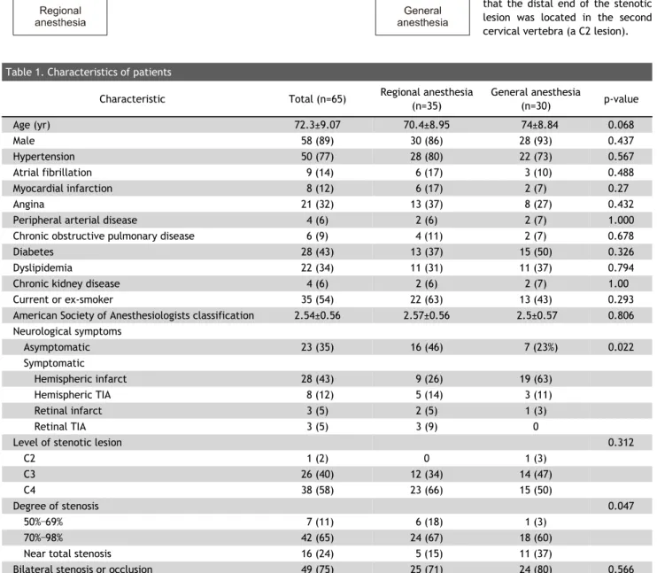
Fig. 1. Operative indication of car- otid endarterectomy and strategy for selection of anesthetic method.
a)
A high level of the lesion indicates that the distal end of the stenotic lesion was located in the second cervical vertebra (a C2 lesion).
Table 1. Characteristics of patients
Characteristic Total (n=65) Regional anesthesia
(n=35)
General anesthesia
(n=30) p-value
Age (yr) 72.3±9.07 70.4±8.95 74±8.84 0.068
Male 58 (89) 30 (86) 28 (93) 0.437
Hypertension 50 (77) 28 (80) 22 (73) 0.567
Atrial fibrillation 9 (14) 6 (17) 3 (10) 0.488
Myocardial infarction 8 (12) 6 (17) 2 (7) 0.27
Angina 21 (32) 13 (37) 8 (27) 0.432
Peripheral arterial disease 4 (6) 2 (6) 2 (7) 1.000
Chronic obstructive pulmonary disease 6 (9) 4 (11) 2 (7) 0.678
Diabetes 28 (43) 13 (37) 15 (50) 0.326
Dyslipidemia 22 (34) 11 (31) 11 (37) 0.794
Chronic kidney disease 4 (6) 2 (6) 2 (7) 1.00
Current or ex-smoker 35 (54) 22 (63) 13 (43) 0.293
American Society of Anesthesiologists classification 2.54±0.56 2.57±0.56 2.5±0.57 0.806 Neurological symptoms
Asymptomatic 23 (35) 16 (46) 7 (23%) 0.022
Symptomatic
Hemispheric infarct 28 (43) 9 (26) 19 (63)
Hemispheric TIA 8 (12) 5 (14) 3 (11)
Retinal infarct 3 (5) 2 (5) 1 (3)
Retinal TIA 3 (5) 3 (9) 0
Level of stenotic lesion 0.312
C2 1 (2) 0 1 (3)
C3 26 (40) 12 (34) 14 (47)
C4 38 (58) 23 (66) 15 (50)
Degree of stenosis 0.047
50%–69% 7 (11) 6 (18) 1 (3)
70%–98% 42 (65) 24 (67) 18 (60)
Near total stenosis 16 (24) 5 (15) 11 (37)
Bilateral stenosis or occlusion 49 (75) 25 (71) 24 (80) 0.566
Values are presented as mean±standard deviation or number (%). The level of the stenotic lesion was defined as the level at which the distal end of the stenosis was located.
TIA, transient ischemic attack; C2, the second cervical vertebra; C3, the third cervical vertebra; C4, the fourth cervical vertebra.
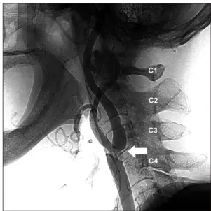
Fig. 2. Level of a stenotic lesion (C3). The level of the stenotic lesion was defined as the level where the distal end of the steno- sis was located (arrow).
(GA) with those of CEA under regional anesthesia (RA) using our center’s strategy.
Methods
The study protocol adhered to the guidelines stipu- lated in the Declaration of Helsinki and was ap- proved as an electronic medical record-based retro- spective study by the Institutional Review Board of Pusan National University Hospital (IRB approval no., H-1901-015-075); as such, the requirement for ob- taining informed consent from the patients prior to study participation was waived.
1) Patient characteristics
Clinical data from 65 patients who had undergone CEA between June 2012 and December 2017 in the Pusan National University Hospital were prospectively collected and retrospectively reviewed. The strategy behind CEA is described in Fig. 1. Based on the type of anesthesia induced, patients were classified into ei- ther those who underwent CEA under RA (the RA group) with selective shunt insertion (n=35) or those who underwent CEA under GA (the GA group) with routine shunt insertion (n=30). Patients’ baseline data and indications of surgery are presented in Table 1.
Symptomatic patients were defined as those with a history of a transient ischemic attack or stroke within a 3-month period prior to surgery. All patients were evaluated by brain magnetic resonance imaging, com- puted tomography perfusion imaging, and computed tomographic or interventional cerebral angiography.
The degree of stenosis was measured according to the method used in the North America Symptomatic Carotid Endarterectomy Trial. The indication for CEA was at least 70% stenosis of the internal carotid ar- tery regardless of symptoms and more than 50%
stenosis with severe contralateral stenosis or occlu- sion in symptomatic patients.
CEA was preferred over carotid artery stenting if
the patient did not have contraindications for CEA, such as previous neck surgery, radiation therapy of the neck, or an extremely high level of the lesion (above the second cervical vertebra). The level of the stenotic lesion was defined as the level where the distal end of the stenosis was located (Fig. 2).
2) Anesthesia
(1) Regional anesthesia: RA was introduced by an ultrasound-guided deep cervical block consisting of 0.75% ropivacaine (20 mL) and 1% lidocaine (20 mL), which was injected by an anesthesiologist into the level of the third to fifth transverse processes of the cervical vertebrae [7]. Then, the patient was lightly sedated while continuously being infused with dexmedetomidine targeted at sedation status level 2 of the Richmond Agitation-Sedation Scale. Prior to CEA clamping for 5 minutes, we performed the awake test, which included speech, grasping a rubber ball, and toe flexion and extension. After CEA clamp- ing, we performed the awake test immediately and every 5 minutes thereafter. If abnormal findings on the awake test were noted during the operation, we inserted a carotid artery shunt (Pruitt-Inahara carotid shunt with T-port; Le maître Vascular Inc., Burlington, MA, USA) to allow patients to recover from the cere- bral ischemic state.
(2) General anesthesia: The induction of anesthesia
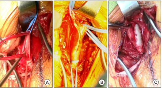
Fig. 3. Operative field view of car- otid endarterectomy. (A) Usage of shunt. (B) Direct closure. (C) Patch closure.
was performed with propofol (2 mg/kg) and rocuro- nium (0.6 mg/kg) followed by tracheal intubation.
Anesthesia was maintained using a 2% sevoflurane concentration with remifentanil (0.05–2.00 μg/kg/min) titrated as required [8]. In patients with a stenotic lesion at the third cervical vertebral level, naso- tracheal intubation was performed for appropriate neck extension of the target lesion.
3) Operative technique
Each patient underwent a cervical incision along the anterior border of the sternocleidomastoid muscle. The common carotid artery, internal carotid artery, external carotid artery, and superior thyroid artery (as necessary) were dissected and dou- ble-looped using a tourniquet. Heparin (50–100 IU/kg) was administered intravenously 5 minutes prior to clamping. An arteriotomy was constructed from the common carotid artery to the internal car- otid artery to expose the entire atheromatous plaque.
In the RA group, no shunt was used except in cases that displayed abnormal findings in the awake test.
In those patients, as well as in all patients in the GA group, the shunt was inserted into the internal car- otid artery first, then into the common carotid artery (Fig. 3A). Endothelium that was thickened with pla- que was dissected using a Freer elevator, and the plaque was removed to ensure that the endothelium was plaque-free. Heparin-mixed saline irrigation was topically applied at the arterial lumen to prevent thromboembolism. In the RA group, the arteriotomy was closed directly, except in patients with a shunt
(Fig. 3B). The 2 main reasons for direct closure were better exposure without interference of the shunt and reduction of clamp time. In the GA group, the arteriotomy was closed using path angioplasty of the bovine pericardium (Vascu-Guard; Biovascular, St.
Paul, MN, USA) (Fig. 3C). The shunt was removed be- fore completely closing the suture with de-airing.
4) Postoperative care
After surgery, the patients under GA were ex- tubated in the operative room and transferred to the neurological intensive care unit (ICU) to stay for at least one day for strict monitoring of their blood pressure (systolic blood pressure <140 mm Hg), any neurological deficits, and any operative wound complications. All patients immediately underwent postoperative computed tomographic angiography of the brain to evaluate the possible presence of anasto- motic site stenosis, acute thrombosis, vascular spasm, embolism, cerebral edema, and hemorrhage. All pa- tients were asked to revisit at 3 months from the discharge date for a neurological examination and carotid duplex ultrasound scanning.
5) Statistical analysis
The categorical variables were assessed using the
Fisher exact test, whereas the continuous variables
were assessed using the Wilcoxon rank-sum test. All
analyses were performed using R software ver. 3.2.2
(R Foundation for Statistical Computing, Vienna,
Austria), and a p-value of less than 0.05 was consid-
ered to indicate statistical significance.
Stroke 0 0 0
Nerve injury 4 (6.2) 3 (8.6) 1 (3.3) 0.618
Use of intravenous painkiller
a)51 (78.5) 27 (77.1) 24 (80.0) 0.780
Use of intravenous blood pressure control drug 37 (56.9) 17 (48.6) 20 (66.7) 0.142
Intensive care unit stay (day) 1.57±0.83 1.54±0.78 1.6±0.89 0.786
Hospital stay (day) 16.62±7.38 14.6±5.05 18.97±8.92 0.022
Values are presented as mean±standard deviation or number (%). Systolic blood pressure was strictly controlled below 140 mm Hg.
a)