pISSN 2288-9272 eISSN 2383-8493 J Oral Med Pain 2016;41(2):41-47 http://dx.doi.org/10.14476/jomp.2016.41.2.41
Cone-Beam Computed Tomographic Assessment of Temporomandibular Joint Morphology in Patients with Temporomandibular Joint Disc Displacement and in Healthy Subjects: A Pilot Study
Hang-Moon Choi
1, Moon-Soo Park
21
Department of Oral and Maxillofacial Radiology, Research Institute of Oral Science, College of Dentistry, Gangneung-Wonju National University, Gangneung, Korea
2
Department of Oral Medicine and Diagnosis, Research Institute of Oral Science, College of Dentistry, Gangneung-Wonju National University, Gangneung, Korea
Received April 16, 2016 Revised June 7, 2016 Accepted June 7, 2016
Purpose: The purpose of this study was to analyze the size and morphology of mandibular condyle and mandibular fossa between temporomandibular joint (TMJ) disc displacement (DD) patients and healthy subjects using cone-beam computed tomography (CBCT).
Methods: Twenty healthy subjects and twenty TMJ DD patients participated in this study re- spectively. We made five measurements in mandibular condyle (medio-lateral dimension, an- tero-posterior dimension, condyle height, intercondylar distance and intercondylar angle) and two measurements in mandibular fossa (mandibular fossa depth and articular eminence angle) using CBCT image.
Results: There was no difference between two groups in medio-lateral dimension. In case of antero-posterior dimension, average of healthy controls was larger than that of TMJ DD pa- tients, but that was not significant statistically. There were no significant differences between two groups in condyle height. Comparing intercondylar distance and intercondylar angle be- tween two groups, there was no significant difference between two groups. In comparison of mandibular fossa depth and articular eminence angle, there was no significant difference be- tween two groups.
Conclusions: We couldn’t find any definite relationship between TMJ morphology and TMJ DD.
Key Words: Cone-beam computed tomography; Disc displacement; Morphology; Temporoman- dibular joint
Correspondence to:
Moon-Soo Park
Department of Oral Medicine and Diagnosis, Research Institute of Oral Science, College of Dentistry, Gangneung-Wonju National University, 7 Jukheon-gil, Gangneung 25457, Korea
Tel: +82-33-640-2466 Fax: +82-33-640-3113 E-mail: [email protected]
This study was supported by Scientific Research (SR1204) of Gangneung- Wonju National University Dental Hospital.
JOMP
Journal of Oral Medicine and PainCopyright
Ⓒ2016 Korean Academy of Orofacial Pain and Oral Medicine. All rights reserved.
CC
This is an open-access article distributed under the terms of the Creative Commons Attribution Non-Commercial License (http://creativecommons.org/licenses/by-nc/4.0/),
INTRODUCTION
The temporomandibular joint (TMJ) is one of the most important joints of the body which comprises the mandibu- lar condyle and the temporal bone.
1)Temporomandibular disorder (TMD) is a symptom complex rather than a sin- gle condition, and it is thought to be caused by multiple factors.
2-4)The successful diagnosis of TMD comes from the
comprehensive examination and the proper diagnostic.
Although the use of radiography for a diagnosis of TMD
has some limitations because of structural variation of TMJ,
the radiography plays an important role in the diagnos-
tic assessment of TMD.
5,6)A variety of imaging modalities
have been used to evaluate the TMJ. Panoramic radiogra-
phy, conventional linear or complex motion tomography
and computed tomography (CT) are used to assess the osse-
ous components of the joints, whereas magnetic resonance
imaging (MRI) is used to assess the soft tissue components.
Traditionally used two-dimensional TMJ projections, such as the transcranial view, are of limited use nowadays. These projections suffer from significant superimposition of the overlying structures, which compromises their ability to de- tect pathological TMJ changes. Panoramic radiography is still useful in evaluating gross TMJ osseous pathology.
7,8)CT has been a valuable aid in the evaluation of the TMJ.
This technique was found to be superior to hypocycloi- dal tomography.
9)However, the high cost, access to equip- ment and the relatively high radiation dose have limited the widespread use of CT for TMJ evaluation. With the advent of cone-beam CT (CBCT), these barriers have been over- come. Today CBCT units are located in dental schools, den- tal radiographic laboratories and private practices, and have provided increased access to CT technology. Furthermore, the cost of imaging patients with CBCT units is generally lower than medical CT.
10,11)CBCT has high dimensional ac- curacy in measuring facial structures including TMJ.
12,13)As a clinician treating TMD patients, author has frequent- ly observed relatively small mandibular condyle in TMJ disc displacement (DD) patients. Therefore the relationship between TMJ DD and TMJ morphology has been a mat- ter of great concern to author. There were some researches on the size and morphology of TMJ, but most of them were based on autopsy or two-dimensional image and their sub- jects were non-Asian.
The aim of this study was to analyze the size and mor- phology of mandibular condyle and mandibular fossa (MF) in Korean healthy controls and TMJ DD patients using CBCT.
MATERIALS AND METHODS
1. Subjects
Twenty healthy subjects participated voluntarily in this study and twenty TMD patients had visited in the Depart- ment of Oral Medicine at Gangneung-Wonju National University Dental Hospital (Gangneung, Korea) from October 2013 to January 2016 were also included. The aver- age age of participants is displayed in Table 1. All subjects were examined clinically by a specialist. A comprehen- sive history and an informed consent were taken from all
subjects. This study protocol was approved by Gangneung- Wonju National University Dental Hospital Ethics Review Committee (IRB2012-25-4). TMD patients with the sign and symptom of TMJ DD such as noise, periodic lock and limi- tation of motion were Included in TMJ DD group. Subjects having muscle disorder and degenerative bony change in TMJ were excluded. Subjects with class II or class III maloc- clusion and missing permanent tooth were also excluded.
2. Methods
All CBCT scans were acquired at position of maximum dental intercuspation with an Alphard Vega unit (Asahi Roentgen, Kyoto, Japan) and set at 80 kV, 5-9 mA, 17 sec- onds scan time, and 0.3 mm voxel size. Scan data were re- positioned along Frankfort horizontal plane using bilateral porions and right orbitale and then three-dimensionally reconstucted by using of Xelis Dental software (Infinitt Healthcare, Seoul, Korea).
This study was designed to access the morphology of condylar head and MF which included condylar medio- lateral (ML) and antero-posterior (AP) dimension, condyle height, intercondylar (IC) distance and angle, MF depth, and articular eminence (AE) angle (Table 2).
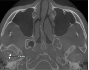
All measurements were performed by one oral and max- illofacial radiologist. For ML dimension, we use oblique coronal plane made by condylar axis and measure the hori- zontal distance between medial and lateral ends at the con- dylar head in the oblique coronal plane (Fig. 1). AP dimen- sion was measured at the axial image showed the longest ML condylar dimension (Fig. 2). For condyle height, we re- constructed an oblique sagittal plane image which showed apex of coronoid process and center of condylar head. We
Table 1. Demographic characteristics of the subjects (n=40)
Characteristic Group
Healthy control Temporomandibular disorder Sex
Male 10 10
Female 10 10
Average age (y)
Male 36.4±9.3 23.0±5.6
Female 32.0±12.8 32.5±12.4
Values are presented as number of patients or mean±standard
deviation.
widened the thickness of image for showing condylar heads and ramus in the same image (Fig. 3). For IC distance, we reconstructed an oblique coronal image which showed right and left condylar lateral pole in the same image, and then measured horizontal distance between right and left lateral end of condyles (Fig. 4). For condylar axis angle, we used axial image which showed the longest ML condylar dimen- sion and measured the angle between the ML plane of the condylar head and the midsagittal plane (Fig. 5). IC angle was defined as sum of both condylar axis angles. MF depth and AE angle were measured at the oblique sagittal image bisecting condylar head (Fig. 6).
Table 2. Definitions of measurements
Measurement Definition
ML dimension Horizontal distance between medial and lateral ends at the condylar head in the coronal plane
AP dimension Linear distance between anterior and posterior ends of the condylar head at the bisecting line of ML dimension in axial plane
Condyle height Vertical distance between the most superior point of the MF and the most inferior point of the AE at the bisecting sagittal plane of condylar head
IC distance Horizontal distance between right and left lateral end of condyles in the coronal plane
IC angle Sum of right and left condylar axis angles (each angle is defined as angle between the ML plane of the condylar head and the midsagittal plane)
AE angle Angle between line formed by the most superior point of the MF and the most inferior point of the AE and horizontal plane at the bisecting sagittal plane of condylar head
MF depth Perpendicular linear distance between the most superior point of condyle and a line passing through the deepest point mandibular notch and perpendicular to tangent of posterior surface of ramus in sagittal plane
ML, medio-lateral; AP, antero-posterior; MF, mandibular fossa; AE, articular eminence; IC, intercondylar.
20.9 mm
Fig. 1. Oblique coronal image shows measurement of medio-lateral dimension.
6.9 mm
Fig. 2. Axial image shows measurement of antero-posterior dimension.
Fig. 3. Oblique sagittal image shows measurement of condyle height.
22.0 mm
3. Statistical Analysis
Independent t-test and Mann-Whitney U test were used to compare the mean values of ML dimension, AP dimen- sion, condyle height, IC distance, IC angle, MF depth and AE angle. p-values less than 0.05 were considered sta- tistically significant. All statistical analyses were per- formed with the IBM SPSS Statistics version 21.0 (IBM Co., Armonk, NY, USA).
RESULTS
We made five measurements in mandibular condyle (ML
dimension, AP dimension, condyle height, IC distance, and IC angle) and two measurements in MF (MF depth and AE angle) in each CBCT image.
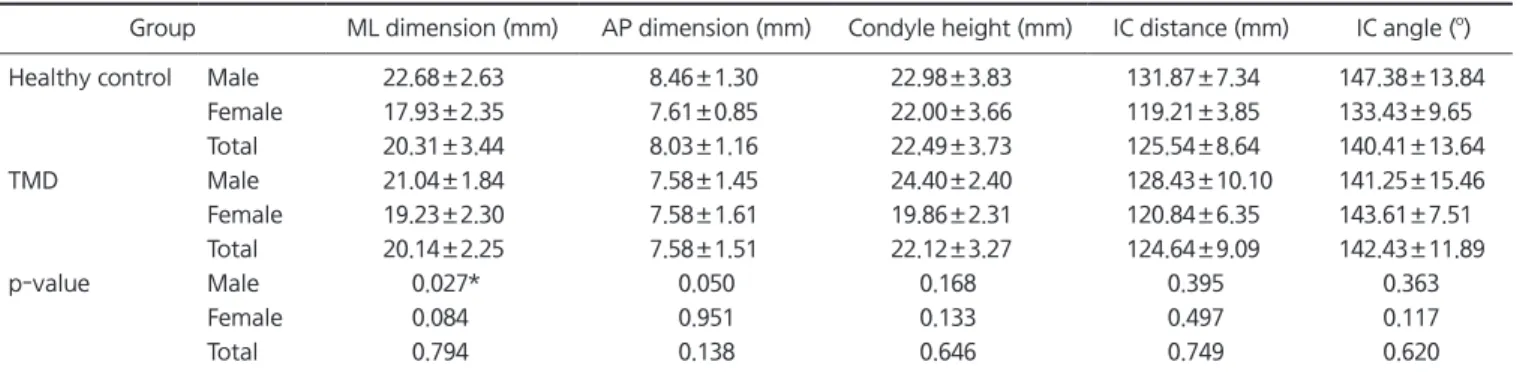
Average ML dimension of healthy controls is 20.31 mm, and average ML dimension of TMD patients is 20.14 mm.
We couldn’t find any difference between two groups. But, comparing ML dimension in males, there was a significant difference in ML dimension (p<0.05). In case of AP dimen- sion, average AP dimension of healthy controls is 8.03 mm, and average AP dimension of TMD patients is 7.58 mm.
Average AP dimension of healthy controls was larger than that of TMD patients, but that was not significant statisti- cally. Comparing condyle height, IC distance and IC angle between healthy controls and TMD patients, there was no significant difference between two groups (Table 3).
Average MF depth of healthy controls is 7.08 mm, and average MF depth of TMD patients is 6.84 mm. And aver- age AE angle of healthy controls is 32.29
o, and average AE angle of TMD patients is 32.74
o. In comparison of MF depth and AE angle, there was no significant difference between two groups (Table 4).
DISCUSSION
CBCT, a recently developed imaging technology, is likely the modality of choice for assessing TMJ osseous
Fig. 4. Oblique coronal image shows measurement of intercondylar distance.
124.5 mm