• 검색 결과가 없습니다.

Titanium Plate Fixation for Sternal Dehiscence in Major Cardiac Surgery

N/A
N/A
Protected

Academic year: 2021

Share "Titanium Plate Fixation for Sternal Dehiscence in Major Cardiac Surgery"

Copied!
6
0
0

로드 중.... (전체 텍스트 보기)

전체 글

(1)

http://dx.doi.org/10.5090/kjtcs.2013.46.4.279 ISSN: 2233-601X (Print) ISSN: 2093-6516 (Online)

Department of Thoracic and Cardiovascular Surgery, Asan Medical Center, University of Ulsan College of Medicine Received: September 21, 2012, Revised: January 31, 2013, Accepted: March 7, 2013

Corresponding author: Joon Bum Kim, Department of Thoracic and Cardiovascular Surgery, University of Ulsan College of Medicine, 88 Olympic-ro 43-gil, Songpa-gu, Seoul 138-736, Korea

(Tel) 82-2-3010-5416 (Fax) 82-2-3010-6966 (E-mail) jbkim1975@amc.seoul.kr

C

The Korean Society for Thoracic and Cardiovascular Surgery. 2013. All right reserved.

CC
This is an open access article distributed under the terms of the Creative Commons Attribution Non-Commercial License (http://creative- commons.org/licenses/by-nc/3.0) which permits unrestricted non-commercial use, distribution, and reproduction in any medium, provided the original work is properly cited.

Titanium Plate Fixation for Sternal Dehiscence in Major Cardiac Surgery

Wan Kee Kim, M.D., Joon Bum Kim, M.D., Gwan Sic Kim, M.D., Sung-Ho Jung, M.D., Suk Jung Choo, M.D., Cheol Hyun Chung, M.D., Jae Won Lee, M.D.

Background: Sternal dehiscence is one of the most troublesome complications following cardiac surgery. Treatment failure and consequent lethal outcomes are very common. The aim of this study was to evaluate titanium plate fix- ation as a treatment for sternal dehiscence following major cardiac surgery. Materials and Methods: Between 2010 and 2012, 17 patients underwent sternal reconstruction using horizontal titanium plating for the treatment of post-cardiac-surgery sternal dehiscence. The plates were cut and shaped, and then were fixed to corresponding costal segments using 2-3 titanium screws per each side. Results: The median age of our patients was 66 years (range, 50 to 78 years) and 9 were female. Indications for sternal reconstruction included aseptic sternal de- hiscence in 3 patients and osteomyelitis in 14 patients including 6 patients who were diagnosed with mediastinitis.

During the operation, sternal resection and autologous flap interposition were combined in 11 patients. One patient died due to sepsis. Two patients required additional soft tissue wound revisions. Another patient presented with a tuberculous wound infection which was resolved using anti-tuberculosis medications. The postoperative course was uncomplicated in the other 13 patients. Conclusion: Titanium plate fixation that combines appropriate debridement and flap interposition is very effective for the treatment of sternal dehiscence following major cardiac surgery.

Key words: 1. Sternum

2. Wound dehiscence 3. Wound infection 4. Reoperation

INTRODUCTION

Sternal dehiscence following major cardiac surgery is a common problem with an incidence rate of 0.5% to 5.0%

[1-4]. However, the management of severe sternal dehiscence that invades the sternal bone remains a challenge for cardiac surgeons [2,5]. Recently, the mortality rate from deep sternal wound infection following major cardiac surgery was reported to be in decline, but still to be as high as 16% to 22% [4].

The classic management of sternal dehiscence includes par- tial or total sternal resection with intravenous antibiotic ther- apy [6], continuous irrigation with saline containing anti-mi- crobial agents [7], and localized negative pressure therapy, al- so known as vacuum-assisted closure [8]. When severe in- flammation cannot be controlled using these methods, de- bridement of the devitalized sternum is necessary [9].

These classic treatments, however, have several limitations,

especially in severe cases. First, massive resection which is

(2)

essectial to prevent recurrence [10,11] brings out small dis- ease-free margin, and it does not provide enough space to hold the sternum in classical treatments [12]. Second, when the defect is large, the remaining bony structure following sufficient resection of the devitalized tissues is not enough to prevent paradoxical chest wall movement using classic management.

Recently, several groups have advocated the use of rigid metal plate fixation to close the sternum and have reported promising results [13-15]. Since our previously published case report [16], we have treated 10 more cases of sternal fixation using titanium plates in patients who suffered sternal compli- cations following major cardiac operations. The aim of this study was to evaluate the effectiveness of horizontal titanium plating for the treatment of post-cardiac-surgery sternal dehiscence.

MATERIALS AND METHODS 1) Patients

In August 2009, we adopted an aggressive strategy that in- cluded radical sternal resection and reconstruction using tita- nium plates for a sternal wound problem involving the sternal bone or the area beneath following major cardiac surgery.

From August 2009 through Dec 2012, a total of 2,769 adult patients (age 18 years or older) underwent major cardiac sur- gery at Asan Medical Center, Seoul, Korea. Of this patient population, 36 cases (1.3%) developed postoperative deep sternal wound complications. Nineteen patients developed wound problems that were limited to the soft tissue layer, and they were completely treated using conventional methods such as vacuum suctioning or soft tissue revision. The other 17 patients who underwent titanium plate fixation with mas- sive debridement of the sternum comprised our current study cohort.

2) Surgical technique

Under general anesthesia, the previous median sternotomy incision was reopened and the wires were cut and removed.

After debridement of all devitalized tissues and broken bone materials, sufficient irrigation with warm saline was performed. Both pectoralis major muscles were laterally un-

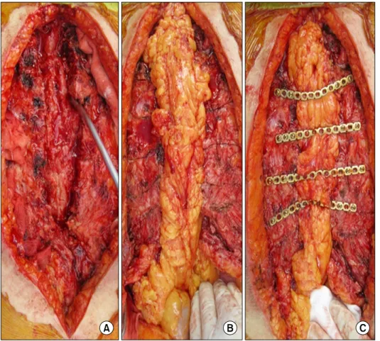
dermined up to the mid-clavicular line in order to secure a sufficient area for titanium plate fixation. Both sides of the remnant thoracic wall were brought together, and the plate’s types and sizes were chosen to fit each patient’s thoracic wall. This plate fixation system (Angled Universal Fracture Plate; AO Foundation, Davos Platz, Switzerland) consists of 2-mm-thick titanium plates that have multiple holes for screw fixation (Table 1). After measurement to determine the appro- priate lengths of the plates, the plates were cut, shaped, and fixed to the corresponding costal segments using 2-3 titanium screws per side (Fig. 1). The pectoralis major muscle or omental flap was plugged to fill the tissue defects following extensive debridement (Fig. 2). After sufficient irrigation with warm saline, a draining catheter was interposed beneath the pectoralis major muscle layer. Over the titanium plates, the undermined pectoralis muscle layer was closed using multiple interrupted sutures. Adequate antibiotic therapy was ad- ministered according to the bacterial sensitivity tests of the wound swab cultures.

RESULTS

Of the 17 patients, 8 patients had diabetes mellitus, 2 had

osteoporosis, and 1 patient was receiving immunosuppressant

medications after liver transplantation (Table 1). Previous car-

diac procedures included coronary arterial bypass grafting

(CABG) in 5 patients, cardiac valve surgery in 8 patients,

and simultaneous coronary and valve surgery (including

Bentall operation) in 4 patients. The reconstructive operation

was performed after a median of 45 days following the initial

operation (range, 0 to 255 days). Several conventional treat-

ments such as soft tissue debridement or application of a vac-

uum suctioning system were used before titanium plate fix-

ation in 8 patients during these periods. Indications for sternal

reconstruction using titanium plate fixation included osteo-

myelitis with (n=6) or without (n=6) mediastinitis in 12 pa-

tients, aseptic sternal dehiscence in 2 patients, and bony de-

struction due to osteoporosis in 1 patient. The latter patient

was a 66-year-old female who had been diagnosed with os-

teoporosis, and the upper part of the sternal body was broken

into several particles during concomitant CABG and mitral

annuloplasty. To prevent sternal instability in this case, 2 hor-

(3)

Table 1. Patient characteristics Patient Age/

gender

Co-morbid

factors Initial operations Resected sternum Osteo- myelitis

Media- stinitis

No. of plates/

screws

Autologous tissue flaps

1 2 3 4 5 6 7 8 9 10 11 12 13 14 15 16 17

76/F 61/M 66/F 52/M 68/M 76/F 56/M 54/M 50/M 53/M 62/M 74/F 78/F 71/F 70/F 68/F 70/F

DM None Osteoporosis Sepsis DM Osteoporosis Transplantation DM

DM DM DM, CRF DM None None None None None

AVR, CABG AVR CABG, MVP

AVR, MVP, TVP, CABG, Dor AVR, Ao-MvR

CABG, MVP MVR OPCAB OPCAB MVP OPCAB OPCAB OPCAB MVP, TVP Bentall procedure Redo MVR AVR

Total sternum Sternal angle Upper sternal body Whole sternal body Mid-sternal body Mid-sternal body Manubrium Mid-sternal body Sternal angle Sternal angle Sternal angle Whole sternal body Whole sternal body Whole sternal body Lower sternal body Total sternum Whole sternal body

+ + - + + - + - + + + + + + + + +

- + - + - - + - - + - + - - + - +

5/19 1/4 2/12 6/24 2/10 2/12 2/4 2/8 2/10 3/14 2/12 4/17 4/18 5/24 5/20 6/14 3/12

None Omentum None Omentum Pectoralis None Pectoralis Pectoralis None Pectoralis Pectoralis Omentum Omentum Omentum None TRAM Omentum DM, diabetes mellitus; AVR, aortic valve replacement; CABG, coronary arterial bypass grafting; MVP, mitral valve repair; TVP, tri- cuspid valve repair; Dor, Dor procedure; Ao-MvR, aorto-mitral reconstruction; MVR, mitral valve replacement; OPCAB, off-pump beating coronary arterial bypass; CRF, chronic renal failure; TRAM, transverse rectus abdominis muscle.

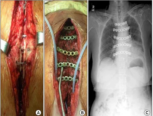
Fig. 1. Fixation of transverse titanium plates for dehisced sternum (patient #1).

(A) Intra-operative findings of sternal de-

hiscence with purulent discharge which

are indicative of sternal osteomyelitis

(following aortic valve replacement and

coronary artery bypass). (B) Horizontal

fixation of the titanium plates was per-

formed on the same patient. Two drain-

age catheters were placed under the

pectoralis muscle layer. (C) Postoperative

simple chest radiograph.

(4)

Fig. 2. Total sternectomy followed by omental flap interposition and titanium plating (patient #13). (A) The dead space occurring after total sternectomy is seen.

(B) The omental flap is interposed to the defect following debridement. (C) Titanium plates are laid over the omental flap, and fixed to the thoracic wall with screws.

izontal titanium plates were placed on the sternal body during initial sternal closure.

For the other 16 patients, segmental (n=13) or total (n=3) sternal resection was performed. Flap interposition of the omental (n=6), the rectus abdominis muscle (n=1), or pector- alis major muscle (n=5) was done to fill in the empty space according to the extent of the tissue defects following sternal resection. The median number of inserted titanium plates and screws was 5 (range, 1 to 6 plates) and 19 (range, 4 to 24 screws), respectively.

One patient in whom overwhelming sepsis and multi-organ failure had already developed following his initial surgery (aortic valve replacement, mitral annuloplasty, CABG, and Dor procedure) died due to profound sepsis. Having been di- agnosed with mediastinitis 1 month after the initial surgery, he underwent massive debridement, total sternectomy, omen- tal flap interposition, and titanium plate fixation. He died of septic shock, which did not respond to high-dose vaso- constrictors for 7 days following surgery.

The in-hospital clinical course was complicated in 2 patients. One patient required additional wound revision due to aseptic wound dehiscence that was confined to the soft tis- sue layer, which was completely resolved by revision. The other patient presented with wound problems at postoperative 5 months, which was determined to be a tuberculous in- fection and was treated with anti-tuberculosis medications without further complications.

The median duration from sternal reconstruction to dis- charge was 37 days (range, 7 to 122 days). The postoperative course was uncomplicated in another 14 patients during a median follow-up of 522 days (range, 50 to 872 days) postoperatively.

DISCUSSION

The present study indicates that fixation of the sternum us-

ing a rigid plate is an effective management strategy to deal

with a severely dehisced sternum, providing thoracic wall sta-

(5)

bility and reducing the recurrence rate. Six of our patients had undergone several wound revisions using classic manage- ment (maximum of 4 revisions per patient) before titanium plate fixation. However, additional wound revision was not needed following titanium plate fixation in most of those patients.

The treatment with titanium plate fixation for dehisced ster- nal wound problem has several definite advantages. The most important benefit of titanium plate fixation is the guarantee of adequate resection of devitalized tissue by offering thoracic wall stabilization even without the sternal bone. If the ster- num maintains stability after sufficient debridement, the ster- nal wound can be closed without using rigid plates [17]. On the other hand, a sternum with poorly qualified bone marrow, insufficient for closure using classic methods [18], may in- crease the rate of recurrence [19-22]. Immobilization and the maintenance of stability are the major principles of bony re-union and wound healing [23]. In this regard, titanium plate fixation may be an ideal option to offer bony re-union and stability.

The second benefit of titanium plate fixation is that the tension of the thoracic wall can be released by bridging both ribs over the sternal defects. Without titanium plates, both sides of the thoracic wall must be pulled toward the midline.

Then the thoracic wall might tense up, disturbing wound healing. We believe that releasing the tension of the thoracic wall might contribute to rapid healing and, thereby, shorten hospitalization. The third benefit is ease of handling.

Titanium plates are modifiable without regard to the varying shapes of each thoracic wall. Fixation of the plate was also performed with simple tightening screws.

On the other hand, some disadvantages have been reported with titanium plate fixation. First, some studies have reported a high incidence of plate removal, ranging from 8% to 25%

[14,19]. This is explained by the fact that sternocostal junc- tions are highly mobile articulations during respiration and this motion is more active in more far location from the ster- num [13]. The size mismatch between the titanium plate and the thoracic wall requires a long plate, and it may result in chest wall pain, which is the main cause of titanium plate re- moval [13]. However, none of our 11 cases needed plate re- moval or readjustment. We made titanium plates shaped and

curved by roller to fit to the adjacent thoracic wall in every case, making efforts to tie the plates to the utmost medial part of the costal cartilage, which could have contributed to our good results.

Second, titanium plate fixation has potential drawbacks, in- cluding bleeding and lung injury. Cicilioni et al. [19] reported an approximately 10% incidence of seroma formation and 4%

incidence of bleeding associated with this technique, but the author stated that these complications were mainly related to pectoralis muscle dissection. Nevertheless, drilling the screws into the ribs should be carefully performed because measuring the actual depth of the ribs is difficult [13]. However, there were no cases of postoperative bleeding or pneumothorax in the present study. To determine the proper length of the screws, we estimated the depth of each patient’s ribs using preoperative computed tomography, which was confirmed in- tra-operatively.

The cost problem is another disadvantage to the use of ti- tanium plate fixation. Other classic methods, including irriga- tion, debridement, and re-wiring using steel wires without rig- id plates, are less expensive options. However, considering its effectiveness, titanium plate fixation might help shorten the overall postoperative course by addressing the troublesome and frequently relapsing problems in a more complete way.

The major limitations of this study include the limited number of patients treated with titanium plates in a relatively short-term follow-up. The titanium plate fixation was not compared with conventional treatments; therefore, the study results require further verification through comparative studies.

In conclusion, fixation of the sternum using a rigid plate is feasible and might be a good alternative solution to treating sternal dehiscence following major cardiac surgery, especially for those requiring aggressive sternal resection. Results from a larger number of patients are required to verify the results of this study.

CONFLICT OF INTEREST

No potential conflict of interest relevant to this article was

reported.

(6)

REFERENCES

1. Losanoff JE, Richman BW, Jones JW. Disruption and in- fection of median sternotomy: a comprehensive review. Eur J Cardiothorac Surg 2002;21:831-9.

2. Douville EC, Asaph JW, Dworkin RJ, et al. Sternal preser- vation: a better way to treat most sternal wound complica- tions after cardiac surgery. Ann Thorac Surg 2004;78:1659- 64.

3. El Oakley RM, Wright JE. Postoperative mediastinitis: clas- sification and management. Ann Thorac Surg 1996;61:1030- 6.

4. Matros E, Aranki SF, Bayer LR, McGurk S, Neuwalder J, Orgill DP. Reduction in incidence of deep sternal wound in- fections: random or real? J Thorac Cardiovasc Surg 2010;

139:680-5.

5. Toumpoulis IK, Anagnostopoulos CE, Derose JJ Jr, Swistel DG. The impact of deep sternal wound infection on long- term survival after coronary artery bypass grafting. Chest 2005;127:464-71.

6. Schroeyers P, Wellens F, Degrieck I, et al. Aggressive pri- mary treatment for poststernotomy acute mediastinitis: our experience with omental- and muscle flaps surgery. Eur J Cardiothorac Surg 2001;20:743-6.

7. Shumacker HB Jr, Mandelbaum I. Continuous antibiotic irri- gation in the treatment of infection. Arch Surg 1963;86:

384-7.

8. Malmsjo M, Ingemansson R, Sjogren J. Mechanisms govern- ing the effects of vacuum-assisted closure in cardiac surgery.

Plast Reconstr Surg 2007;120:1266-75.

9. Sarr MG, Gott VL, Townsend TR. Mediastinal infection af- ter cardiac surgery. Ann Thorac Surg 1984;38:415-23.

10. Mansour KA, Thourani VH, Losken A, et al. Chest wall re- sections and reconstruction: a 25-year experience. Ann Thorac Surg 2002;73:1720-5.

11. Robicsek F, Fokin A, Cook J, Bhatia D. Sternal instability after midline sternotomy. Thorac Cardiovasc Surg 2000;48:1- 8.

12. Schimmer C, Reents W, Berneder S, et al. Prevention of sternal dehiscence and infection in high-risk patients: a pro-

spective randomized multicenter trial. Ann Thorac Surg 2008;86:1897-904.

13. Voss B, Bauernschmitt R, Will A, et al. Sternal reconstruc- tion with titanium plates in complicated sternal dehiscence.

Eur J Cardiothorac Surg 2008;34:139-45.

14. Hallock GG, Szydlowski GW. Rigid fixation of the sternum using a new coupled titanium transverse plate fixation system. Ann Plast Surg 2007;58:640-4.

15. Plass A, Grunenfelder J, Reuthebuch O, et al. New trans- verse plate fixation system for complicated sternal wound in- fection after median sternotomy. Ann Thorac Surg 2007;83:

1210-2.

16. Kim WK, Kim JB, Jung SH, Choo SJ, Chung CH, Lee JW.

Titanium plate fixation for a dehisced sternum following cor- onary artery bypass grafting: a case report. Korean J Thorac Cardiovasc Surg 2012;45:127-30.

17. Song DH, Lohman RF, Renucci JD, Jeevanandam V, Raman J. Primary sternal plating in high-risk patients prevents mediastinitis. Eur J Cardiothorac Surg 2004;26:367-72.

18. Losanoff JE, Jones JW, Richman BW. Primary closure of median sternotomy: techniques and principles. Cardiovasc Surg 2002;10:102-10.

19. Cicilioni OJ Jr, Stieg FH 3rd, Papanicolaou G. Sternal wound reconstruction with transverse plate fixation. Plast Reconstr Surg 2005;115:1297-303.

20. Sargent LA, Seyfer AE, Hollinger J, Hinson RM, Graeber GM. The healing sternum: a comparison of osseous healing with wire versus rigid fixation. Ann Thorac Surg 1991;52:

490-4.

21. Snyder CW, Graham LA, Byers RE, Holman WL. Primary sternal plating to prevent sternal wound complications after cardiac surgery: early experience and patterns of failure.

Interact Cardiovasc Thorac Surg 2009;9:763-6.

22. Mitra A, Elahi MM, Tariq GB, Mir H, Powell R, Spears J.

Composite plate and wire fixation for complicated sternal closure. Ann Plast Surg 2004;53:217-21.

23. Claes L, Eckert-Hubner K, Augat P. The effect of mechan-

ical stability on local vascularization and tissue differ-

entiation in callus healing. J Orthop Res 2002;20:1099-105.

수치

Fig. 1. Fixation of transverse titanium plates for dehisced sternum (patient #1).
Fig. 2. Total sternectomy followed by omental flap interposition and titanium  plating (patient #13)

참조

관련 문서

Autho: Effectiveness of surgery for lumbar stenosis and degenerative spondylolisthesis in the octogenarian population: analysis of the Spine Patient Outcomes Research

Purpose: Calcaneal fracture is a rare fracture, which accounts for about 2% of all fractures, but is one of the most common fractures in the ankle bone.. There is

Lumbar discogenic radiculopathy (LDR) is one of the most common spinal disorders which can lead to high medical expenses for the patient, occupational impairment,

The purpose of this study was to identify the frequency and related factors of advanced airway management for patients with cardiac arrest by the

This study prepares an educational program which helps the patients reduce anxiety on surgery which is considered as one of the biggest emotional crises

Result: During the study period, 482 patients had elevated cardiac troponin-T levels. The positive predictive value for the.. The diagnostic importance of

Approved clinical use of bone marrow stem cells for myocardial infarction treatment... Cardiac

– A fixed lump-sum price based on the quantities provided by the owner for the major components of the project. – Most infrastructure projects and