• 검색 결과가 없습니다.

Erdheim-Chester Disease Presenting as an Anterior Mediastinal Tumor without Skeletal Involvement

N/A
N/A
Protected

Academic year: 2021

Share "Erdheim-Chester Disease Presenting as an Anterior Mediastinal Tumor without Skeletal Involvement"

Copied!
4
0
0

로드 중.... (전체 텍스트 보기)

전체 글

(1)

ISSN: 2233-601X (Print) ISSN: 2093-6516 (Online)

− 223 −

Received: September 13, 2017, Revised: September 29, 2017, Accepted: October 7, 2017, Published online: June 5, 2018

Corresponding author: Hyeong Ryul Kim, Department of Thoracic and Cardiovascular Surgery, Asan Medical Center, University of Ulsan College of Medicine, 88 Olympic-ro 43-gil, Songpa-gu, Seoul 05505, Korea

(Tel) 82-2-3010-3580 (Fax) 82-2-3010-6966 (E-mail) scena@dreamwiz.com

© The Korean Society for Thoracic and Cardiovascular Surgery. 2018. All right reserved.

This is an open access article distributed under the terms of the Creative Commons Attribution Non-Commercial License (http://creativecommons.org/

licenses/by-nc/4.0) which permits unrestricted non-commercial use, distribution, and reproduction in any medium, provided the original work is properly cited.

Erdheim-Chester Disease Presenting as an Anterior Mediastinal Tumor without Skeletal Involvement

Kanghoon Lee, M.D. 1 , Hyeong Ryul Kim, M.D., Ph.D. 2 , Jin Roh, M.D. 3 , You Jung Ok, M.D. 2 , Bo Bae Jeon, M.D. 2 , Young Woong Kim, M.D. 2

1
Department of Thoracic and Cardiovascular Surgery, Korea University Guro Hospital,

Korea University College of Medicine, Departments of

2

Thoracic and Cardiovascular Surgery and

3

Pathology, Asan Medical Center, University of Ulsan College of Medicine

Erdheim-Chester disease (ECD) is a form of non–Langerhans cell histiocytosis that most commonly involves the skeletal system. We report an unusual case of ECD presenting as an anterior mediastinal tumor without skeletal involvement. A 60-year-old man with no remarkable medical history was referred for evaluation of a mediastinal mass. The patient underwent surgical excision of the tumor via video-assisted thoracoscopic surgery. Histologic examination revealed marked proliferation of atypical histiocytes with sclerosis, and the results of immunohistochemical staining were suggestive of ECD.

Key words: 1. Erdheim-Chester disease 2. Histiocytosis

3. Non-Langerhans cell 4. Mediastinum

5. Video-assisted thoracic surgery

Case report

1) Introduction

Erdheim-Chester disease (ECD), a rare form of non-Langerhans cell histiocytosis with an unknown cause, was first described by Chester [1] in 1930.

Histopathologically, ECD is characterized by xanthog- ranulomatous infiltration of numerous “lipid-laden”

macrophages surrounded by fibrosis. Immunohisto- chemically, ECD histiocytes are typically positive for CD68, CD163, and factor XIIIa, and negative for CD1a [2-4]. Although ECD is regarded as a multisystemic and heterogeneous disease, skeletal involvement is present in 96% of cases [3]. Herein, we report an unusual case of ECD presenting as an anterior media-

stinal tumor without skeletal involvement that was treated surgically.

2) Case description

A 60-year-old man with no remarkable medical history was referred to Asan Medical Center in Seoul for the evaluation of an anterior mediastinal mass.

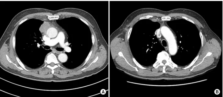
He had presented to a local hospital with a 4-month history of moderate cough. His laboratory findings were nonsignificant, but computed tomography showed a 4.0-×6.5-cm irregular anterior mediastinal mass with heterogeneous enhancement (Fig. 1A). A percutaneous needle biopsy was nondiagnostic, but was suggestive of an inflammatory myofibroblastic tumor. Positron emission tomography–computed to-

Korean J Thorac Cardiovasc Surg 2018;51:223-226 □ CASE REPORT □

https://doi.org/10.5090/kjtcs.2018.51.3.223

(2)

Kanghoon Lee, et al

− 224 −

Fig. 1. (A) Contrast-enhanced chest computed tomography scan, showing a 2.6-cm mass in the right anterior mediastinum. (B) Contrast-enhanced chest computed tomography scan, showing enlarged intrathoracic lymph nodes presumed to indicate recurrence.

mography demonstrated increased fluorodeoxyglucose uptake without any other hypermetabolism sugges- tive of metastasis. Brain magnetic resonance imaging and technetium-99m bone scintigraphy revealed no significant abnormal findings.

Under suspicion of a thymic neoplasm, the patient underwent surgical excision of the tumor via vid- eo-assisted thoracoscopic surgery (VATS). Two 11.5-mm ports and one 5-mm port were created, with the patient in the left semilateral decub itus position. An approximately 5.5-cm firm irregular mass was observed in the anterior mediastinum, and was excised completely via VATS. Because it was strongly suspected that the mass had invaded the right upper lobe of the lung and pericardium, pulmo- nary wedge resection and partial pericardiectomy were also performed. One of the 11.5-mm ports was extended, and the tumor was pulled out using an en- doretrieval pouch. On frozen biopsy, an inflammatory lesion with lymphoplasmacytic infiltration and fib- rosis was observed, with no residual tumor on the resection margins. Because there was no clinical find- ing suggesting malignancy, no further resection was attempted. As the operation was primarily performed to obtain a biopsy sample, the pericardial resection was very small (<2 cm); this rarely results in car- diac herniation, so the pericardial resection space was left open. After hemostasis, a Jackson-Pratt drain

was inserted into the mediastinal side of the pleural cavity, and the patient was transferred to the post- anesthetic care unit.

The specimen included a well-demarcated, lobu- lated, firm mass (5.5×4.2×4 cm) in the mediastinal soft tissue. The cut surface showed pinkish-white fib- rosis with multifocal necrosis, and extension to the lung parenchyma was observed (Fig. 2). Histologic examination revealed marked proliferation of atypical histiocytes with sclerosis, and the results of im- munohistochemical staining were positive for CD68 and factor XIIIa, and negative for CD1a. Although a BRAF V600E mutation test was negative, the overall findings were suggestive of ECD. The mass was densely adherent to the pericardium with micro- scopic invasion, and extension to the visceral pleura of the lung was also observed. These histologic find- ings were confirmed by 2 expert pathologists through an intradepartmental consultation.

The patient’s postoperative course was uneventful,

and he was discharged on postoperative day 3 with-

out any complications. Three months after discharge,

follow-up computed tomography revealed enlarged

internal thoracic lymph nodes in the right first and

second intercostal spaces (Fig. 1B). The patient un-

derwent repeated VATS for excisional biopsy using

the previous incisions. The lymph nodes were ex-

cised en bloc with adjacent soft tissue, and pathology

(3)

An Unusual Presentation of Non-Langerhans Cell Histiocytosis

− 225 −

Fig. 2. (A) Cross-section of the resected anterior mediastinal mass, with the edge connecting to the area of lung invasion, showing pinkish-white fibrosis with multifocal necrosis. (B) Histopathologic section demonstrating the proliferation of atyp- ical histiocytes with sclerosis (H&E stain, ×200). (C) Positive im- munohistochemical staining of histiocytes with CD68 (×400).

confirmed recurrence of ECD. The patient showed no further disease recurrence during the following 22 months.

Discussion

Although the total numb er of reported cases of ECD has increased in the most recent decade, only 500–550 cases have been reported worldwide [2].

The mean age at diagnosis is 53–55 years [3], and most published data show a male predominance [3].

The pathogenesis of ECD is uncertain, b ut it has b een reported that more than half of cases are associated with the BRAF V600E mutation [5].

ECD is diagnosed based on clinical, radiologic, and histopathologic findings. Although ECD is a multi- systemic disease, the most commonly affected organ is the skeletal system. Symmetric diaphyseal and metaphyseal osteosclerosis of the long bones of the lower extremities is present on technetium-99m bone scintigraphy in nearly all patients [2-4]. According to a report by Arnaud et al. [3], only 4% of patients with ECD lack radiographic findings of skeletal involvement. Central nervous system involvement usually is asymptomatic; it is relatively common

(25%–50%), and has been reported to be a crucial prognostic factor [2,3].

The most important diagnostic hallmark of ECD is its histopathologic characteristics, because xanthoma- tous or xanthogranulomatous infiltration of tissue by foamy histiocytes is a typical finding. Furthermore, ECD has a unique inflammatory cytokine signature consisting of elevated levels of interferon alpha, in- terleukin (IL)-12, and monocyte chemotactic pro- tein-1 and decreased levels of IL-4 and IL-7 [2].

Furthermore, immunohistochemical staining of ECD histiocytes is positive for CD68, CD163, and factor XIIIa, and negative for CD1a, whereas staining of Langerhans cell histiocytosis is positive for CD1a and negative for CD68 [2-4].

The patient in this report presented with an ante- rior mediastinal tumor that mimicked a locally in- vasive thymic neoplasm without distant metastasis.

Although intrathoracic manifestations of ECD are

rather common, no previous case has been published

of ECD presenting as a mediastinal tumor without

skeletal involvement. Most intrathoracic manifes-

tations of ECD include skeletal lesions, and may have

cardiovascular involvement or nonspecific patterns of

pulmonary disease. Only a few cases of ECD have

(4)

Kanghoon Lee, et al

− 226 − presented as a mass, usually as an infiltrative lesion in the right atrium [4].

The prognosis of ECD has b een reported to b e poor; in one study, fewer than half of patients (43%) were alive after a mean follow-up of 32 months [2].

The initial treatment is based on interferon alpha therapy, with a recent report demonstrating an im- proved 5-year survival rate of 68% [3]. In patients with the BRAF V600E mutation, treatment also may include a mutant BRAF inhibitor (vemurafenib).

In conclusion, ECD without skeletal involvement is rare, and can be diagnosed only by a histopathologic examination. Therefore, awareness of this disorder and expert pathologic review are important for man- aging patients with ECD. Although surgical excision may help to confirm the diagnosis, its role in treat- ment is uncertain. More observational data are need- ed to clarify the prognosis of patients who undergo surgical treatment of ECD.

Conflict of interest

No potential conflict of interest relevant to this ar- ticle was reported.

References

1. Chester W. Uber lipoidgranulomatose. Virchows Arch Pathol Anat Physiol Klin Med 1930;279:561-602.

2. Diamond EL, Dagna L, Hyman DM, et al. Consensus guide- lines for the diagnosis and clinical management of Erdheim-Chester disease. Blood 2014;124:483-92.

3. Arnaud L, Hervier B, Neel A, et al. CNS involvement and treatment with interferon- α are independent prognostic factors in Erdheim-Chester disease: a multicenter survival analysis of 53 patients. Blood 2011;117:2778-82.

4. Haroche J, Arnaud L, Cohen-Aubart F, et al. Erdheim- Chester disease. Curr Rheumatol Rep 2014;16:412.

5. Haroche J, Charlotte F, Arnaud L, et al. High prevalence of BRAF V600E mutations in Erdheim-Chester disease but not in other non-Langerhans cell histiocytoses. Blood 2012;

120:2700-3.

수치

Fig. 1. (A) Contrast-enhanced chest computed tomography scan, showing a 2.6-cm mass in the right anterior mediastinum

참조

관련 문서

 As a affiliated company of Green Cross in the field of genome analysis, we provide disease diagnosis services, such as prenatal genetic test, oncogenomic analysis and

머리뼈를 구성화는 뼈를 확인하고, 각 척추뼈의 구조와 기능의 차이를 설 명하며, 가슴우리의 기능을 설명한다.. 팔이음뼈, 다리이음뼈, 팔다리뼈대를

Although most often this energy is released in the form of an emitted photon, the energy can also be transferred to another electron, which is ejected from the atom;

oceans and all seas and adjacent coastal areas - forms an integrated whole that is an essential component of the global life-support system and a

1 John Owen, Justification by Faith Alone, in The Works of John Owen, ed. John Bolt, trans. Scott Clark, "Do This and Live: Christ's Active Obedience as the

The foreign policy triumphs with China and the Soviet Union and the administration’s announcement that peace “is at hand” in Vietnam helped reelect Nixon as

7. Identification [pursuant to Family relation registration procedure No. certification of an ID is required if the reporters sign the application form

Finally this thesis provides an pragmatic analysis of the as constructions, which argues that the emergence of functional marking of as is attributable