Pseudogout is a condition where crystals of calcium pyrophos- phate dihydrate (CPPD) are deposited within a joint1). Calcifica- tion of cartilage in the knee is the most common finding as a result of CPPD deposition2). Pseudogout classically presents with symptoms of acute or chronic inflammation of a joint3), but pa- tients can often be asymptomatic1). In a postoperative patient, the presentation of an attack of pseudogout can be identical to that of a septic arthritis1). We present a report of a rare case of pseudog- out attack in a patient after anterior cruciate ligament (ACL) re- construction. The common features, presentation and diagnostic difficulties are discussed.
Case Report
A 35-year-old man underwent ACL reconstruction of the right knee with hamstring tendons and partial lateral meniscectomy for a meniscal tear. Magnetic resonance imaging scan showed moderate osteoarthritic changes and a subchondral cyst in the central portion of the lateral femoral condyle (Fig. 1). At arthros- copy, he was found to have degenerative chondral changes in the lateral compartment, mainly on the tibial side. The medial meniscus showed evidence of previous partial meniscectomy that had been performed six years previously. He responded well ini- tially and underwent a physiotherapy rehabilitation program.
At six weeks after surgery, he presented with a two-day history of pain and swelling in the knee. He did not report any locking or giving way, or a new injury. Clinically, he had a severe effusion in the knee, but he was able to bear weight with some discom- fort. He had a limited range of movement with only 10o–90o of knee flexion; further movements were restricted by pain. The arthroscopic portals were well healed. Plain radiographs showed no chondrocalcinosis. Although he was apyrexial, the inflam- matory markers were raised, with the erythrocyte sedimentation rate and C-reactive protein levels at 110 mm/hr (normal range,
Pseudogout: A Rare Cause of Acute Arthritis
Following Arthroscopic Anterior Cruciate Ligament Reconstruction
Mahvash Zaman, MBChB
1, Numaera Sabir, MBChB
1, Simon Peter Mills, MBChB
1, and Charalambos P.
Charalambous, FRCS
1-31Department of Orthopaedic, Blackpool Victoria Hospital, Blackpool; 2School of Medicine and Dentistry, University of Central Lancashire, Preston; 3Institute of Inflammation and Repair, Faculty of Medical and Human Sciences, University of Manchester, Manchester, UK
We report a case of an acute pseudogout attack following single-bundle anterior cruciate ligament (ACL) reconstruction in a 35-year-old man.
At the initial reconstruction surgery, he was found to have early degenerative changes mainly in the lateral compartment. He presented with acute onset pain and swelling following reconstruction of the ACL. Arthroscopic irrigation was performed and the synovial fluid was positive for calcium pyrophosphate crystals. A pseudogout attack must be considered in the differential diagnosis in cases of acute onset pain and swelling after arthroscopic surgery, especially with the background of degenerative knee changes, and this may signify a poorer long-term outcome.
Keywords: Knee, Arthritis, Pseudogout, Anterior cruciate ligament, Reconstruction
Case Report
Knee Surg Relat Res 2015;27(3):194-196 http://dx.doi.org/10.5792/ksrr.2015.27.3.194 pISSN 2234-0726 · eISSN 2234-2451
Knee Surgery & Related Research
Received August 7, 2013; Revised (1st) March 24, 2014;
(2nd) November 10, 2014; (3rd) February 21, 2015; Accepted June 1, 2015 Correspondence to: Simon Peter Mills, MBChB
Department of Orthopaedic, Blackpool Victoria Hospital, 38 Whinney Heys Road, Blackpool, Lancashire FY3 8NR, UK
Tel: +44-01253-655983, Fax: +44-01253-303530 E-mail: [email protected]
194
This is an Open Access article distributed under the terms of the Creative Commons Attribution Non-Commercial License (http://creativecommons.org/licenses/by-nc/4.0/) which permits unrestricted non-commercial use, distribution, and reproduction in any medium, provided the original work is properly cited.
Copyright © 2015 KOREAN KNEE SOCIETY www.jksrr.org
Knee Surg Relat Res, Vol. 27, No. 3, Sep. 2015
195
1 to 7 mm/hr) and 130 mg/L (normal range, 0.1 to 6 mg/L), re- spectively. He underwent an arthroscopic joint washout of the knee. The graft was found to be intact (Fig. 2), and there was no chondrocalcinosis of the menisci. He had no new arthroscopic findings in terms of degenerative changes. The synovial fluid was straw-colored and grew no organisms or culture but was positive for calcium pyrophosphate crystals on microscopic examina- tion. The fluid was obtained at the time of washout, as given the amount of swelling and raised inflammatory markers, it was con- sidered more appropriate to improve the patient’s symptoms even if the diagnosis proved not to be septic arthritis.
He was treated with non-steroidal anti-inflammatory drugs (Diclofenac) and his pain improved. Unfortunately, he did not follow the full postoperative physiotherapy rehabilitation and did not attend clinic appointments. One year later, he presented with
continuing pain in the knee. A further arthroscopy at that time was performed to debride the knee and alleviate the pain, which showed the graft had incorporated well but degenerative changes had progressed to involve both the lateral and patellofemoral compartments (Fig. 3).
Discussion
Acute onset of pain following an ACL reconstruction can raise the possibility of infective arthritis. In our case, the knee was washed out and the synovial fluid was positive for pseudogout crystals.
Pseudogout is characterised by the deposition of CPPD crystals in the synovial fluid or menisci. It usually presents in one or mul- tiple large joints, with severe joint pain, redness and swelling due
Fig. 1. Coronal magnetic resonance imaging scan of the knee showing abnormal bone in the lateral femoral condyle and the adjacent tibial pla- teau.
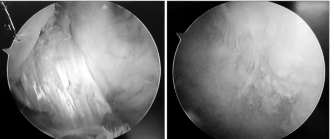
Fig. 2. Arthroscopic image obtained during knee washout after anterior cruciate ligament reconstruction showing the intact graft.
Fig. 3. Arthroscopic images obtained one year after anterior cruciate ligament (ACL) reconstruction showing the ACL graft fully incorporated and intact, and extensive car- tilage loss over the trochlear region.
196
Zaman et al. Pseudogout Following Arthroscopic ACL Reconstructionto effusion1-4).
Several causes of CPPD deposition have been proposed, with its occurrence being classified as either primary (non-traumatic) or secondary (traumatic). Primary deposits are thought to be due to a metabolic cause, such as hyperparathyroidism and haemochro- matosis where crystallization occurs within the synovial fluid2,5,6). Secondary deposits are more likely to occur in the presence of degenerative knee changes as a result of trauma and surgery2). It has been proposed that the mechanism for this is either that the crystals are liberated or ‘shed’ from preformed deposits within the joint or alternatively that an intra-articular insult may trigger their formation2,5).
Symptoms of acute pseudogout are similar to that of septic ar- thritis1), which poses diagnostic difficulties. Investigations such as joint aspiration, synovial fluid microscopic examination and microbiological culture as well as radiological imaging may help in the diagnosis1). Where doubt exists, arthroscopic washout of the knee is performed.
Pseudogout attack following an ACL reconstruction is a very rare event with only one previous report having been described (Table 1). Minezaki et al.2) described a 34-year-old female who was found to have calcification of the medial and lateral meniscus due to CPPD at 17 months after ACL reconstruction. Our report seems to be the first presentation of an acute pseudogout attack in the early postoperative period following ACL reconstruction.
One can only postulate why such an attack occurs. We suggest that it may be that the trauma of surgery (including notch de- bridement and drilling of the femoral and tibial tunnels), when superimposed on pre-existing degenerative changes, can precipi- tate an acute attack, and that pseudogout attack may signify the progression of degenerative changes and a poorer outcome in the
long-term.
In summary, pseudogout must be considered as a differential diagnosis of acute onset pain following ACL reconstruction, es- pecially in a knee with pre-existing degenerative changes.
Conflict of Interest
No potential conflict of interest relevant to this article was re- ported.
References
1. Hirose CB, Wright RW. Calcium pyrophosphate dihydrate deposition disease (pseudogout) after total knee arthroplas- ty. J Arthroplasty. 2007;22:273-6.
2. Minezaki T, Tomatsu T, Hanada K. Calcium pyrophosphate dihydrate crystal deposition disease after anterior cruciate ligament reconstruction. Arthroscopy. 1998;14:634-6.
3. Ellman MH, Krieger MI, Brown N. Pseudogout mimick- ing synovial chondromatosis. J Bone Joint Surg Am. 1975;
57:863-5.
4. Longmore M, Wilkinson I. Rheumatology: crystal arthropa- thies. In: Longmore M, Wilkinson I, Turmezei T, Cheung CK, eds. Oxford handbook of clinical medicine. 7th ed. Ox- ford: Oxford University Press; 2007. p534.
5. Doherty M, Dieppe PA. Acute pseudogout: “crystal shed- ding” or acute crystallization? Arthritis Rheum. 1981;24:954-7.
6. Resnik CS, Resnick D. Calcium pyrophosphate dihydrate crystal deposition disease. Curr Probl Diagn Radiol. 1982;
11:1-40.
Table 1. Comparison of Our Case with a Previously Reported Case of Pseudogout Following Anterior Cruciate Ligament Reconstruction
Variable Minezaki et al.2) Our case
Age (yr) 34 35
Sex Female Male
Knee observations Medial meniscal tear Lateral meniscal tear, chondral changes in lateral
Graft used Leeds-Keio artificial ligament Hamstring tendons
Time of presentation Seventeen months post-operative Six weeks post-operative
Presentation Pain and occasional effusion in the right knee Sudden onset of pain and swelling in the knee Diagnosis method Microscopic joint fluid examination, radiographs Microscopic joint fluid examination
Treatment - Non-steroidal anti-inflammatory medications