PGHN
Original Article
Neonatal Late-onset Hypocalcemia: Is There Any Relationship with Maternal Hypovitaminosis D?
Hyun Jeong Do, Ji Sook Park, Ji-Hyun Seo, Eun Shin Lee*, Chan-Hoo Park, Hyang-Ok Woo and Hee-Shang Youn
Departments of Pediatrics and *Rehabilitation Medicine, Gyeongsang Institute of Health Science, Gyeongsang National University School of Medicine, Jinju, Korea
Purpose: Neonatal late-onset hypocalcemia is defined as hypocalcemia developed after postnatal 3 days and asso- ciated with hypoparathyroidism, high phosphate diets and vitamin D deficiency. We experienced the increment of neonatal late onset hypocalcemia over 1 year. We tried to evaluate the relationship between late onset hypocalcemia and maternal hypovitaminosis D.
Methods: The medical records in the neonates with late-onset hypocalcemia during January 2007 to July 2008 were retrospectively reviewed. Among those patients, 17 paired sera of mothers and neonates had collected. The levels of 25-OH vitamin D (25OHD) and intact parathyroid hormone (iPTH) were measured and were compared with neo- nate and the mother.
Results: The mean gestational age was 38+1 weeks, and the mean body weight was 2,980 g. The onset time of hypo- calcemia was 5.9 days of age. Most of them (88.2%) were feeding with formula and no one was only breast milk feeding. Of the 17 patients, 13 were born in spring or in winter. The median levels of calcium, phosphorus, alkaline phosphatase, iPTH and 25OHD were 7.0 mg/dL, 8.6 mg/dL, 191.0 U/L, 57.2 pg/mL and 24.0 ng/mL in neonates.
The levels of 25OHD of 6 neonates were <20 ng/mL. A total of 16 mothers were considered vitamin D-deficient (<20 ng/mL), and vitamin D insufficient (20<25OHD<30 ng/mL).
Conclusion: Neonatal late-onset hypocalcemia in our study seems to be influenced by maternal vitamin D deficiency and insufficiency. Sun tanning and vitamin D supplements from winter to spring would be helpful to prevent maternal vitamin D deficiency, one of the causes of neonatal late-onset hypocalcemia.
Key Words: Vitamin D deficiency, Mother-newborn pair, Neonatal late-onset hypocalcemia
Received:November 26, 2013, Revised:December 19, 2013, Accepted:December 31, 2013
Corresponding author: Chan-Hoo Park, Department of Pediatrics, Gyeongsang National University Hospital, 79, Gangnam-ro, Jinju 660-702, Korea. Tel: +82-55-750-8161, Fax: +82-55-752-9889, E-mail: [email protected]
Copyright ⓒ 2014 by The Korean Society of Pediatric Gastroenterology, Hepatology and Nutrition
This is an openaccess article distributed under the terms of the Creative Commons Attribution NonCommercial License (http://creativecommons.org/licenses/by-nc/3.0/) which permits unrestricted noncommercial use, distribution, and reproduction in any medium, provided the original work is properly cited.
INTRODUCTION
Neonatal late-onset hypocalcemia is defined as
hypocalcemia developed after postnatal 3 days. The causes of neonatal late-onset hypocalcemia are hy- poparathyroidism, hypomagensemia, high phos-
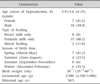
Table 1. Characteristics of Neonates Who Had Late-onset Hypocalcemia
Charateristic Value
Age (onset of hypocalcemia, d) Gender
Female Male
Type of feeding Breast milk only Formula milk only Mixed feeding Season of birth time
Spring (March-May) Summer (June-August) Autumn (September-November) Winter (December-February) Birth weight (wk)
Gestational age (g) Maternal age (y)
5.9±3.6 (4-15)
7 (41.2) 10 (58.8)
0 (0) 15 (88.2)
2 (11.8)
7 (41.2) 4 (23.5) 0 (0) 6 (35.3) 38+1 (35+5-40+5) 2,980 (2,350-3,900)
30.5
Values are presented as mean±standard deviation (range), number (%), median (range), or number only.
phate intakes, maternal hyperparathyroidisms, or vitamin D deficiency [1-3].
Recently, maternal vitamin D deficiency is a com- mon issue around the world [4]. Newborn-serum vi- tamin D concentrations depend on the maternal vi- tamin D status [5]. Exclusive breast milk feeding without exposure to sunlight is another risk factor of vitamin D deficiency for the newborns [6]. We expe- rienced the increments of neonatal late onset hypo- calcemia over 1 year, especially in neonates who were born in winter and spring time. The purpose of the present study is to investigate the relation of late onset hypocalcemia in newborns and maternal vita- min D levels by measuring the level of 25-OH vitamin D (25OHD) and intact parathyroid hormone (iPTH) in neonate-mother pairs.
MATERIALS AND METHODS
The medical records of newborn with late-onset hypocalcemia (serum total Ca<7.5 mg/dL) admitted at Gyeongsang National University Hospital from January 2007 to July 2008 were reviewed after in- stitutional review board-approved protocols (GNUHIRB-2012-07-014). They were admitted at our hospital because of several mild illnesses such as suspected sepsis, jaundice and tachypnea. They did not have any history of maternal diabetes mellitus, neonatal asphyxia, renal insufficiency, blood trans- fusions, or use of diuretics, anticonvulsants, Di George syndrome or ventilator care. Hypocalemia was detected by initial blood chemistry or follow up.
No one presented hypomagnesemia, hypoalbumi- nemia or renal dysfunction.
The serum of newborn and mother were obtained after diagnosis of late-onset hypocalcemia and other laboratory findings were collected by chart reviews.
The serum 25OHD levels of newborn and mother were measured using an enzyme immunoassay (Roche Modular E170; Roche, Basel, Switzerland) at the same time. The normal range for 25OHD using this kit was 10-80 ng/mL in the newborn and 20-200 ng/mL in the mother. The normal range for iPTH was defined as 15-65 pg/mL.
RESULTS
In total, 17 neonates from our hospital were en- rolled in this study (Table 1). They consisted of 7 fe- males (41.2%) and 10 males (58.8%) and the mean onset age was 5.9 days (range, 4-15 days). The mean age of mother was 30.5 years. Mean gestational age was 38+1 weeks (range, 35+5-40+5) and mean birth weight was 2,980 g (range, 2,350-3,900 g). Type of feeding was almost formula-fed (88.2%) and mixed-fed (11.8%). Nobody was exclusively breastfed.
Most of them was born in the winter (35.3%) and the spring (41.2%).
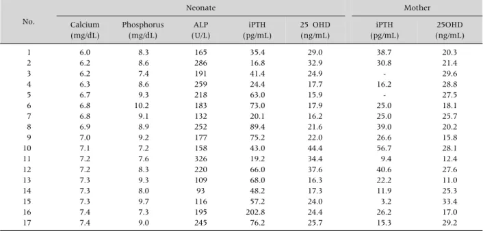
Table 2 showed the laboratory findings of 17 neo- nate-mother pairs. Their mean serum calcium level was 6.9±0.5 mg/dL, serum phosphorus was 8.6±0.9 mg/dL, serum alkaline phosphatase (ALP) was 196±65 U/L, and plasma iPTH level was 60±43 pg/mL. Their serum 25OHD was 24.8±8.4 ng/mL.
Maternal serum 25OHD was 23.0±6.6 ng/mL and plasma iPTH level was 26±14 pg/mL. Vitamin D defi- ciency (25OHD<20 ng/mL) was observed in 29.4%
of the subjects. Insufficient vitamin D status (20
<25OHD<30 ng/mL) was 58.9% (Fig. 1). All 17 ne- onates were managed with oral calcium gluconate
Fig. 1. Vitamin D status of mothers of hypocalcemic babies.
Most of them showed vitamin D insufficiency and deficiency.
Table 2. The Serum or Plasma Levels of Calcium, Phosphorus, Alkaline Phosphatase, Intact Parathyroid Hormone, and 25-OH Vitamin D3 of Babies with Late-onset Hypocalcemia and Intact Parathyroid Hormone and 25-OH Vitamin D3 of Their Mothers
No.
Neonate Mother
Calcium (mg/dL)
Phosphorus (mg/dL)
ALP (U/L)
iPTH (pg/mL)
25 OHD (ng/mL)
iPTH (pg/mL)
25OHD (ng/mL) 1
2 3 4 5 6 7 8 9 10 11 12 13 14 15 16 17
6.0 6.2 6.2 6.3 6.7 6.8 6.8 6.9 7.0 7.1 7.2 7.2 7.3 7.3 7.3 7.4 7.4
8.3 8.6 7.4 8.6 9.3 10.2 9.1 8.9 9.2 7.2 7.6 8.3 9.3 8.0 9.7 7.3 9.0
165 286 191 259 218 183 132 252 177 158 326 220 109 93 116 195 245
35.4 16.8 41.4 24.4 63.0 73.0 20.1 89.4 75.2 43.0 19.2 66.0 68.0 48.2 57.2 202.8 76.2
29.0 32.9 24.9 17.7 15.9 17.9 16.2 21.6 22.0 44.4 34.4 37.6 16.3 17.3 24.0 24.4 25.7
38.7 30.8 - 16.2
- 25.0 25.0 39.0 26.6 56.7 9.4 40.6 22.2 11.9 3.2 26.2 15.3
20.3 21.4 29.6 28.8 27.5 18.1 25.7 20.2 15.8 28.1 12.4 27.6 11.0 25.3 33.4 17.0 29.2 ALP: alkaline phosphatase, iPTH: intact parathyroid hormone, 25OHD: 25-OH vitamin D.
(calcium gluconate; Daihan Pharm Co., Ltd., Seoul, Korea) replacements for hypocalcemia and phos- phate scavenger. Twelve neonates were followed up until 4 months and were regularly checked on body weight, height, and serum level of calcium, phospho- rus, ALP and albumin levels were in normal range.
DISCUSSION
Maternal hypovitaminosis D is one of common causes of neonatal late-onset hypocalcemia [7,8].
Rickets is an uncommon problem in children and ne- onates as Korea is a developed country. Only patients with risk factors of cholestasis, mal-absorption syn- dromes, vitamin-D resistant diseases are considered for the risk of vitamin D deficiency.
This is the first study to evaluate the relationship between neonatal late onset hypocalcemia and ma- ternal vitamin D status in Korea using paired neo- natal-mother sera. In this study, neonatal late-onset hypocalcemia seems to be related with the maternal vitamin D deficiency and insufficiency, on the bases of their birth seasons and maternal vitamin D status.
The fact is supported that most of the neonates born in spring and winter are short of sunlight exposures due to the late pregnancy periods of mothers [9]. No neonates born at autumn were diagnosed as late on- set hypocalcemia, and it may be related with long and high sunlight exposures of mother during late preg- nancy during summers [9]. In Iran, 100% of neonates
with delayed hypocalcemia were born by mothers with vitamin D deficiency [10]. Previous studies showed same results that neonate who were born from mother of vitamin D deficiency or insufficiency have vitamin D deficiency and late onset hypo- calcemia due to vitamin D deficiency [11,12]. In the present study, 76.5% of neonates who had late-onset hypocalcmia were born by mothers with vitamin D deficiency or insufficiency. Exclusive breast milk feeding without vitamin D supplementations is an- other risk factor of vitamin D deficiency for neonates and infants. Our report of neonatal vitamin-D defi- ciency indicates that the newborns intake the entire milk. Late onset hypocalcemia is usually caused by high phosphate intakes [13]. Late-onset hypo- calcemia caused by excessive phosphate load is usu- ally accompanied by raised PTH and ALP levels [12].
However, most of our patients with hypocalcemia and hyperphosphatemia represented normal or near nor- mal ranges of iPTH and ALP. Only case 16 showed hy- pocalcemia, hyperphosphatemia and remarkably raised PTH level but serum ALP level was relatively low despite of hyperphosphatemia. Practically, vita- min D insufficiency or deficiency was identified in the mothers with hypocalcemic babies according to the preliminary data at our hospital. Therefore, we tenta- tively considered that maternal vitamin D in- sufficiency could be associated with the occurrence of late-onset hypocalcaemia even in the formula-fed ne- onates, although there was no statistically significant relationship (r=−0.131, Pearson correlation by PASW Statistics 18.0 [IBM Co., Armonk, NY, USA]).
Studies in adults have demonstrated that para- thyroid hormone concentrations are at their ideal physiologic concentrations when 25OHD concen- trations are above 32 ng/mL [14,15]. Similar data in children are unavailable [16]. According to Avery's disease of the newborn, iPTH levels are usually de- pendent on the serum calcium status. The normal ranges of iPTH are suggested as 10-60 pg/mL under normocalcemia, but hypocalcemia can stimulate iPTH maximally up to 100-150 pg/mL and hyper- calcemia can supress iPTH to 2-5 pg/mL [17]. In Korea, there were several studies on the levels of PTH
and hypocalcemia: One study was that serum PTH was elevated in the hypocalcemic infants with in- fectious diseases [16]. The other was that iPTH ele- vated in infants with subclinical rickets [18]. In the present study, most iPTH levels of neonates with late-onset hypocalcemia was not remarkably ele- vated except for only 1 case based on Avery’s diseases of the newborn. The response of parathyroid hor- mone might be less sensitive in neonates as com- pared to the study in infants with subclinical rickets (range of iPTH: 74.7 to 462 pg/mL) [18]. In our study, no elevation of ALP or iPTH was observed in neo- nates with late-onset hypocalcemia despite maternal vitamin D insufficiency or deficiency. Given the re- sults, we deduced that there might be an another cause for late-onset hypocalcaemia in the subject ne- onates, besides the maternal vitamin D deficiency and insuffiency. This might be caused by delayed re- sponses of parathyroid hormones to hypocalcemia in neonates [18] and associated with transient hypoparathyroidism. However, it is difficult to make results clear due to the lack of studies regarding the responses of parathyroid gland in neonates.
Therefore, more studies should be followed in the near future regarding the effects of vitamin D or hy- pocalcemia on the functions of parathyroid in neonates.
We could stop calcium supplementations to the hypocalcemic infants around the age of 3-4 months.
Currently, 45 IU of vitamin D is contained in 100 mL of formula which is commercially available in Korea, indicating that about 1 L of milk should be ingested to meet the recommended vitamin D daily intakes (400 IU/day). In most cases, they could ingest around 1 L of milk when they are 3-4 months.
Considering that calcium, phosphorus, and ALP val- ues were observed to be normal in the examination around the fourth month after birth, the authors as- sumed that vitamin D insufficiency or deficiency would be recovered as the recommended vitamin D daily intakes was met due to an increase of milk intakes. On the basis of their clinical course, neo- natal late onset hypocalcemia in this study might be affected by maternal vitamin D status rather than
delayed response of parathyroid hormone.
There are several limitations in this study. First, the number of newborns with late-onset hypo- calcemia was small. Second, we did not obtain in- formation on each mother’s amount of sunlight ex- posures, sunscreen uses and diets. Third, whether vi- tamin D concentration was normalized was not con- firmed as vitamin D concentration was not exam- ined around 3-4 months of age. Fourth, this study is not a case-control study and maternal vitamin D in- sufficiency or deficiency is difficult to determine a definitive cause of neonatal late-onset hypocalcemia in the present study. Regardless of these limitations, the present study is likely to be meaningful because it is the first study to evaluate the relationship be- tween neonatal late onset hypocalcemia and mater- nal vitamin D status in Korea using paired neo- natal-mother sera and late-onset hypocalcaemia could occur under the conditions of maternal hypo- vitaminosis D or transient hypoparathyroidism in formula-fed neonates besides phosphorus overload.
REFERENCES
1. Salle BL, Delvin E, Glorieux F, David L. Human neo- natal hypocalcemia. Biol Neonate 1990;58(Suppl 1):22-31.
2. Lee CT, Tsai WY, Tung YC, Tsau YK. Transient pseudo- hypoparathyroidism as a cause of late-onset hypo- calcemia in neonates and infants. J Formos Med Assoc 2008;107:806-10.
3. Thomas TC, Smith JM, White PC, Adhikari S.
Transient neonatal hypocalcemia: presentation and outcomes. Pediatrics 2012;129:e1461-7.
4. Bodnar LM, Catov JM, Simhan HN, Holick MF, Powers RW, Roberts JM. Maternal vitamin D deficiency in- creases the risk of preeclampsia. J Clin Endocrinol Metab 2007;92:3517-22.
5. Thomas SD, Fudge AN, Whiting M, Coates PS. The cor- relation between third-trimester maternal and new- born-serum 25-hydroxy-vitamin D in a selected South Australian group of newborn samples. BMJ Open
2011;1:e000236.
6. Specker BL, Valanis B, Hertzberg V, Edwards N, Tsang RC. Sunshine exposure and serum 25-hydroxyvitamin D concentrations in exclusively breast-fed infants. J Pediatr 1985;107:372-6.
7. Greer FR. 25-Hydroxyvitamin D: functional outcomes in infants and young children. Am J Clin Nutr 2008;88:529S-33S.
8. Bowyer L, Catling-Paull C, Diamond T, Homer C, Davis G, Craig ME. Vitamin D, PTH and calcium levels in pregnant women and their neonates. Clin Endocrinol (Oxf) 2009;70:372-7.
9. Halicioglu O, Aksit S, Koc F, Akman SA, Albudak E, Yaprak I, et al. Vitamin D deficiency in pregnant wom- en and their neonates in spring time in western Turkey.
Paediatr Perinat Epidemiol 2012;26:53-60.
10. Khalesi N, Bahaeddini SM, Shariat M. Prevalence of maternal vitamin D deficiency in neonates with de- layed hypocalcaemia. Acta Med Iran 2012;50:740-5.
11. Dawodu A, Saadi HF, Bakdache G, Altaye M, Hollis BW. Extraordinarily high prevalence and lack of sea- sonal variationof vitamin D deficiency in pregnant Arab women. Vancouver, 1-4 May 2010: Pediatric Academic Societies Annual Meeting; E-PAS 2010:1451.
12. Kim HS. Calcium and phosphate metabolism and dis- orders in the newborn. Korean J Pediatr 2007;50:230-5.
13. Jain A, Agarwal R, Sankar MJ, Deorari A, Paul VK.
Hypocalcemia in the newborn. Indian J Pediatr 2010;77:1123-8.
14. Holick MF. The vitamin D epidemic and its health consequences. J Nutr 2005;135:2739S-48S.
15. Heaney RP. Functional indices of vitamin D status and ramifications of vitamin D deficiency. Am J Clin Nutr 2004;80(6 Suppl):1706S-9S.
16. Rovner AJ, O'Brien KO. Hypovitaminosis D among healthy children in the United States: a review of the current evidence. Arch Pediatr Adolesc Med 2008;
162:513-9.
17. Lewis PR. Disorders of calcium and phosphorus metabolism. In: Gleason CA, Devaskar SU, eds. Avery's diseases of the newborn. 9th ed. Philadelphia:
Saunders, 2012:1257.
18. Park SY, Park SW, Kang SK, Jun YH, Kim SK, Son BK, et al. Subclinical rickets in breastfed infants. Korean J Pediatr 2007;50:1188-93.