• 검색 결과가 없습니다.

Cathepsin B Is Implicated in Triglyceride (TG)-Induced Cell Death of Macrophage

N/A
N/A
Protected

Academic year: 2021

Share "Cathepsin B Is Implicated in Triglyceride (TG)-Induced Cell Death of Macrophage"

Copied!
8
0
0

로드 중.... (전체 텍스트 보기)

전체 글

(1)

Corresponding author: Yoon Suk Kim

Department of Biomedical Laboratory Science, College of Health Science, Yonsei University, 1 Yeonsedae-gil, Heungeop-myeon, Wonju 26493, Korea

E-mail: yoonsukkim@yonsei.ac.kr

ORCID: https://orcid.org/0000-0002-1956-0126

The first two authors contributed equally to this work.

ORIGINAL ARTICLE

Cathepsin B Is Implicated in Triglyceride (TG)-Induced Cell Death of Macrophage

Byung Chul Jung 1,2,† , Jaewon Lim 2,3,† , Sung Hoon Kim 2,4 , Yoon Suk Kim 2

1

Department of Nutritional Sciences and Toxicology, University of California, Berkeley, United States

2

Department of Biomedical Laboratory Science, College of Health Sciences, Yonsei University, Wonju, Korea

3

Department of Biomedical Laboratory Science, College of Medical Sciences, Daegu Haany University, Gyeongsan, Korea

4

Department of Biomedical Laboratory Science, Korea Nazarene University, Cheonan, Korea

중성지방에 의한 대식세포 사멸 과정에서 Cathepsin B의 영향

정병출 1,2,† , 임재원 2,3,† , 김성훈 2,4 , 김윤석 2

1

캘리포니아대학교 버클리캠퍼스 영양과학 및 독성학과,

2

연세대학교 보건과학대학 임상병리학과,

3

대구한의대학교 의과학대학 임상병리학과,

4

나사렛대학교 임상병리학과

ARTICLE INFO ABSTRACT

Received August 24, 2020 Revised August 27, 2020 Accepted August 28, 2020

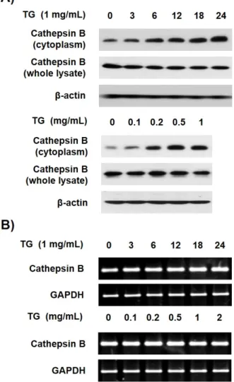
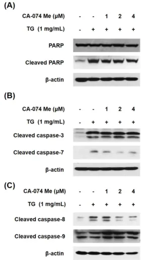
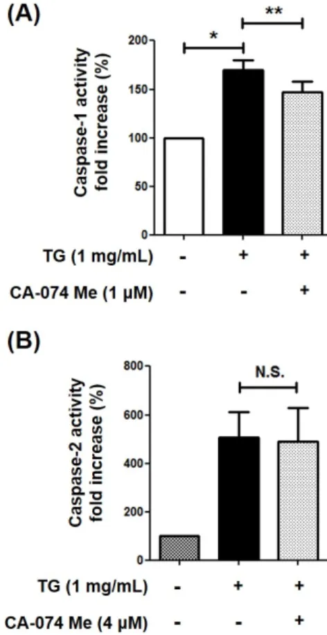
Macrophage cell death contributes to the formation of plaque, leading to the development of atherosclerosis. The accumulation of triglyceride (TG) is also associated with the pathogenesis of atherosclerosis. A previous study reported that TG induces the cell death of macrophages. This study examined whether the cytoplasmic release of cathepsin B from lysosome is associated with the TG-induced cell death of macrophage. The release of cathepsin B was increased in the TG-treated THP-1 macrophages, but the TG treatment did not affect cathepsin B expression. Furthermore, the inhibition of cathepsin B by its inhibitor, CA-074 Me, partially inhibited the TG-induced cell death of macrophage. TG-triggered macrophage cell death is mediated by the activation of caspase-1, -2, and apoptotic caspases. Therefore, this study investigated whether cathepsin B is implicated in the activation of these caspases. The inhibition of cathepsin B blocked the activation of caspase-7, -8, and -1 but did not affect the activity of caspase-3, -9, and -2. Overall, these results suggest that TG-induced cytoplasmic cathepsin B causes THP-1 macrophage cell death by activating caspase-1, leading to subsequent activation of the extrinsic apoptotic pathway.

Copyright 2020 The Korean Society for Clinical Laboratory Science. All rights reserved.

Key words Caspase-1 Cathepsin B Cell death THP-1 macrophage Triglyceride

INTRODUCTION

Atherosclerosis is a prevalent disease caused by clogged arteries with lipid-rich plaque, which, in turn, progress myocardial ischemia [1]. The prevalence of clinical atherosclerosis per 1,000 individuals was 101.11 in 2015 in South Korea [2]. While a variety of cell

types contribute to the pathogenesis of atherosclerosis, macrophages play a pivotal role in terms of plaque formation and development [3]. In the initial stage of atherosclerosis, macrophages engulf cholesterol-rich lipoprotein via specific receptors and subsequently are converted to foam cells, which is a crucial step for the development of plaque formation [4]. As plaques develop, macrophage cell death increases, which contributes to the development of vulnerable plaques [5]. Previously, our group have reported that TG induces cell death of macrophages implying the implication of TG in the

Korean Society for Clinical Laboratory Science

(2)

pathogenesis of atherosclerosis [6, 7].

Cathepsins are cysteine proteases which are localized in the early endosome and lysosomes of immune cells such as macrophages [8]. Among 11 human cathepsins, cathepsin B is the most well-studied protease regarding cell death. The released cathepsin B in the cytosol cleaves proapoptotic molecule, BID, which in turn leads to the release of cytochrome c from mitochondria and subsequently induces apoptosis in a variety of cell types [9]. Interestingly, multiple investigations have supported that cathepsin B is associated with the pathogenesis of atherosclerosis. For example, upregu- lation of cathepsin B was reported in atherosclerosis animal model, apoE −/− mice [10]. In addition, cathepsin B can degrade the extracellular matrix in the arterial intima, which promotes inflammatory response as well as the development of vulnerable plaques [11].

In the present study, we aimed to understand the role of cathepsin B in TG-induced macrophage cell death using differentiated human THP-1 macrophages. We found that challenge of TG induced release of cathepsin B from the lysosome to the cytoplasm, which subsequently activates extrinsic apoptotic pathways. Moreover, we showed cathepsin B is a prerequisite for caspase-1 activation, which is the same observance with the very recent report [12]. Our results show that TG-induced macrophage cell death is mediated by the cytosolic release of cathepsin B from the lysosome.

MATERIALS AND METHODS

1. Materials

Lipofundin MCT/LCT 20% was obtained from B.

Braun Melsungen AG (Melsungen, Germany). Lipofundin MCT/LCT 20% was used to deliver TG into cells as described in previous study [13]. Hereafter, Lipofundin MCT/LCT 20% will be referred to as TG for convenience.

The caspase-1 substrate, Ac-YVAD-p-nitroanilide (Ac- YVAD-pNA) was purchased from Biomal (Plymouth Meeting, PA, USA). The caspase-2 substrate, Ac-VAVAD- pNA was obtained from Sigma-Aldrich (St. Louis, MO,

USA). CA-074 Me (cathepsin B specific inhibitor) was purchased from Calbiochem (Darmstadt, Germany).

Antibodies specific for caspase-3, -7, -8 and -9, as well as PARP, were purchased from Cell Signaling tech- nology (Danvers, MA, USA). Antibody specific for cathepsin B was obtained from Santa Cruz biotechnology (Santa Cruz, CA, USA).

2. Cell culture

The THP-1 human acute monocytic leukemia cell line (ATCC, Manassas, VA, USA) was cultured in RPMI 1640 media supplemented with 10% fetal bovine serum (FBS) and penicillin-streptomycin and maintained at 37°C in a humidified atmosphere with 5% CO 2 . To induce differentiation of THP-1 monocytic cells into macrophages, cells were plated in 6-well plates at a density of 1×10 6 cells/well and incubated with 200 nM phorbol 12-myristate 13-acetate (PMA) for 48 h [14, 15].

3. Oil red-O staining and assessment

THP-1 cells were seeded on cover-glass (24×24 mm) and incubated with 200 nM PMA for differentiation into macrophages. Then, the cells were incubated with or without TG for 24 h. Cell monolayers were washed twice with Dulbecco’s phosphate buffered saline (DPBS) and fixed in 10% formaldehyde for 5 min. 1 mL of 0.2% Oil red-O solution (Sigma-Aldrich) was added to well and incubated for 30 min at room temperature. Cell monolayers were washed with 60% isopropanol for 5 sec and washed twice with DPBS. Cells were examined under a microscope. For quantification, 1 mL of 100%

isopropanol was added to the stained cell monolayers.

After 5 min, the absorbance of the extract was assayed by UV/Vis spectrophotometer at 510 nm (Beckman Coulter, Fullerton, CA, USA).

4. Trypan blue dye exclusion assay

Method were previously described in detail [7]. In brief, THP-1 cells were trypsinized and 10 µL of 0.4%

trypan blue stain solution was mixed with 10 µL of the

trypsinized cells at a 1:1 ratio. Viable cells (unstained

(3)

cells) were counted on a hemocytometer (Marienfeld, Lauda-Königshofen, Germany).

5. Subcellular fractionation

Method were previously described in detail [16]. The THP-1 cells were plated at a density of 6×10 6 cells on each culture plate (100 mm×20 mm) and incubated with 200 nM PMA for 48 h. Thereafter, differentiated THP-1 macrophage were incubated with TG for an additional 24 h. Scraped THP-1 cells were washed with cold PBS and cells were pelleted by centrifugation at 1,000 g for 5 min. All samples were resuspended in 350 μL of buffer containing 250 mM sucrose, 50 mM Tris-HCl pH 7.4, 5 mM MgCl 2 , 1% (v/v) protease inhibitor cocktail and 1% (v/v) phosphatase inhibitor cocktail and incubated on ice for 30 min. Then, samples were vortexed at maximum speed for 15 sec and centrifuged at 7,500 g for 15 min. The supernatant was used as cytosolic fractions.

6. Western-blot analysis

THP-1 cells were washed with PBS and lysed at 4°C with lysis buffer including 1% Triton X-100, protease inhibitor cocktail (Sigma-Aldrich), phosphatase inhibitor cocktail (Roche, Mannheim, Germany), and PBS. Cell lysates were clarified and the supernatants subjected to Western blotting as described previously [17].

7. RNA extraction and semi-quantitative reverse transcriptase PCR (RT-PCR)

Total RNA was extracted from THP-1 cells using Trizol reagent according to the manufacturer’s inst- ructions (Invitrogen, Thermo Fisher Scientific, Waltham, MA, USA). Reverse transcriptase PCR (RT-PCR) was performed as previously described [6]. Complementary DNA (cDNA) was synthesized by reverse transcription with 2 µg of total RNA, 0.25 µg of random hexamer (Invitrogen) and 200 U of MMLV-RT (Invirogen) for 10 min at 25°C, 50 min at 37°C and 15 min at 70°C. cDNA was PCR amplified using Prime Taq premix PCR kit (Genet Bio, Chungnam, Korea) for 25∼40 cycles using

specific primers. Primer sequences are as follows:

Cathepsin B; 5’-ACA GTG TCC CAC CAT CAA AG-3’

(forward), 5’-CAC CAT TAC AGC CGT CCC-3’ (reverse) and GAPDH; 5’-CGG GAA GCT TGT CAT CAA TGG-3’

(forward), 5’-GGC AGT GAT GGC ATG GAC TG-3’

(reverse). GAPDH was used as an internal control. PCR amplicons were electrophoresed on 2% (w/v) agarose gels including 0.5 μg/mL ethidium bromide and the amplicon size determined by comparison to 100 bp DNA ladder marker (Intron, Gyeonggi, Korea). Gel images were obtained using Gel Doc TM XR+ system (Bio-Rad, Hercules, CA, USA). The PCR amplicon band intensity was measured and normalized against GAPDH using Image Lab TM software (version 4.1, Bio-Rad).

8. Measurement of caspase-1 and caspase-2 activities The activity of caspase-1 and caspase-2 was mea- sured as previously described [18]. Briefly, THP-1 cells were lysed with PBS buffer containing 1% Triton X-100 and then centrifuged at 19,000 g for 10 min at 4°C. The supernatant was collected and the total protein con- centration was quantified. To measure caspase-1 activity, 90 μg of protein sample was mixed with 200 μM of the caspase-1 substrate Ac-YVAD-pNA in 150 μL of PBS. To measure caspase-2 activity, protein was mixed with the caspase-2 substrate Ac-VAVAD-pNA in PBS.

Mixtures were incubated for 3 h at 37°C, and the activity was determined by measuring the absorbance at 405 nm.

9. Statistical analysis

Statistical analysis was performed with student’s t-test using GraphPad Prism 5 software (GraphPad Software Inc., San Diego, CA, USA). Values are ex- pressed as the mean and standard error of the mean (SEM). Each experiment was conducted three times and the date were pooled for analysis. Differences were considered to be statistically significant at *P<0.05,

**P<0.01, or ***P<0.001.

(4)

Figure 1. The accumulation of TG in THP-1 macrophages forms foam cells. (A) THP-1 cells were differentiated with 200 nM PMA for 48 h and incubated with or without 1 mg/mL of TG for additional 24 h. Oil red-O staining was performed to detect lipid droplets in cells (scale bars, 100 µm). (B) Quantitative analysis of Oil red-O uptake was performed after cells were cultured with TG and then stained with Oil red-O. The dye was extracted and the absorbance was measured by spectrophotometry at 510 nm. All data are expressed as the mean±SEM of three independent experiments. P-values were determined with Student’s t-test. * P <0.05, ** P <0.01.

Figure 2. TG induces cytoplasmic release of cathepsin B. (A) THP-1 macrophages were incubated with the indicated concentration of TG for the indicated times, and the cytosol and residues were separated using gradient centrifuges. The cathepsin B was detected by Western blotting. (B) THP-1 macrophages were incubated with the indicated concentration of TG for the indicated times, and expression of cathepsin B mRNA was assessed by RT-PCR.

RESULTS

1. TG accumulation induces cytoplasmic release of cathepsin B from lysosome

We previously reported that TG-induced macrophage cell death is caused by caspase-1 [6]. Several studies showed that the activation of caspase-1 can also occur via lysosomal rupture and subsequent cytosolic release of cathepsin B [19, 20]. Therefore, we first investigated whether the release of cathepsin B is involved in TG-induced THP-1 macrophage cell death. We first reconfirmed whether TG accumulates in THP-1 macrophages when it is added in a cell culture media.

PMA-differentiated THP-1 macrophages were incubated with TG for 24 h and then stained with Oil-red-O

solution. Unlike in macrophages without TG treatment, stained droplets were more prominently found in the cytoplasm of TG-treated macrophages (Figure 1). Next, to elucidate whether the cytoplasmic protein level of cathepsin is influenced by TG treatment, the cyto- plasmic fraction which do not include lysosomes was obtained through the process of subcellular fraction.

TG treatment increased the cytoplasmic protein level of

cathepsin B in a dose- and time-dependent manner,

but the total protein level of cathepsin B in THP-1

macrophages was unchanged in response to TG (Figure

(5)

Figure 3. TG-stimulated increase of cytoplasmic cathepsin B is associated with TG-triggered macrophage cell death. THP-1 macro- phages were incubated with 1 mg/mL of TG in the presence of cathepsin B inhibitor, CA-074 Me for 24 h. Then, trypan blue ex- clusion assay was performed to enumerate viable cells. The number of viable cells in THP-1 macrophages without TG treatment was set as 100%. All data are expressed as the mean±SEM of three independent experiments. P -values were determined with Student’s t-test. ** P <0.01.

Figure 4. TG-induced cytoplasmic cathepsin B affects THP-1 macrophage cell death via the extrinsic apoptotic pathway. THP-1 macrophages were incubated with of TG in the presence of cathepsin B inhibitor, CA-074 Me for 24 h. (A) The cleaved form of PARP, (B) cleavage of caspase-3 and -7, and (C) cleavage of caspase-8 and caspase-9 were detected by Western blotting.

2A). Furthermore, when the RNA level of cathepsin B was examined by RT-PCR analysis, the expression level of cathepsin B was unchanged in the presence of TG in THP-1 macrophages (Figure 2B). These results imply that the increase of cathepsin B in the cytoplasm of TG-treated THP-1 macrophages is caused by TG- triggered cytosolic release of cathepsin B from lyso- some and not by the increased expression of cathepsin B.

2. TG-stimulated cytoplasmic release of cathepsin B involves in THP-1 macrophage cell death

Next, to elucidate the role of cathepsin B in TG-induced macrophage cell death, it was examined whether cell viability was influenced by treatment with a cathepsin B inhibitor, CA-074 Me. THP-1 macro- phages were differentiated with PMA and incubated with TG in the absence or presence of CA-074 Me for 24 h and viable cells were enumerated. We found that it partially recovered cell viability of TG-treated THP-1 macrophages in an inhibitor dose-dependent manner (Figure 3). This result suggests that the cytosolic release of cathepsin B by the accumulation of TG leads to THP-1 macrophage cell death.

3. TG-induced cytoplasmic cathepsin B release leads to THP-1 macrophage cell death via the extrinsic apoptotic pathway

TG induced-macrophage cell death is reportedly mediated by classical apoptotic caspases [7]. Therefore, we examined the association of cathepsin B with the activation of caspases in TG-induced THP-1 cell death.

When THP-1 macrophages were incubated with TG and

cathepsin B inhibitor, CA-074 Me for 24 h, the cleavage

of PARP, which was increased by TG treatment, was

partially reduced (Figure 4A). In addition, CA-074 Me

blocked the cleavage of caspase-7, which is known to

cleave PARP, however, caspase-3 was not affected

(6)

Figure 5. Cathepsin B is an upstream molecule of caspase-1 in TG-triggered macrophage cell death, but not caspase-2. (A) THP-1 macrophages were incubated with TG in the absence or presence of the CA-074 Me for 24 h, after which caspase-1 activity was assessed. Absorbance of THP-1 macrophages without treatment with TG was set as 100%. (B) THP-1 macrophages were incubated with TG in the absence or presence of the CA-074 Me for 24 h and caspase-2 activity was assessed. All data are expressed as the mean±SEM of three independent experiments. P-values were determined with Student’s t-test. * P <0.05, ** P <0.01.

(Figure 4B). Caspase-8 and -9 are upstream molecules of caspase-7, and therefore, we examined their activity in the presence or absence of CA-074 Me. The cleavage of caspase-8 was decreased by CA-074 Me in a dose-dependent manner, whereas caspase-9 showed no significant changes in response to CA-074 Me in TG-treated macrophages (Figure 4C). These results suggest that TG-induced cytoplasmic cathepsin-B release leads to THP-1 macrophage cell death via the extrinsic apoptotic pathway.

4. Cathepsin B is associated with activation of caspase-1 in TG-triggered macrophage cell death Previous studies have reported that caspase-1 and -2 is implicated in TG-induced THP-1 macrophage cell death [6, 7]. To determine whether the increase of cytoplasmic cathepsin B is involved in the activation of caspase-1 and -2 in TG-treated THP-1 macrophages, PMA-differentiated THP-1 macrophages were incu- bated with TG in the presence or absence of CA-074 Me for 24 h, and the activation of the caspase-1 and -2 was analyzed. Ca-074 Me reduced the activity of caspase-1, which was increased by TG treatment (Figure 5A).

However, the inhibition of cathepsin B did not affect the TG-induced activation of caspase-2 in THP-1 macrophages (Figure 5B). These data suggest that released cytosolic cathepsin B is an upstream molecule of caspase-1 in TG-treated THP-1 macrophages.

DISCUSSION

Multiple lines of evidence support that raised TG concentration in blood can be accumulated in the arterial wall and cause atherosclerosis [21]. In a similar context, the role of cathepsin B in the development of atherosclerosis is well documented, such as involve- ment in apoptosis of macrophage and degradation in fibrous cap of atherosclerotic plaque [22]. However, the link between TG and cathepsin B, which contributes to the pathogenesis of atherosclerosis, has not been studied. In this study, we have elucidated that TG-

induced macrophage cell death is mediated cytosolic release of cathepsin B from lysosome. This is the first report to demonstrate that TG can liberate cathepsin B from lysosome to cytosol, followed by activation of extrinsic caspase cascade in the human macrophage cell line.

Macrophages are crucial cells contributing to the

formation of plaques in atherosclerosis, and its apoptosis

promotes the conversion from stable to unstable

plaques [23]. It has been reported that cathepsin B is

implicated in extracellular degradation, which is highly

associated with the development of vulnerable plaque

[24]. In addition, cathepsin B is highly expressed and

appears in the cytoplasm of apoptotic macrophages

(7)

located in the atherosclerotic lesion [25]. So far, the well-known mechanism by which cathepsin B mediates macrophage cell death is highly dependent on the intrinsic caspase cascade, including Bid and caspase-9 [26]. However, Results in the present study showed that TG-mediated cytosolic release of cathepsin B leads to cell death of macrophages is an extrinsic caspase cascade instead of an intrinsic caspase cascade.

In this study, we observed that TG treatment induces the translocation of cathepsin B to cytosol in macro- phage. Previously, we have shown that TG treatment results in increase of TNF-α in the THP-1 cells [27].

Since it has been reported that TNF-α triggers the cellular redistribution of the proteins including cathepsin B, it is probably that TG-induced cytosolic release of cathepsin B can be mediated via TG-sti- mulated increase of TNF-α[28]. In conclusion, we report for the first time that cathepsin B plays a role in TG-stimulated cell death of macrophages. This sug- gests that TG-induced macrophage death occurs in part via cytosolic release of cathepsin B from lysosome.

Our results will likely provide clues in understanding the mechanisms by which TG implicates in the development of atherosclerosis.

요 약

대식세포사멸은 죽상판 형성에 영향을 미쳐 죽상동맥경화증 발병에 관여하는 것으로 알려져 있다. 중성지방 역시 죽상동맥 경화 발병에 기여한다고 알려져 있는데 최근 본 연구팀에서는 중성지방이 대식세포사멸을 유발한다는 결과를 확인하였다. 본 연구에서는 cathepsin B가 중성지방에 의해 유발되는 대식세 포사멸 과정에 관여하는지 확인하고자 연구를 진행하였다.

THP-1 대식세포에 중성지방 처리 시 cathepsin B의 발현량에 는 변화가 없고 리소좀에 있던 cathepsin B가 세포질로 방출되 어 세포질의 cathepsin B가 증가한 것을 확인하였다. 다음으로 cathepsin B 억제제인 CA-074 Me를 처리 시 중성지방에 의 해 유도되는 대식세포사멸이 일부 회복되는 것을 확인하였다.

본 연구팀의 이전 연구에서 중성지방에 의한 대식세포사멸이 caspase-1, -2 및 apoptotic caspase 활성화를 매개로 일어 남을 확인하였기 때문에 본 연구에서는 이러한 caspase 활성

경로와 cathepsin B와의 연관성에 대해 연구하였다. cathepsin B 억제시 caspase-7, -8 및 -1의 활성은 억제되었으나, caspase-3, -9 및 -2는 활성에는 영향을 미치지 않음을 알 수 있었다. 정리하면, 중성지방에 의해 세포질로 방출된 cathepsin B는 caspase-1 활성화에 기여하고, 활성화된 caspase-1은 외 인성 apoptotic caspase 경로를 활성화하여 THP-1 대식세포 사멸을 유발한다는 것을 알 수 있다.

Acknowledgements: This research was supported by a grant from Daegu Haany University Ky-lin Foundation in 2018.

Conflict of interest: None

Author’s information (Position): Jung BC 1,2 , Researcher;

Lim J 2,3 , Professor; Kim SH 2,4 , Professor; Kim YS 2 , Professor.

REFERENCES

1. Ndrepepa G. Atherosclerosis and ischaemic heart disease: Here to stay or gone tomorrow. Indian J Med Res. 2017;146:293-297.

https://doi.org/10.4103/ijmr.IJMR_1668_17

2. Kim H, Kim S, Han S, Rane PP, Fox KM, Qian Y, et al. Prevalence and incidence of atherosclerotic cardiovascular disease and its risk factors in korea: A nationwide population-based study. BMC Public Health. 2019;19:1112. http://doi.org/10.1186/s12889-019- 7439-0

3. Moore KJ, Sheedy FJ, Fisher EA. Macrophages in atherosclerosis:

A dynamic balance. Nat Rev Immunol. 2013;13:709-721. https://

doi.org/10.1186/s12889-019-7439-0

4. Rader DJ, Pure E. Lipoproteins, macrophage function, and athe- rosclerosis: Beyond the foam cell? Cell Metab. 2005;1:223-230.

https://doi.org/10.1016/j.cmet.2005.03.005

5. Tabas I. Apoptosis and plaque destabilization in atherosclerosis:

The role of macrophage apoptosis induced by cholesterol. Cell Death Differ. 2004;(11 Suppl 1):12-16. https://doi.org/10.1038/

sj.cdd.4401444

6. Son SJ, Rhee KJ, Lim J, Kim TU, Kim TJ, Kim YS. Triglyceride-in- duced macrophage cell death is triggered by caspase-1. Biol Pharm Bull. 2013;36:108-113. https://doi.org/10.1248/bpb.b12- 00571

7. Lim J, Kim HK, Kim SH, Rhee KJ, Kim YS. Caspase-2 mediates triglyceride (TG)-induced macrophage cell death. BMB Rep.

2017;50:510-515. https://doi.org/10.5483/bmbrep.2017.50.10.106 8. Perisic Nanut M, Sabotic J, Jewett A, Kos J. Cysteine cathepsins as

regulators of the cytotoxicity of NK and T cells. Front Immunol.

2014;5:616. https://doi.org/10.3389/fimmu.2014.00616 9. Liu CL, Guo J, Zhang X, Sukhova GK, Libby P, Shi GP. Cysteine

protease cathepsins in cardiovascular disease: From basic re- search to clinical trials. Nat Rev Cardiol. 2018;15:351-370.

https://doi.org/10.1038/s41569-018-0002-3

10. Chen J, Tung CH, Mahmood U, Ntziachristos V, Gyurko R,

(8)

Fishman MC, et al. In vivo imaging of proteolytic activity in atherosclerosis. Circulation. 2002;105:2766-2771. https://do- i.org/10.1161/01.cir.0000017860.20619.23

11. Zhao CF, Herrington DM. The function of cathepsins B, D, and X in atherosclerosis. Am J Cardiovasc Dis. 2016;6:163-170.

12. Chevriaux A, Pilot T, Derangere V, Simonin H, Martine P, Chalmin F, et al. Cathepsin B is required for NLRP3 inflam- masome activation in macrophages, through NLRP3 in- teraction. Front Cell Dev Biol. 2020;8:167. https://doi.org/10.

3389/fcell.2020.00167

13. Aronis A, Madar Z, Tirosh O. Lipotoxic effects of triacylglycerols in j774.2 macrophages. Nutrition. 2008;24:167-176. https://do- i.org/10.1016/j.nut.2007.10.017

14. Tsuchiya S, Kobayash Y, Goto Y, Okumura H, Nakae S, Konno T, et al. Induction of maturation in cultured human monocytic leu- kemia cells by a phorbol diester. Cancer Res. 1982;42:1530-1536.

15. Auwerx J. The human leukemia cell line, THP-1: A multifacetted model for the study of monocyte-macrophage differentiation.

Experentia. 1991;47:22-31. https://do.org/10.1007/BF02041244 16. Dimauro I, Pearson T, Caporossi D, Jackson MJ. A simple protocol

for the subcellular fractionation of skeletal muscle cells and tissue. BMC Res Notes. 2012;5:513. https://doi.org/10.1186/1756- 0500-5-513

17. Jo HS, Kim DS, Ahn EH, Kim DW, Shin MJ, Cho SB, et al.

Protective effects of TAT-NQO1 against oxidative stress-induced HT-22 cell damage, and ischemic injury in animals. BMB Rep.

2016;49:617-622.

18. Joo D, Woo JS, Cho KH, Han SH, Min TS, Yang DC, et al. Biphasic activation of extracellular signal-regulated kinase (ERK) 1/2 in epidermal growth factor (EGF)-stimulated SW480 colorectal can- cer cells. BMB Rep. 2016;49:220-225. https://doi.org/10.5483/

bmbrep.2016.49.11.117

19. Guicciardi ME, Leist M, Gores GJ. Lysosomes in cell death.

Oncogene. 2004;23:2881-2890. https://doi.org/10.1038/sj.onc.

1207512

20. Bruchard M, Mignot G, Derangere V, Chalmin F, Chevriaux A, Vegran F, et al. Chemotherapy-triggered cathepsin B release in

myeloid-derived suppressor cells activates the NLRP3 in- flammasome and promotes tumor growth. Nat Med. 2013;19:

57-64. https://doi.org/10.1038/nm.2999

21. Han SH, Nicholls SJ, Sakuma I, Zhao D, Koh KK. Hypertriglyceri- demia and cardiovascular diseases: Revisited. Korean Circ J.

2016;46:135-144. https://doi.org/10.4070/kcj.2016.46.2.135 22. Wuopio J, Hilden J, Bring C, Kastrup J, Sajadieh A, Jensen GB, et

al. Cathepsin B and s as markers for cardiovascular risk and all-cause mortality in patients with stable coronary heart disease during 10 years: A claricor trial sub-study. Atherosclerosis.

2018;278:97-102. https://doi.org/10.1016/j.atherosclerosis.2018.

09.006

23. Tabas I. Macrophage apoptosis in atherosclerosis: Consequences on plaque progression and the role of endoplasmic reticulum stress. Antioxid Redox Signal. 2009;11:2333-2339. https://doi.

org/10.1089/ars.2009.2469

24. Premzl A, Turk V, Kos J. Intracellular proteolytic activity of cathe- psin B is associated with capillary-like tube formation by endo- thelial cells in vitro. J Cell Biochem. 2006;97:1230-1240. https://

doi.org/10.1002/jcb.20720

25. Li W, Yuan XM. Increased expression and translocation of lysoso- mal cathepsins contribute to macrophage apoptosis in athe- rogenesis. Ann N Y Acad Sci. 2004;1030:427-433. https://doi.

org/10.1196/annals.1329.053

26. Hentze H, Lin XY, Choi MS, Porter AG. Critical role for cathepsin B in mediating caspase-1-dependent interleukin-18 maturation and caspase-1-independent necrosis triggered by the microbial toxin nigericin. Cell Death Differ. 2003;10:956-968. https://doi.

org/10.1038/sj.cdd.4401264

27. Lim J, Kim YS, Kim SH, Cho Y, Lee MH, Jung BC. et al.

Triglyceride enhances susceptibility to TNF-α-induced cell death in THP-1 cells. Genes & Genomics. 2013;36:87-93. https://do- i.org/10.1007/s13258-013-0144-y

28. Foghsgaard L, Wissing D, Mauch D, Lademann U, Bastholm L,

Boes M, et al. Cathepsin B acts as a dominant execution protease

in tumor cell apoptosis induced by tumor necrosis factor. J Cell

Biol. 2001;153:999-1010. https://doi.org/10.1083/jcb.153.5.999

수치

Figure 1. The accumulation of TG in THP-1 macrophages forms foam  cells.  (A) THP-1 cells were differentiated with 200 nM PMA for  48  h  and  incubated  with  or  without  1  mg/mL  of  TG  for  additional  24  h
Figure  3.  TG-stimulated  increase  of  cytoplasmic  cathepsin  B  is  associated with TG-triggered macrophage cell death
Figure  5.  Cathepsin  B  is  an  upstream  molecule  of  caspase-1  in  TG-triggered macrophage cell death, but not caspase-2

참조

관련 문서

PI3K inhibition decreased antioxidants/GD-induced apoptosis in A549 cells, and PI3K inhibitor LY294002 had inhibitory effect on antioxidants/GD-induced caspase-3

또한 지질 개선 효과에 대한 실험에서도 혈중 TG(triglyceride)와 간 조직의 TG 수치를 비교하였을 때 감비산 투여군이 고지혈증 관련 수치들의 상승을 유의하게

Taylor Series

interaction with anti-apoptotic Bcl-2 family members, we investigated whether Mcl-1 can inhibit the Noxa-induced cell death.. As expected, Mcl-1 significantly inhibits

However, recent reports suggested that dynamin has been implicated in altering cell membrane shape during cell migration associated with cytoskeleton

In this study, we found that Pin1 plays a pivotal role in insulin- induced AP-1 activation through its interaction with p70S6K and prolongs activity of

저탄소원의 전력 생산량을 기존 탄소중립 공표안보다 확대해야 하며 이를 위해 재생 에너지, 원자력 활용과 수소 생산 확대를 언급함.. ■ 보고서에서는 2050

Glutamate excitotoxicity induced by excessive activation of NMDA receptor causes various damage to cells, which leads to cell death.. In previous studies, increased ROS