대흉외지 2004;37:925-928 □ 증례보고 □
- 925 -
증 례
증례 1
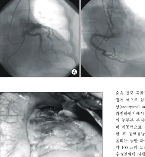
환자는 65세 여자로 8년 전 타 병원에서 심혈관 질환으 로 관상동맥 조영술을 받은 경력이 있었고 당시 조영술은 정상 소견을 보였으며 고혈압과 당뇨병 및 20년의 흡연력 을 가지고 있었다. 흉통을 주소로 내원하여 시행한 심초 음파상 관상동맥-폐동맥루가 의심되어 심도자술 및 관상 동맥 조영술을 시행하였고 좌전하행지에 90%의 협착증과 양측성 관상동맥-폐동맥루를 진단받았다(Fig. 1). 수술은 정중 흉골절개로 체외순환하에 시행하였다. 주폐동맥 근
위부에 혈관종양성(angiomatous) 관상동정맥루가 있었으며 (Fig. 2) 우선 좌전하행지 및 우관상동맥의 누두부가지에 서 오는 기형혈관을 박리 후 4-0 prolene으로 결찰하였다.
그 후 심박동상태에서 주 폐동맥을 종 절개해 보니 아직 도 출혈되는 관상동정맥루 입구를 확인할 수 있었다. 페 동맥내로 유입되는 입구 2곳을 6-0 prolene으로 봉합결찰 한 후 폐동맥을 봉합하였다. 심정지유도 후 좌측내유동맥 을 좌전하행지에 문합 후 수술을 종료하였다. 술 후 8일째 에 시행한 심초음파상 관상동정맥루는 보이지 않았으며 환자는 술 후 10일째에 퇴원하였고 현재 5개월째 외래 추 적관찰 중이나 별다른 문제점은 없었다.
양측성 관상동맥 -폐동맥루
-2예 보고-
김 혁*․박지권*․김영학*․강정호*․정원상*․전석철**․김경수***
Bilateral Coronary Artery to Pulmonary Artery Fistula
-Two case report-
Hyuck Kim, M.D.*, Ji Kwon Park, M.D.*, Young Hak Kim, M.D.*, Jung Ho Kang, M.D.*
Won Sang Chung, M.D.*, Seok Chol Jeon, M.D.**, Kyung Soo Kim, M.D.***
Bilateral coronary artery to pulmonary artery fistulas are very rare anomaly. Echocardiography, cardiac catheteriz- ation and coronary angiography of two patients having chest pain and dyspnea showed bilateral coronary to pul- monary artery fistulas. One patient had left anterior descending coronary artery stenosis and the other patient had cystic tumor. We report the good results of the surgical treatment of two patients with bilateral coronary to pulmonary artery fistulas.
(Korean J Thorac Cardiovasc Surg 2004;37:925-928) ꠏꠏꠏꠏꠏꠏꠏꠏꠏꠏꠏꠏꠏꠏꠏꠏꠏꠏꠏꠏꠏꠏꠏꠏꠏꠏꠏꠏꠏꠏꠏꠏꠏꠏꠏꠏꠏꠏꠏꠏꠏꠏꠏꠏꠏꠏꠏꠏꠏꠏꠏꠏꠏꠏꠏꠏꠏꠏꠏꠏꠏꠏꠏꠏꠏꠏꠏꠏꠏꠏꠏꠏꠏꠏꠏꠏꠏꠏꠏꠏꠏꠏꠏꠏꠏꠏꠏꠏꠏꠏꠏꠏ Key words: 1. Coronary artery fistula
2. Fistula
*한양대학교 의과대학 흉부외과학교실
Department of Thoracic and Cardiovascular Surgery, College of Medicine, Hanyang University **한양대학교 의과대학 진단방사선과학교실
Department of Radiology, College of Medicine, Hanyang University ***한양대학교 의과대학 내과학교실
Department of Internal Medicine, College of Medicine, Hanyang University 논문접수일:2004년 8월 20일, 심사통과일:2004년 9월 17일
책임저자 : 김영학 (133-792) 서울특별시 성동구 행당동 산 17번지, 한양대학교 의과대학 흉부외과학교실 (Tel) 02-2290-8461, 8470, (Fax) 02-2290-8462, E-mail: [email protected]
본 논문의 저작권 및 전자매체의 지적소유권은 대한흉부외과학회에 있다.
대흉외지 2004;37:925-928
- 926 - 증례 2
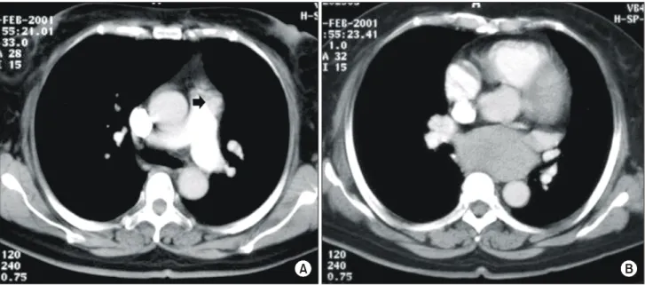
환자는 53세 여자로 10년간 당뇨병으로 치료 받은 병력 이 있으며 2달 전부터 시작된 간헐적인 흉통을 주소로 내 원하였다. 심전도검사상 심근 허혈 소견이 있어 협심증 의심하에 심초음파검사와 심도자술 및 관상동맥 조영술 을 시행하였다. 검사상 동맥류성 확장을 동반한 다발성 양측성 관상동맥-폐동맥루가 관찰되었으며 좌심방을 누르 고 있는 기관지성 낭종이 의심되었다. 흉부 전산화 단층 촬영상 기관지성 낭종(8×5 cm)이 기관분지부 밑에서 좌 심방을 누르고 있었으며 양측성 관상동맥-폐동맥루가 주 폐동맥의 전방으로 유입되는 것을 확인하였다(Fig. 3). 수
술은 정중 흉골절개로 체외순환하에 시작하였다. 혈성 심 정지 액으로 심정지 유도 후 주 폐동맥 근위부의 동맥류 낭(aneurysmal sac: 1.5×1.5 cm in size)을 종절개하여 보니 좌전하행지에서 오는 관상동정맥루 입구 및 우관상동맥 의 누두부 분지에서 오는 관상동정맥루의 입구가 보였으 며 폐동맥으로 유입되고 있었다. 각각의 입구를 봉합결찰 한 후 동맥류낭을 역시 봉합결찰로 제거하였다. 체온을 올리는 동안 좌심방을 누르고 있는 낭종을 1 cm 절개 후 약 100 cc의 노란색의 겔 액체를 흡입 후 봉합하였다. 술 후 8일째에 시행한 심초음파상 관상동정맥루는 보이지 않 았으며 낭성 종양도 보이지 않았다. 환자는 술 후 2주째에 퇴원하였으며 현재 2년 6개월째 외래 추적 중이나 별다른 문제점은 없었다.
고 찰
관상동정맥루는 드문 심기형으로 1865년 Krause[1]에 의 해 처음 기술되었다. 전체 인구 중 선천성 관상동맥 기형 은 1∼2%를 차지하고 있으며 그중 관상동정맥루는 48.7%
를 차지한다. 이러한 관상동정맥루 중 관상동맥-폐동맥루 는 15∼30%이며 다발성인 경우가 10.7∼16%이나 양측성 인 경우는 약 5%이다[2-4]. 증상으로는 60%에서 피로감 및 운동 시 호흡곤란을 호소하며 3∼7%에서는 coronary artery steal에 의한 협심증 및 심근경색이 나타난다. 19%
에서는 울혈성 심부전증이 나타나고 20%에서 심내막염이 발생하며 드물게 자연적으로 파열하여 혈성심낭 및 심낭 압전을 초래한다[5]. 대부분의 관상동정맥루는 선천성이 나 성인 환자의 약 30%에서는 관상동맥협착과 동반되 Fig. 1. (A) Left coronary artery angiogram in the right anterior obligue view shows coronary to pulmonary artery fistulas origi- nating from left anterior descen- ding coronary artery to main pulmonary artery. A severe ste- nosis is seen in the middle of the left anterior descending cor- onary artery. (B) Right cor- onary arteriogram in the left an- terior oblique view shows dila- ted and tortuous coronary to pulmonary artery fistulas.
A B
Fig. 2. Operative photograph after longitudinal opening of the pericardium shows bilateral coronary to pulmonary artery fis- tulas. The tortuous fistulas were originated from the proximal right coronary artery and the left anterior descending coronary artery.
김 혁 외 양측성 관상동맥-폐동맥루
- 927 - 며[6] 이들 간의 병리학적인 관련은 입증되지 않았다. 본 증례 1의 환자도 좌전하행지 협착이 동반되었으며 8년 전 타병원에서 관상동맥조영술 상 정상소견을 보였다고 하 나 기록이 소실된 상태로 후천성일 가능성이 있으나 입증 되지 못했다. 또 증례 2는 좌심방 위에서 좌심방을 압박하 는 낭성 종양을 동반하였는데 관상동정맥루와의 연관성 은 없는 것으로 보이며 기관지성 낭종으로 생각된다. 수 술적 처치로는 폐동맥이나 심방 혹은 심실 등을 절개하여 내측에서 봉합결찰하는 방법, 측부에서 나오는 경우 접촉 동맥봉합(tangential arteriorrhaphy)하는 방법, 원위부 결찰 만 시행하는 방법, 근위부와 원위부 모두 결찰하는 방법, 관상동맥결찰 및 우회술을 시행하는 방법, 동맥류성 관상 동맥을 통해서 봉합하는 방법 등이 있다[2,4,5]. 증례 1에 서는 혈관종양성 관상동정맥루였으며 좌전하행지 및 우 관상동맥에서 유입되는 근위부를 결찰한 후 확인을 위해 주폐동맥을 절개하였는데 계속 출혈되는 2곳의 입구가 있 어 봉합결찰하였다. 증례 2에서는 동맥류성 관상동정맥루 였고 동맥류를 절개한 후에 좌전하행지 및 우관상동맥에 서의 유입구를 각각 확인할 수 있었고 이들이 다시 주폐 동맥으로 들어가는 곳을 확인 후 봉합하였고 다시 동맥류 벽은 봉합결찰(aneurysmorrhaphy)하였다. 양측성 관상동맥
폐동맥루 환자에서 1예는 좌전하행지 협착을, 다른 1예는 낭종을 동반하였으며 수술적 치료를 하여 좋은 결과를 얻 었기에 보고하는 바이다.
참 고 문 헌
1. Krause W. Ueber den ursprung einer akzessorischen A. coro- naria aus der A. pulmonalis. Z Ratl Med 1865;24:225-9.
2. Fernandes ED, Kadivar H, Hallmann GJ, et al. Congenital malformations of the coronary arteries: the Texas Heart Institute experience. Ann Thorac Surg 1992;54:732-40.
3. Kirklin JW, Barratt-Boyes BG. Congenital anormalies of the coronary arteries. In: Kirklin JW, Barratt-Boyes BG, eds.
Cardiac surgery. 1st ed. New york: Dhurchill Livingstone.
1993; 1167-93.
4. Bauer EP, Piepho A, Klovekorn WP. Coronary arteriovenous fistula: surgical correction of a rare form. Thorac Cardio- vasc Surg 1994;42:237-9.
5. Bauer HH, Allmendinger PD, Flaherty J, et al. Congenital coronary arteriovenous fistula: spontaneous rupture and car- diac tamponade. Ann Thorac Surg 1996;62:1521-3.
6. Vavuranakis M, Bush CA, Boudoulas H. Coronary artery fistulas in adults: incidence, angiographic characteristics, natural histiry. Cathet Cardiovasc Diagn 1995;35:116-20.
Fig. 3. Chest CT scan showed dilated and tortuous fistulas tract (black arrow) ending anterior aspect of the main pulmonary artery (A) and subcarinal large cystic mass (B).
A B
대흉외지 2004;37:925-928
- 928 -
=국문 초록=
관상동맥-폐동맥루는 관상동맥 기형 중 드문 질환으로 그중에서도 양측성은 매우 드물다. 흉통 및 호 흡곤란을 주소로 내원한 두 환자에서 심도자술 및 관상동맥 조영술로 양측성 관상동맥-폐동맥루를 진단하였고 이들 중 한 환자에서는 좌전하행지 협착증, 다른 환자에서는 낭성 종양을 동반하였다. 이 두 환자에 대해 수술적 교정을 시행하여 좋은 결과를 얻었기에 보고하는 바이다.
중심 단어:1. 관상동맥-폐동맥루 2. 루