수소양 삼초경근의 해부학적 연구
박경식1
1상지대학교 한의과대학 해부학교실
Anatomical observation on the Triple Energizer Meridian Muscle in human
Kyoung-Sik Park1
1Dept. of Anatomy, College of Oriental Medicine, Sangji University Abstract
목 적 : 본 硏究는 手少陽三焦經筋의 理論的 根據를 解剖學的으로 提供하고 臨床에 經筋의 正確한 適用을 위함이다.
방 법 : Cadaver에 經筋을 表示하고 各各의 經穴部位에 標識와 pore 작업을 수행하고 각 經穴部를 皮膚, 筋膜, 그리고 筋肉의 淺層, 中間層, 그리고 深層部를 順序的으로 解剖하여 筋肉, 神經, 血u 등을 觀察한다.
결 과 및 결 론 : 手少陽三焦經筋의 解剖學的 考察 結果는 다음과 같다.
1) 筋 肉 : 천층에 근막(TE1), 근막확장대(TE2), 근막과 근간결합(TE3), 근막과 신근지대(TE4), 근막과 총지신근건(TE5), 근막및 총지신근과 소지신근간(TE6), 근막과 소지신근(TE7), 총지신근(TE8), 척측수근신근 과 소지신근간(TE9), 상완삼두근건(TE10, 11), 상완삼두근(TE12), 삼각근(TE13), 삼각근및 극하근과 극상근 간(TE14), 승모근(TE15), 흉쇄유돌근(TE-16, 17, 18), 후이개근(TE19, 22), 상이개근(TE20), 전이개근및 이하선근막(TE21), 안륜근(TE23), 중층에 소지신근건과 총지신근건간(TE4), 측두근막과 측두근(TE20, 22, 23), 심층에 배측골간근(TE3), 시지신근과 골간막(TE5) 장모지신근(TE6), 시지신근(TE7), 장지신근과 장모지 외전근간(TE8, 9), 상완삼두근(TE13), 견갑거근(TE15), 두판상근(TE16), 경상설골근과 하악이복근간 (TE17), 이복근(TE18).
2) 神 經 : 천층에 척골신경의 배측지(TE1, 2, 3), 후전완피신경(TE4, 5, 6, 8, 9, 10, 11), 내측전완피신 경(TE5, 6, 7, 8, 9, 10, 11), 후상완피신경(TE12, 13), 상외측상완피신경(TE13), 외측쇄골상신경(TE14, 15), 대이개신경(TE16, 17, 18, 19), 소후두신경(TE19, 20), 이개측두신경(TE20, 21, 22), 안면신경측두지(TE22, 23), 관골측두신경(TE23), 중층에 견갑상신경(TE15), 견갑배신경(TE15), 경상설골근신경(TE17), 후이개신경 (TE18, 19, 20), 안면신경측두지(TE20, 21, 22), 심층에 후골간신경(TE5, 6, 7), 요골신경심지(TE8, 9, 12, 13), 견갑상신경(TE14), 액와신경가지(TE14), 부신경(TE16), 안면신경과 부신경가지(TE17), 설인신경 (TE17), 설하신경(TE17), 경신경고리(TE17), 미주신경(TE17), 안면신경 (TE18).
3) 血 u : 천층에 척측정맥배측지(TE1, 2), 고유수장지동맥배측지(TE1), 배측중수골동맥배측지(TE2), 배 측중수골정맥(TE3), 척측피정맥(TE4, 5, 6, 7, 8, 9, 10, 11), 배측정맥궁(TE4), 부요측피정맥(TE6, 8, 9),요 측피정맥(TE10, 11), 후견봉정맥가지(TE13, 14), 후이개동·정맥(TE16, 17, 18, 19, 20), 전이개동·정맥 (TE20), 천측두동·정맥(TE22, 23), 중층에 후상완회선동맥(TE14), 견갑배동맥(TE15), 견갑상동맥(TE15), 천측두동·정맥(TE21), 관골측두동·정맥(TE23), 심층에 배측중수골동맥(TE3), 배측수근동맥궁(TE4), 후골간
⋅교신저자 : 박경식, 강원도 원주시 우산동 산 660번지, 상지대학교 한의과대학 해부학교실, Tel. 033-730-0667, Fax. 033-730-0653, E-mail: [email protected]
⋅본 연구는 2005학년도 교내연구비 지원에 의하여 수행되었음.
⋅투고 : 2007/02/27 심사 : 2007/03/09 채택 : 2007/03/13
동맥(TE4, 5, 6, 7, 8, 9), 전골간동맥(TE6, 7, 9), 심상완동맥(TE10, 11), 상완동맥측부지(TE10, 11), 중간 측부동맥(TE12), 요측측부동맥(TE12), 심상완동맥가지(TE13), 후상완회선동맥(TE13), 견갑상동맥(TE14), 후 두동·정맥(TE16, 17), 내경정맥(TE17).
결 론 : 1. 手少陽三焦經筋은 筋肉, 그리고 關聯 神經, 血u으로 구성된다.
2. 본 硏究는 經筋에 관한 旣存의 硏究와 比較하여 볼 때에 經筋의 構成要素에 있어서 若干의 差異를 보여준다.
3. 解剖學的 硏究結果, 經筋 筋肉을 支配하는 神經․血u의 槪念과 經筋을 스쳐 지나가는 神經․血u의 槪念은 區分된다.
Key words : 手少陽三焦經筋, 經穴(TE1~TE23), 筋肉, 神經, 血u
Ⅰ. Introduction
The concept of Meridian Muscle shown in Ling Shu (Miraculous Pivot) of HUANDI NEIJING (The Yellow Empero r′s Classic of Medicine : A bible in tra- ditional chinese medicine for about two thousand years) is close connected with The Twelve Main Meridian (TMM). Main Meridian or Meridian Muscle (MM) is a general term of muscular system distributed in circulation of The Twelve Main Meridian, is classified into 3Yin (The Negative)-3Yang (The Positive) of upper
& low limb1) and is composed of muscular tissue such as muscle (involving tendon), fascia, ligament2), which Ch'i (Gie : Life energy) in TMM is collected or concluded or translated3). TMM is distributed in the body surface of limb, trunk or head part, and in most case it's way is made in the opposite direction to the tip of limb.
The term of MM means a lot to myol- ogy, arthrology, rehabilitation, and the oth- er clinics. Since anatomical, constituent ele-
ments of individual MM are wrongly known to the academic world of oriental medicine, it bring about a mistaken clinical application or a wrong diagnosis.
This study was carried out in order to investigate correct elements of TMM and to support the meridianology or oriental clinics.
At this time we report The Triple Energizer Meridian Muscle (TEMM) in human, following The Lung MM4), The Large Intestine MM5), The Spleen MM6), The Small Intestine MM7), The Heart MM8), The Pericardium MM9).
II. Material and method 1. Reagents and injection
1) The preparation of a preservative
Phenol weighing one kilogram is dissolved in one litre methylalcohol (The 1st sol- ution).
The 500 ml of glycerin is dissolved in 2 L of methylalcohol and thereafter the addi-
tional 500 ml of glycerin is dissolved in this solution(The 2nd solution). The 1st and 2nd solution is well mixed, and made warm(30 min, 20℃). The 1 L of methyl- alcohol is added to this mixed solution, is stirred for 10 minutes. For the last time 1.5 L of formalin is added to mixed sol- ution of this.
2) Injection
The sheath of femoral artery & vein is exposed by vertical incision at the medial third of inguinal ligament, and femoral ar- tery carefully is separated from femoral vein.
A preservative is injected into femoral artery at the speed of 150 ml per minute.
After 6 L of preservative is injected, A needle inserted part is ligated, subsequently injector needle is inserted downwards for the preservation of the leg.
2. Embalmment of cadaver and experimental procedure
1) Cadaver is pending in the embalm- ment system for 40 hrs at 40℃.
2) Cadaver is exposed for 1hr at the normal temperature, and after that, is kept in refrigerated storage(3℃, 30% humidity).
3) TEMM is labelled by latex at the sur- face of cadaver, subsequently photographed.
4) Pore is made by drill in the vertical
direction at each meridian point.
5) Skin and superficial fascia are strip- ped off in order and thereafter is labelled by latex at the exposed deep fascia surface, once more is photographed.
6) Deep fascia is also removed.
7) Subsequently muscle, tendon, nerve, blood vessels are investigated, photographed, being divided into three layers(outer, mid- dle, and inner or deep layer).
III. Results
TEMM was marked at the surface of cadaver, and also constituent elements was divided into three layer (outer, middle, in- ner or deep layer), being dissected. The results were identified as follows
1) A schema of TEMM (Fig. 1, 5, 10, 12, and referred to 14).
2) Muscle, and related blood vessels, nerve constituting TEMM.
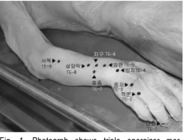
1. Gwanchung (TE1)
As shown in fig. 1, 2, 3 muscle group in case of superficial dissection is recognized as muscle fascia.
Dorsal digital vein, dorsal branch of proper palmar digital artery are distributed at this meridian muscle.
As the result of dissection, there are dor- sal digital nerve from ulnar nerve at super-
ficial fascia.
2. Aengmun (TE2)
Muscle group related to Aengmun is con- sidered as dorsal expansion of fascia at su- perficial dissection(Fig. 1, 2, 3).
This meridian are made up dorsal digital nerve from ulnar nerve dorsal digital vein, dorsal digital artery from dorsal metacarpal artery as Gwanchung are.
3. Jungjeo (TE3)
As gathering from this observation(Fig.
1, 2, 3, 4) muscle group constituting this meridian muscle(mm) are intertendinous connection at outer layer and dorsal inter- osseous muscle at inner layer. Dorsal branch of ulnar nerve exists at the outer layer of this meridian. As for blood vessel, dorsal metacarpal vein lies at outer layer, dorsal metacarpal artery at inner layer.
4. Yangji (TE4)
Muscle group related to Yangji are com- posed of fascia and extensor retinaculum at outer layer(Fig. 2, 3) and exists at space between extensor digiti minimi muscle and extensor digitorum muscle at middle layer.
Posterior antebrachial cutaneous nerve from radial nerve lies at outer layer as con- stituent nerve. As to constituent blood ves-
sels basilic vein and dorsal venous arch ex- ists at outer layer, dorsal carpal arterial arch, posterior interosseous artery at deep layer(Fig. 4).
5. Oegwan (TE5)
Constituent elements of this mm are fas- cia and extensor digitorum muscle tendon, medial antebrachial cutaneous nerve from ulnar nerve, posterior antebrachial cuta- neous nerve from radial nerve, branch of basilic vein at outer layer(Fig. 2, 3), and extensor indicis muscle, interosseous mem- brane, posterior interosseous nerve from ra- dial nerve, posterior interosseous artery at deep layer.
6. Jigu (TE6)
Constituent muscle(m) are fascia, space between extensor digiti minimi m. and extensor digitorum m. at outer layer(Fig. 2, 3), extensor pollicis longus m. at inner lay- er(Fig. 4).
As to nerve the composition is same to that of Oegwan. that is, medial antebrachial cutaneous nerve from ulnar nerve, posterior antebrachial cutaneous nerve from radial nerve, posterior interosseous nerve(Fig. 4) from radial nerve.
In relation to blood vessels, outer layer are composed of basilic vein and accessory cephalic vein, and inner layer, posterior in-
terosseous artery and anterior interosseous artery(Fig. 4).
7. Hoejong (TE7)
Outer layer are composed of fascia and extensor digiti minimi muscle(Fig. 3), and deep layer, extensor indicis muscle(Fig. 4).
There is medial antebrachial cutaneous nerve from ulnar nerve at outer layer, pos- terior interosseous nerve from radial nerve like Oegwan at inner layer(Fig. 4). As to blood vessels basilic vein exists and at in- ner layer(Fig. 4), posterior interosseous ar- tery, anterior interosseous artery like Jigu.
8. Samyangnak (TE8)
Muscle constituent at outer layer(Fig. 2, 3) is extensor digitorum muscle, nerve are medial antebrachial cutaneous nerve from ulnar nerve, posterior antebrachial cuta- neous nerve from radial nerve, blood ves- sels are basilic vein, accessory cephalic vein.
Constituents at inner layer(Fig. 4) are space between extensor pollicis longus mus- cle and abductor pollicis longus muscle in case of muscle, deep branch of radial nerve in case of nerve, posterior interosseous ar- tery in case of blood vessel.
9. Sadok (TE9)
Constituent elements of outer layer(Fig.
3) are composed of space between extensor carpi ulnaris muscle and extensor digiti minimi muscle, in case of nerve and blood vessels, like Samyangnak, medial antebrachial cutaneous n. from ulnar nerve, posterior antebrachial cutaneous nerve from radial nerve, basilic vein, accessory cephalic vein. Those of inner layer(Fig. 4) are ex- tensor pollicis longus muscle and abductor pollicis longus muscle, and nerve, deep branch of radial nerve, blood vessel, poste- rior interosseous artery like Samyangnak.
10. Cheonjeong (TE10)
This mm is composed of triceps brachii muscle tendon, constituent nerve is same as that of Samyangnak, that is, medial antebrachial cutaneous nerve from ulnar nerve, posterior antebrachial cutaneous nerve from radial nerve. constituent blood vessels are cephalic vein and basilic vein at outer layer, deep brachial artery and col- lateral artery from brachial artery at inner layer(Fig. 3, 6, 7).
11. Cheongnaengyeon (TE11)
The composition of Cheongnaengyeon mm is nearly same as Cheonjeong(Fig. 6, 7), that is, triceps brachii muscle tendon, medial antebrachial cutaneous nerve from ulnar nerve, posterior antebrachial cuta-
neous nerve from radial nerve, cephalic vein and basilic vein, deep brachial artery and collateral artery from brachial artery.
12. Sorak (TE12)
The constituents of outer mm(Fig. 6) are triceps brachii muscle, posterior brachial cutaneous nerve from radial nerve, and those of inner mm(Fig. 7), deep branch of radial nerve, middle collateral artery, branch of radial collateral artery.
13. Nohoe (TE13)
Muscle constituent at outer layer(Fig. 6) is deltoid muscle, nerve group are composed of superior lateral brachial cutaneous nerve from axillary nerve, posterior brachial cuta- neous from radial nerve, blood vessel is branch of thoracoacromial vein.
Constituents at inner layer(Fig. 7) are triceps brachii muscle, deep branch of radi- al nerve, branch of deep brachial artery, posterior circumflex humeral artery.
14. Gyeollyo (TE14)
Muscle group related to Gyeollyo are composed of deltoid muscle, space between infraspinatus muscle and supraspinatus muscle at outer layer. Nerve group are made up lateral supraclavicular nerve at outer layer, and suprascapular nerve,
branch of axillary nerve at deep layer.
Blood vessel group are composed of acro- mial branch of thoracoacromial vein, poste- rior circumflex humeral vein at outer lay- er, posterior circumflex humeral artery at middle layer, suprascapular artery at deep layer.
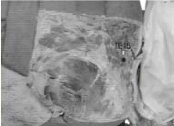
15. Cheollyo (TE15)
As a result of investigation(Fig. 8, 9) there are trapezius muscle, lateral supra- clavicular nerve at outer layer, supra- scapular nerve, dorsal scapular nerve, dor- sal scapular artery, suprascapular artery at middle layer, levator scapular muscle at deep layer.
16. Cheonyu (TE16)
Muscle constituent at outer layer is ster- nocleidomastoid muscle, nerve group are greater auricular nerve and lesser occipital nerve, blood vessel group are posterior au- ricular artery and vein.
Constituents at inner layer are splenius capitis muscle, accessory nerve, occipital ar- tery and vein(Fig. 11).
17. Yepung (TE17)
Muscle group related to Yepung are made up sternocleidomastoid muscle at outer layer, space between stylohyoideus
muscle and venter digastricus muscle at deep layer. Nerve group are composed of greater auricular nerve at outer layer, Styloyhyoideus m. from Facial n. at middle layer, branchs of facial nerve and accessory nerve, glossopharyngeal nerve, hypoglossal nerve, ansacervicalis, vagus nerve at deep layer. Blood vessel group are made up pos- terior auricular artery and vein at outer layer, Occipital artery and vein, internal jugular vein at deep layer(Fig. 11).
18. Gyemaek (TE18)
Muscle group related to Gyemaek are composed of sternocleidomastoid muscle at outer layer and digastricus muscle at in- ner layer. Nerve group are composed of greater auricular nerve and posterior auric- ular nerve from facial nerve at outer layer, facial nerve at inner layer. Blood vessel group are made up posterior auricular ar- tery and vein at outer layer(Fig. 11).
19. Nosik (TE19)
The outer mm of Nosik are composed of auricularis posterior muscle, greater auric- ular nerve and lesser occipital nerve, and posterior auricular artery & vein.
At middle layer there is posterior auric- ular nerve from facial nerve(Fig. 11).
20. Gakson (TE20)
Muscle group related to Gakson are composed of auricularis superior muscle at outer layer, temporalis fascia and tempora- lis muscle at inner layer. Nerve group are composed of auriculotemporal nerve, lesser occipital nerve at outer layer, and posterior auricular nerve from facial nerve, temporal branch of facial nerve at inner layer. In case of blood vessels there are anterior au- ricular artery from superficial temporal ar- tery & vein, branch of posterior auricular artery & vein(Fig. 11).
21. Imun (TE21)
Muscle group related to Imun are com- posed of auricularis anterior muscle and parotid fascia, and nerve group, composed of auriculotemporal nerve from Mandibular nerve(Trigerminal nerve) at outer layer, temporal branch of facial nerve at inner layer. Blood vessels are made up super- ficial temporal artery & vein at inner lay- er(Fig. 11).
22. Hwaryo (TE22)
The outer mm of Hwaryo are auricularis superior muscle, auriculotemporal nerve, temporal branch of facial nerve, and super- ficial temporal artery & vein.
At inner layer there are temporalis fascia and temporalis muscle(Fig. 13).
23. Sajukgong (TE23)
Muscle group related to Sajukgong are composed of orbicularis oculi muscle at outer layer, temporalis fascia and tempora- lis muscle at inner layer. This mm involves zygomaticotemporal nerve from maxillary nerve (Trigerminal nerve) and temporal
branch of facial nerve. Blood vessels are made up frontal branch of superficial tem- poral artery & vein, and supraorbital ar- tery & vein from ophthalmic artery &
vein at outer layer, zygomaticotemporal ar- tery & vein from middle temporal artery
& vein at inner layer(Fig. 13).
Fig. 1. Photogrph shows triple energizer mer- idianpoints(TE 1-TE 9) at body surface of forearm.
Fig. 2. Photograph shows triple energizer mer- idianpoints(TE 1-TE 8) at deep fascia of forearm.(△ : Extensor digitorum m.)
Fig. 3. Photograph shows triple energizer mer- idianpoints(TE 1-TE 10) in case of su- perficial dissection of forearm. (A : Extensor digitorum muscle(m), B : Extensor minimi m., C: Extensor carpi ulnaris m., D: Extensor retinaculum, E : Extensor carpi radialis brevis m., F : Triceps brachii m.)
Fig. 4. Photograph shows triple energizer mer- idianpoints(TE 3-TE 9) in case of deep dissection of forearm.(A : Extensor digitorum m., B : Extensor carpi radialis longus m., C : Abductor pollicis longus m., D : Extensor pollicis brevis m., E,F : Posterior interosseous nerve, artery, G, H : Ulnar nerve, ar- tery, I : Extensor carpi ulnaris m., J : Extensor minimi m.)
Fig. 5. Photograph shows triple energizer mer- idianpoints(TE 10-TE 13) at body sur- face of arm.
Fig. 6. Photograph shows triple energizer mer- idianpoints(TE 10-TE 13) at deep fascia of arm.(A : Triceps brachii m. tendon, B : Deltoid m.)
Fig. 7. Photograph shows triple energizer mer- idianpoints(TE 10-TE 13) in case of deep dissection of arm.(A : Deep bra- chial artery, B : Radial nerve, C : Posterior circumflex humeral artery, D : Deltoid m., E : Axillary artery, F : Long head of triceps brachii m.)
Fig. 8. Photograph shows triple energizer mer- idianpoint(TE 15) at deep fascia level of upper back.
Fig.9. Photograph shows triple energizer mer- idianpoint(TE 15) in case of superficial dis- section of upper back.(△ : Trapezius m.)
Fig. 10. Photograph shows triple energizer mer- idianpoints(TE 15-TE 21) at body sur- face of head.
Fig. 11. Photograph shows triple energizer mer- idianpoints(TE 16-TE 21) at deep fas- cia level of head.
Fig. 12. Photograph shows triple energizer mer- idianpoints(TE 22-TE 23) at body sur- face of head.
Fig. 13. Photograph shows triple energizer mer- idianpoints(TE 22-TE 23) at deep fas- cia level of head.(△ : Temporalis m.)
IV. Discussion
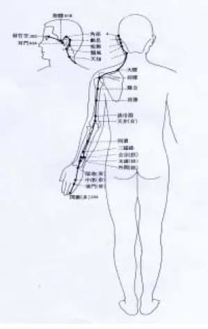
TEMM originates in the ulnad tip, Gwanchung of 4th finger, go up via 4th, 5th metacarpals, covers the dorsal part of antebrachium and the posterior part of brachium, continuously via the posterior part(Nohoe, Gyeollyo) of shoulder goes up the auricle, subsequently goes round it(Yepung, Gyemaek, Nosik, Gakson, Imun, Hwaryo)(Fig. 14).
Fig. 14. The scheme of the triple energizer meridian in human.
MM in oriental medicine means a concept comprising soft tissue such as muscle, fas-
cia ligament, and nerve on the out skirts of them10). But we mean to comprice blood vessel additionally in this concept.
It is possible to know the mode of ac- tion of MM if we analyze the distribution of MM in connection with human anat- omy11-13). In the view of clinical applica- tion, MM plays an important role in the flexion & extention of muscle or joint or limb, since the abnormality of MM is ex- pressed as the abnormalities of MM-pierc- ing part, such as stretching, convulsion, re- lax, rigidity, displacement.14-17) Referring to the disability of MM, the chapter MM of Ling Shu(Miraculous Pivot) explains the following meaning "if Yang is over, the muscle extended and so long as Yin is over, then the muscle flexed. Cold brings about the muscle contraction, and hot, muscle slackness."18) This means the symp- tom of disease induced by abnormal meri- dian muscle subsquent to Yang or Yin over.
As mentioned above, the anatomical knowledge of muscle is essentially required for the clinical application of MM. And al- so at the same time such a knowledge must be exact. Such a knowledge guaran- tees the exact and effective application of MM to clinics.
This study shows some differences from already established study1,19) on mm ; that is, constituent elements of MM such as
muscle, nerve, blood vessels, ligament, fas- cia, and assay method. Above all the structure of each meridian point inves- tigated in this study was divided into three layers according to depth from body sur- face but on the other hand we came across that it may be wide differences in opinion according to the disparity of real meridian point or the angle of acupunctuation20).
V. Conclusion
This study was carried to identify the component of TEMM in human, dividing into outer, middle, and inner part. Upper extremity and head skin were opened wide- ly to demonstrate muscles, nerve, blood vessels and the others, displaying the inner structure of TEMM. We obtained the con- clusions as follows;
1. TEMM is composed of the muscle, and related nerve, blood vessels.
2. This study shows some differences from already established study from the view- point of constituent elements of MM.
3. In human anatomy, it is present the difference between a term of nerve or blood vessels which control the muscle of MM and those which pass near by MM.
VI. Reference
1. 전국한의과대학침구경혈학회. The allied department of acupuncture & mox- ibustion, oriental medicine. Acupuncture
& moxibustion(the 1st volume). 3rd ed.
Seoul : Gypmundang. 1991 : 581-608.
2. Song CH. Study on The 12 Meridian Muscle and The 12 Skin. The Journal of Korean Acupuncture & Moxibustion Society. 1989 ; 6(1) : 179-86.
3. Giovanni Maciocia. The foundations of chinese medicine. USA : Churchill Livingstone. 1989 : XIII-XV.
4. Lee JM, Park KS. Anatomy of The Lung Meridian Muscle in Human. J.
Kor. AM-Meridian & Pointology Soc.
2001 ; 18(2) : 19-25.
5. Lee JM, Sim Y. Park KS. Anatomy of The Large Intestine Meridian Muscle in Human. J. Kor. AM-Meridian &
Pointology Soc. 2002 ; 19(1) : 15-23.
6. Park KS. Anatomy of The Spleen Meridian Muscle in Human. J. Kor.
AM-Meridian & Pointology Soc. 2003 ; 20(4) : 65-76.
7. Park KS. Anatomical Study on The Small Intestine Meridian Muscle in Human. J. of Kor. Inst. of Herbal Acupuncture. 2004 ; 7(2) : 57-64.
8. Park KS. Anatomical Study on the Heart Meridian Muscle in Human. J. of Kor. Orient. Med. Soc. 2005 ; 26(1) : 11-7.
9. Park KS. Study on the Anatomical
Pericardium Meridian Muscle in Human. Kor. J. of Meridian &
Acupoint. 2005 ; 22(1) : 67-74.
10. The allied department of acupuncture
& moxibustion, oriental medicine.
Acupuncture & moxibustion (the 1st volume). 3rd ed. Seoul : Gypmundang.
1991 : 159-70.
11. Abrahams PH, Hutchings RT, Marks SC Jr. Mc. Minn's Color Atlas of Human Anatomy. 4th ed. London : Mosby-Wolfe Publishing. 1999 : 105-41.
12. Ferner H, Stubesand J. Sobotta's Atlas of Human Anatomy (Anatomy 1). 10th ed. Urban & Schwarzenberg. 1983 : 318-59, 135-47.
13. Frank H. Netter. Atlas of Human Anatomy. CIBA. 1987 : 401-52, 17-65.
14. Butler P. Imaging of the Nervous System. Springer-Verlag. 1990 : 231-40.
15. Snell RS. Neuroanatomy. 3rd ed. Little
& Brown. 1992 : 163-260.
16. Liebenson C. Rehabilitation of the Spine. Williams & Wilkins Inc. 1996 : 159-67.
17. Juhl JH. Roentgen Interpretation. 5th Ed. Harper & Row. 1981 : 1171-3, 178-9, 159-62.
18. Jang MH. HUANDI NEIJING (A Korean version). Seoul : Sungbosa.
1975 : 145-54.
19. Ahan YK. Acupuncture & Moxibustion.
Seoul : Sungbosa. 1991 : 497-535.
20. Choi HS. A Search on New Direction
of Meridian Study. J. Kor.
AM-Meridian & Pointology Soc. 2004
; 21(3) : 1-20.