pISSN 1738-3544 eISSN 2288-1662
Detection of Vancomycin-Resistant Enterococci and Related Genes Using VITEK 2 System and Multiplex Real-time PCR Assay
Min-Kyung Jeong 1 , Young-Bin Yu 2 , Sang-Ha Kim 3 , Sunghyun Kim 4 , Young-Kwon Kim 2
1
Department of Laboratory Medicine, Chonnam National University Hospital, Gwangju, Korea
2
Department of Biomedical Laboratory Science, College of Medical Sciences, Konyang University, Daejeon, Korea
3
Department of Laboratory Medicine, Konyang University Hospital, Daejeon, Korea
4
Department of Clinical Laboratory Science, College of Health Sciences, Catholic University of Pusan, Busan, Korea
VITEK 2 시스템과 Multiplex Real-time PCR을 이용한 반코마이신 내성 장알균(VRE)과 내성관련 유전자 검출
정민경 1 , 유영빈 2 , 김상하 3 , 김성현 4 , 김영권 2
1
전남대학교병원 진단검사의학과,
2건양대학교 의과학대학 임상병리학과,
3건양대학교병원 진단검사의학과,
4부산가톨릭대학교 보건과학대학 임상병리학과
In this study, using the VITEK 2 system, 74 samples (22.6%) out of 327 specimens were identified by the growth of Enterococcosel media (EV6 agar) supplemented with 6 g/mL of vancomycin.
Enterococcus faecium was identified as 55 strains (74.3%), Enterococcus casseliflavus as 2 strains (2.7%), Enterococcus avium as 1 strain (1.4%), and Enterococcus gallinarum as 16 strains (21.6%).
Among the 55 phenotypes of Enterococcus faecium , 42 (76.4%), 9 (16.4%), and 4 strains (7.3%) showed the vanA, vanB, and vanC phenotype, respectively. The 16 strains of Enterococcus gallinarum and 2 strains of Enterococcus casseliflavus showed the vanC phenotype and the 1 strain of Enterococcus avium had the vanB phenotype. The one strain of Enterococcus faecium propagated only in EV4 and was susceptible to both vancomycin and teicoplanin according to the antimicrobial susceptibility test using the VITEK 2 system. The vancomycin resistance phenotype gene was not detected by PCR. A total of 327 specimens were cultured in Enterococcosel broth supplemented with 6 g/mL of vancomycin (EV6 broth), and 120 strains (36.7%) were isolated. These 120 strains were subjected to vancomycin resistant genotyping by a multiplex real-time polymerase chain reaction and 51 strains (42.5%) showed van A; 5 strains (4.2%) showed van A and van C; and 18 strains (15%) showed van C. Vancomycin resistance genotypes were not detected in the remaining 46 strains (38.3%).
Key words: Antimicrobial susceptibility test, Enterococcosel broth, Multiplex real-time PCR, Vancomycin resistant enterococci, VITEK 2 system
Corresponding author: Young-Kwon Kim Department Biomedical Laboratory Science, College of Medical Sciences, Konyang University, 158 Gwanjeodong-ro, Seo-gu, Daejeon 35365, Korea
Tel: 82-42-660-6371 Fax: 82-42-543-6370
E-mail: ykkim3245@konyang.ac.kr Co-Corresponding author: Sunghyun Kim Department of Clinical Laboratory Science, College of Health Sciences, Catholic University of Pusan, 57 Oryundae-ro, Geumjeong-gu, Busan 46252, Korea
Tel: 84-51-510-0560 Fax: 82-51-510-0568 E-mail: shkim0423@cup.ac.kr
This is an Open Access article distributed under the terms of the Creative Commons Attribution Non-Commercial License (http://creativecommons.org/licenses/by-nc/4.0) which permits unrestricted non-commercial use, distribution, and reproduction in any medium, provided the original work is properly cited.
Copyright © 2017 The Korean Society for Clinical Laboratory Science. All rights reserved.
Received: November 3, 2017
Revised 1
st: November 15, 2017
Revised 2
nd: November 21, 2017
Revised 3
rd: November 28, 2017
Revised 4
th: December 5, 2017
Accepted: December 6, 2017
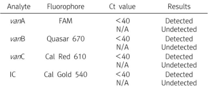
Table 1. Ct value interpretation of Anyplex VanR Real-Time PCR assay
Analyte Fluorophore Ct value Results
van A FAM <40
N/A
Detected Undetected
van B Quasar 670 <40
N/A
Detected Undetected van C Cal Red 610 <40
N/A
Detected Undetected
IC Cal Gold 540 <40
N/A
Detected Undetected Abbreviation: N/A, not amplified.
서 론
반코마이신 내성 장알균(vancomycin resistant enterococci, VRE)은 의료관련 감염의 대표적인 병원체로 1986년 유럽에서 처음 보고된 이래 현재에 이르기까지 30여년 동안 전 세계적으 로 분리 빈도가 증가하여 중요한 의료관련 감염균으로 자리 잡 고 있다[1]. VRE의 신속한 검출은 환자의 치료뿐 아니라 다른 환 자에게로의 전파를 예방할 수 있게 한다[2].
미국 National Nosocomial Infections Surveillance보고에 따르면 2003년 중환자실 환자에서 분리된 장알균 중 VRE 빈도 를 28.5%로 보고하였다[3]. 국내에서도 1992년 처음으로 vanA 유전자를 가지는 Enterococcus durans가 분리되었으며 국내 의료기관 내의 VRE 분리 비율이 1998년 이후 빠르게 증가 하고 있다[4,5]. 특히 중환자실의 경우 환자에서 분리된 장알균 중 반코마이신 내성 장알균 분리 비율이 낮았으나 2004년 29%, 2006년에는 43%, 최근 51%로 급격하게 증가 하고 있다[6].
현재 국내 병원 시험실에서 실시하는 VRE 감시 배양법으로 는 대변 검체를 평판 배지 배양법인 반코마이신 6 g/mL 첨가된 Enterococcosel agar에 접종 후 검게 변한 집락을 혈액우무배 지에 계대배양 후 동정 시험과 항균제 감수성 시험을 실시하는데 보고하기까지 3∼4일이 소요되어 정확하지만 시간이 오래 걸리 고 다중 중합효소 연쇄반응법(multiplex real-time PCR)을 이 용하여 VRE를 검출할 수 있는데, 이는 1∼2일이 소요되어 빠르 지만 비용이 많이 든다는 단점이 있다[7].
이에 본 연구에서는 VRE 감시 배양으로 의뢰된 대변 검체 327개를 대상으로 보다 신속하고 경제적인 VRE 감시배양을 실 시하고자 저자들이 고안한 반코마이신 첨가 배지(enterococco- sel media with vancomycin, EV)와 VITEK 2를 이용하여 VRE 의 vancomycin 내성 유전자의 표현형과 유전형을 분석하여 국 내에서 분리된 VRE의 항균제 내성 양상과 관련 유전자와의 상 관성을 알아보고자 연구를 실시하였다.
재료 및 방법
1. 연구대상
2016년 1월부터 4월까지 전남 소재 C 종합병원에서 VRE 감 시 배양이 의뢰된 대변 검체 총 327개를 대상으로 연구를 실시 하였다.
2. 연구방법
1) Enterococcosel 선택배지에서 VRE의 분리
Enterococcosel agar와 Enterococcosel broth (Beckton Dickinson, sparks, MD, USA)에 vancomycin (Sigma Chemical Company, St. Louis, MO, USA)을 각각 4 g/mL (EV4), 6
g/mL (EV6), 12 g/mL (EV12), 24 g/mL (EV24)씩을 첨가 하여 대변 검체 총 327개에 접종 한 후 35°C, CO
2
5∼7% 배양 기에서 48시간 동안 배양하였다. Enterococcosel agar에서 검 게 변한 집락을 catalase시험 음성, 그람양성알균을 VRE로 잠 정 동정하였다.2) VITEK 2 시스템을 이용한 항균제 감수성 시험
Enterococcosel agar에서 검게 변한 집락을 VITEK 2 system ID GP card (bioMe'rieux, France)를 이용하여 VITEK 2 System (bioMe'rieux, France)으로 동정하였다. E. gallinarum와 E.
casseliflavus 및 E. faecium을 구별 동정하기 위해 methyl D-glucopyranoside (MGP) test를 실시하였다. Vancomycin 과 teicoplanin의 내성 여부를 알아보기 위해 VITEK-2 system AST 600 card (bioMe'rieux, France)를 사용하여 항균제 감수 성 시험을 하였다. 세균 부유액의 탁도를 McFarland tube 0.5 에 맞춘 후 Mueller-Hinton agar (MHA)에 접종하고, 배지의 중앙에 vancomycin (30 g) 항균제 디스크와 teicoplanin (30
g) 항균제 디스크를 20 mm 간격으로 접종하였다. 접종한 배 지는 37°C 항온기에 18시간 배양 후 각 항균제에 대한 억제대를 확인하였다.
3) Anyplex VanR real-time PCR을 이용한 VRE 유전자 검출 EV6 Enterococcosel broth (Beckton Dickinson, USA)에 접종 한 후 35°C, CO
2
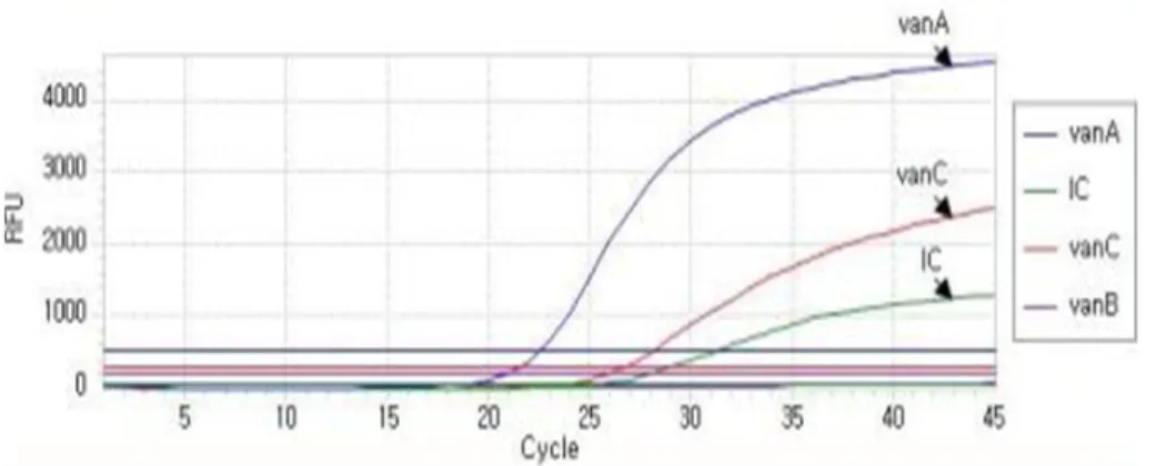
5∼7% 배양기에서 16∼24시간 동안 배 양하였다. 검게 변한 액체배지 배양액 50 L를 DNA extractionFigure 1. Amplification curve of Anyplex VanR Real-Time PCR assay.
Table 2. Growth on different concentration EV agar from stool cultures 327 clinical isolates
Growth on different concentration of EV agar
EV agar EV4 EV6 EV12 EV24 No. (%) of isolates
All EV 56 56 56 56 56 (17.1)
EV4 and EV6 18 18 - - 18 (5.5)
Only EV4 10 - - - 10 (3.1)
No growth on EV 243 243 243 243 243 (74.3)
Total 327 317 299 299 327 (100.0)
Abbreviation: EV. enterococcsel agar with vancomycin.
solution 200 L와 혼합한 뒤 heat block 100°C에서 10분간 비동화 시킨 후 13,000 rpm에서 5분간 원심 분리하여 DNA를 추출하였다. vanA와 vanB, vanC 유전자에 특이적인 프라이머 쌍을 이용하는 Anyplex VanR real-time Detection kit (Seegene, Korea)를 사용하여 추출한 DNA 3 L에 VanR 시험 시약에 포함된 5x VanR Oligo Mix (primer, template of internal control) 4 L, 8-methoxypsoralen 3 L, 2x Detection Mix (DNA polymerase, buffer containing dNTPs) 10 L를 혼합한 뒤 다중 실시간 중합 효소 연쇄반응시 험을 수행하였다. PCR 반응 방법은 CFX 96TM Real-time PCR System (Bio-Rad)을 이용하여 95°C에서 2분간 반응 한 후 95°C 30초, 60°C 60초 반응을 44회 반복하였고, 형광물질 FAM, Cal Gold 540, Cal Red 610, Quasar 670을 이용하여 Anyplex VanR Real-Time PCR의 Ct value 판독기준에 따라 분석하였다(Table 1, Figure 1).
결 과
1. Enterococcosel 배지에서 VRE의 분리
대변 검체 327개를 vancomycin을 첨가한 Enterococcosel EV4, EV6, EV12, EV24 선택배지에 각각 배양한 결과 84주 (25.7%)가 증식하였으며, EV4, EV6, EV12, EV24 배지 모두에 서 증식한 경우가 56건(17.1%), EV4와 EV6 배지에서만 증식한 경우가 18건(5.5%), EV4 배지에서만 증식한 경우는 10건 (3.1%)이었다(Table 2).
2. VITEK 2 시스템을 이용한 VRE 항균제 감수성 시험 VITEK 2 시스템을 이용하여 총 327검체 중 74검체(22.6%) 가 6 g/mL vancomycin이 첨가된 Enterococcosel (EV6agar)에 서 증식한 집락을 동정하였다. 이 중 55주(74.3%)는 E. faecium, 16주(21.6%)는 E. gallinarum, 2주(2.7%)는 E. casseliflavus, 1주
(1.4%)는 E. avium으로 동정되었다.
표현형 유전자 시험 결과, E. faecium 55주 중 42주(76.4%)가 vanA 표현형을 나타냈으며, 9주(16.4%)는 vanB 표현형을 나타 냈고, 4주(7.3%)는 vanC 표현형을 나타냈다. E. gallinarum 16 주와 E. casseliflavus 2주는 모두 vanC 표현형을 나타냈으며, E.
avium 1주는 vanB 표현형을 나타냈다. EV4에서만 증식한 E.
faecium 1주는 VITEK 2 시스템을 이용한 항균제 감수성 시험 을 한 결과 vancomycin과 teicoplanin 모두 감수성이었고 PCR 로 확인한 결과 vancomycin 내성 표현형 유전자가 검출되지 않 았다(Table 3).
3. Anyplex VanR real-time PCR을 이용한 Van 유전자의 검출
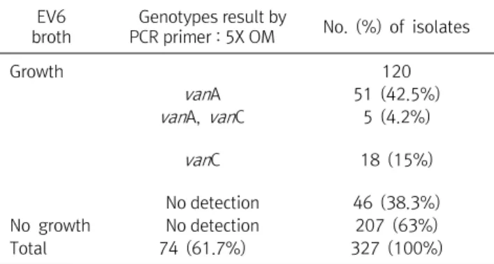
327개의 검체를 vancomycin 6 g/mL (EV6)이 첨가된 Enterococcosel 액체배지(EV broth)에서 배양한 결과 120주 (36.7%)가 분리되었으며, 120주를 다중 중합 효소 연쇄반응으 로 vancomycin 내성 유전형 시험을 한 결과 51주(42.5%)가 vanA 유전형을 나타냈으며, 5주(4.2%)에서 vanA와 vanC 유전 형을 나타냈고, 18주(15%)에서 vanC 유전형을 나타냈으며, 나 머지 46주(38.3%)에서는 vancomycin 내성 유전형 유전자가 검출되지 않았다(Table 4).
Table 3. Distribution of phenotypes and antimicrobial susceptibility test results of 74 VRE clinical isolates by Vitek 2 system
Result by VITEK 2 ID (No. of isolates)
No. of isolates (74)
Vancomycin Teicoplanin
Phenotype MIC
(g/mL)
Disc inhibition (mm)
MIC (g/mL)
Disc inhibition (mm)
E . faecium (42) 37 32 ≤14 32 ≤10 vanA
5 32 ≤14 32 11∼13 vanA
E . faecium (9) 5 32 ≤14 1 ≥14 vanB
3 32 ≤14 1 11∼13 vanB
1 32 ≤14 1 ≤10 vanB
E . avium (1) 1 32 ≤14 2 ≤10 vanB
E . faecium (4) 1 32 ≤14 4 11∼13 vanC
3 32 ≤14 16 ≤10 vanC
E . gallinarum (16) 15 4 ≥17 0.5 ≥14 vanC
1 8 ≥17 0.5 ≥14 vanC
E . casseliflavus (2) 2 4 ≥17 0.5 ≥14 vanC
Table 4. Genotype distribution of 120 VRE clinical isolates from EV6 broth
EV6 broth
Genotypes result by
PCR primer : 5X OM No. (%) of isolates
Growth 120
van A 51 (42.5%)
van A, van C 5 (4.2%)
van C 18 (15%)
No detection 46 (38.3%)
No growth No detection 207 (63%)
Total 74 (61.7%) 327 (100%)
고 찰
장알균(VRE)은 병독성이 높은 세균은 아니지만 중요한 의료 관련 감염균으로 국내 중환자실을 비롯하여 의료환경에 토착화 되어가고 있는 의료관련 감염균이다[8,9]. VRE의 유행과 전파 를 막기 위한 감염관리지침이 있으나 현실적으로 국내의 모든 병원에서 적용하기 어려울 뿐 아니라 시행하여도 확실히 근절 하기 힘든 의료관련 감염의 주요 다제내성균이다[9].
이전 연구에서는 임상 검체에서 분리한 장알균 279주 중 26주 에서 중합 효소 연쇄반응법과 16S rDNA의 염기순서분석법을 사 용한 분자생물학적 동정 결과와 생화학적 동정 결과가 불일치함 을 보고하여[10] VRE의 정확하고 신속한 동정과 항균제 감수성 시험의 결과의 필요성을 강조하였다. 특히 VRE는 메티실린 내성 황색 포도알균(methicillin-resistant Staphylococcus aureus, MRSA)으로 내성 유전자를 전달할 수 있기 때문에, MRSA가 정착 화되어 있는 국내 병원에서 VRE의 빠르고 정확하고 효율적인 동 정과 항균제 감수성시험의 결과는 매우 중요하다[11].
본 연구에서는 총 327개의 검체를 전통적인 방법인 EV6 평
판배지와 EV6 액체배지와 저자들이 새롭게 고안한 다양한 농도 의 vancomycin이 첨가된 EV agar를 이용하여 VITEK 2 시스 템에서의 동정과 PCR을 통해 VRE의 유전형과 표현형의 관계 를 비교하여 보았다. 본 실험에서는 VRE의 감시 배양을 위한 327개의 검체를 EV4, EV6, EV12, EV24로 vancomycin 농도 를 다르게 하여 제조한 배지에서 vancomycin 농도에 따른 세 균 증식 여부를 관찰하고, 주로 병원에서 이용 되고 있는 6
g/mL vancomycin이 첨가된 배지의 정확성을 검증하기 위해 서 실시하였다. 이 중 84검체(25.7%)가 vancomycin을 첨가한 Enterococcosel 선택배지에서 증식하였으며, EV4, EV6, EV12. EV24 선택배지 모두에서 증식한 경우가 56건(17.1%), EV4, EV6 선택배지에서만 증식한 경우가 18건(5.5%), EV4 선 택배지에서만 증식한 경우는 10건(3.1%)이었다. Vancomycin 농도에 따른 세균 증식에서는 기존의 vancomycin 6 g/mL (EV6)배지가 VRE 검출에 효과적임을 확인 하였다. 또한 The multiplex PCR, Bruker MS와 VITEK MS의 결과를 비교한 결 과 E. faecalis, E. faecium, E. casseliflavus, E. gallinarum은 모두 일치 하였다. Fang H, Ohlsson AK 등의 보고에 의하면 VITEK 2 시스템과 비교한 결과는 E. spp.에서 92.3%의 일치율 을 나타낸다는 보고가 있다[12,13]. EV6 배지에 증식하여 혈액 우무배지에 계대배양할 때 서로 다른 두 집락이 증식하였는데 E. faecium이 우세하게 증식하였고 E. faecalis와 E. casseliflavus 는 소수의 집락으로 증식하여 우세하게 증식한 E. faecium만 VITEK 2 시스템으로 동정과 항균제 감수성시험을 실시하였으 며, 함께 증식한 E. faecalis와 E. casseliflavus 2주는 분리 할 수 없었다.
VRE는 주로 vanA, vanB, vanC형이 임상검체에서 검출되며 이 중 획득 내성의 주된 vanA와 vanB 유전자형은 주로 E.
faecium과 E. faecalis가 대부분을 차지하고 있다[14-17]고 보
고한 바 있으며, E. faecium과 E. faecalis는 임상에서 90% 이상 분리되는 종이며, 나머지 E. casseliflavus, E. gallinarum, E.
flavescens, E. avium은 5% 이하로 드물게 분리된다는 보고도 있다[18]. VRE는 vanA, vanB, vanC, vanD, vanE ,vanG ,vanL 7종류의 표현형이 있으며 해당 유전자에 의해 발현된다[19]. 본 연구에서도 EV 액체 배지에 배양 후 다중 PCR법을 실시 한 결과 56주 중 55주가 vanA 유전형을 나타냈으며 E. faecium으로 동 정되었고, 1주는 E. avium으로 동정되어 E. faecium이 대부분 임을 알 수 있었다. 본 연구에서는 최근 3∼4년 동안 vanB 유전 형의 검출 결과가 없어서 추후 타 기관의 분리 빈도를 함께 역학 조사하여 지역적인 분포를 확인할 필요가 있을 것으로 생각된 다. 한편 glycopeptides계 항균제 내성 유전형과 표현형이 일 치하지 않는 VRE 균주도 검출되었다. 본 연구에서 분리된 VRE 균주들 중 vanA의 유전형을 나타낸 51주의 항균제 감수성 시험 결과에 따른 표현형 분석 결과 vanA (40주), vanB (7주), vanD (4주)로 확인되었다. vanA의 유전자형에 vanB의 표현형을 가 진 VRE가 증가하는 추세를 보이고[13,20] 있으며, 국내에서는 vanS 유전자의 코돈에 IS1216V를 가짐으로써 teicoplanin에 대 한 내성 변이가 일어난 것으로 설명하는 보고도 있다[13]. 본 연 구에서도 vanA의 유전자형에 vanB, vanD 등 다른 표현형을 가 진 VRE가 꾸준히 분리되고 있어서 추후 이러한 균주들을 대상 으로 분자역학적인 연구가 필요할 것으로 생각된다.
요 약
이 연구에서 VITEK 2 시스템을 사용하여 6 g/mL van- comycin이 첨가된 Enterococcosel (EV6 agar에서 배양한 327개의 검체 중 74개의 분리균(22.6%)으로 확인하였다. E.
faecium은 55주(74.3%), E. gallinarum 16주(21.6%), E.
casseliflavus 2주(2.7%) 및 E. avium 1주(1.4%)로 확인되었다.
E. faecium의 55가지 표현형 중 vanA가 42주(76.4%), vanB가 9주(16.4%), vanC 표현형이 4주(7.3%)로 나타났다. E.
gallinarum 16주와 E. casseliflavus 2주 모두 vanC 표현형을 보였으며 E. avium 1주는 vanB 표현형을 나타내었다. EV4에서 만 증식된 E. faecium 1주는 VITEK2 시스템을 이용한 항균제 감수성 검사 결과 vancomycin과 teicoplanin에 모두 감수성이 었고 vancomycin 내성 표현형 유전자는 PCR에 의해 검출되지 않았다. 총 327 검체를 6 g/mL vancomycin (EV6 broth)을 첨가 한 Enterococoscosel broth에서 배양하여 120 균주 (36.7%)가 분리되었다. 120균주에서 다중 중합 효소 연쇄반응 에 의한 반코마이신 내성 유전형 실험을 실시한 결과, vanA가
51주(42.5%), vanA와 vanC가 5주(4.2%), vanC가 18주(15%), 나머지 46주(38.3%)에서는 vancomycin 내성 유전형 유전자 는 검출되지 않았다.
Acknowledgements:
NoneFunding:
NoneConflict of interest:
NoneREFERENCES