© 2015 Korean Breast Cancer Society. All rights reserved. http://ejbc.kr | pISSN 1738-6756
INTRODUCTION
Neoadjuvant chemotherapy (NAC) has been the accepted standard of care for patients with operable or inoperable breast cancers. The benefits of NAC performed prior to sur- gery are as follows: (1) reduction of mortality; (2) improve- ment of surgical options, such as conversion to breast-con- serving surgery (BCS) in operable patients, as well as surgery in previously inoperable patients; and (3) early collection of information on the treatment response and tumor biology of the breast cancer [1-4].
The placement of radiopaque markers has proven to be helpful and safe for tumor localization in patients undergoing NAC and BCS [2]. Many types of commercial breast markers have been developed and are widely used prior to NAC, espe- cially in the United States. Until recently, none of these markers
had been legally allowed for use in South Korea due to the lack of approval from the Korean Food and Drug Administration (KFDA), although clinicians have long advocated for a reliable, safe, and less expensive radiopaque marker. As new chemo- therapeutic agents have been developed, patients who have undergone NAC have shown a positive response (approxi- mately 80%). Sometimes a dramatic pathologic complete re- sponse (pCR) can be achieved, but results vary with the treat- ment regimen (6.0%–32.9%) [3,5,6]. For these reasons, inter- national breast cancer specialist panels in 2006 and 2010 re- ferred to the importance of the radiopaque clip [7]. The place- ment of radiopaque markers are essential for patients with NAC and BCS because a dramatic pCR in a patient with a nonradiopaque marker would not allow the surgeon to accu- rately locate and excise any residual cancerous tissue, or recon- struct the breast with a satisfactory cosmetic result [2,6].
Titanium surgical clips are readily available and have been proven to be safe in patients. Titanium clips are relatively less expensive than commercial breast markers, and both are composed mainly of titanium. Surgical clips are considered safer than commercial breast markers because they are re- moved after surgery. For this reason, we studied the use of surgical clips for tumor localization in breast cancer patients
Ultrasonography-Guided Surgical Clip Placement for Tumor Localization in Patients Undergoing Neoadjuvant Chemotherapy for Breast Cancer
Inyoung Youn, Seon Hyeong Choi, Shin Ho Kook, Yoon Jung Choi, Chan Heun Park1, Yong Lai Park1, Dong Hoon Kim2
Departments of Radiology, 1Surgery, and 2Pathology, Kangbuk Samsung Hospital, Sungkyunkwan University School of Medicine, Seoul, Korea ORIGINAL ARTICLE
Purpose: We investigated the feasibility of using surgical clips as markers for tumor localization and their effect on the imaging evaluation of treatment responses after neoadjuvant chemother- apy (NAC). Methods: A total of 16 breast cancers confirmed by needle biopsy in 15 patients were included in this study from October 2012 to June 2014. Under ultrasonography (US)-guid- ance, the surgical clips were placed prior to NAC. Additional mammography, breast US, and breast magnetic resonance ex- aminations were performed within 10 days before surgery. The time period from marker insertion to operation date was docu- mented. Images acquired via the three modalities were evalu- ated for the following parameters: location of clip, clip migration (>1 cm), the presence of complications from clip placement, and the effect of clips on the assessment of treatment. Results:
The mean time period was 128.6±34.4 days (median, 132.0 days) from the date of clip insertion to the date of surgery. The mean number of inserted clips was 2.3±0.7 (median, 2.0). Clip migration was not visualized by imaging in any patient, and there were no complications reported. Surgical clips did not negatively affect the assessment of treatment responses to NAC. Conclu- sion: Surgical clips may replace commercial tissue markers for tumor localization in breast cancer patients undergoing NAC without migration. Surgical clips are well tolerated and safe for the patient, easily visualized on imaging, do not interfere with treatment response, and are cost-effective.
Key Words: Breast neoplasms, Image-guided biopsy, Neoadjuvant therapy, Surgical instruments, Ultrasonography
Correspondence to: Seon Hyeong Choi
Department of Radiology, Kangbuk Samsung Hospital, Sungkyunkwan University School of Medicine, 29 Saemunan-ro, Jongno-gu, Seoul 110-746, Korea
Tel: +82-2-2001-1880, Fax: +82-2-2001-1182 E-mail: [email protected]
Received: December 30, 2014 Accepted: February 27, 2015
Cancer
who were scheduled for surgery after preoperative NAC.
The purpose of our study was to investigate the feasibility of using surgical clips as markers for tumor localization and their influence on the imaging assessment of treatment responses after NAC.
METHODS
The Institutional Review Board approved this retrospective study (KBSMC 2014-01-123-002) and required neither patient approval nor patient informed consent for the review of ima- ges and records. However, all patients in this study had agreed to the procedure and prior informed consent for the proce- dure was obtained.
Patient selection
Between October 2012 and June 2014, 94 patients at our in- stitution received NAC for breast cancer. The decision to per- form clip placement was made subjectively by the attending surgeon, and patients who agreed to the procedure were en- rolled in this study. A total of 21 patients underwent preopera- tive ultrasonography (US)-guided surgical clip insertion to ac- curately localize the malignant lesion before their scheduled preoperative NAC. After a course of NAC, patients underwent mammography, US, and magnetic resonance imaging (MRI) to evaluate chemotherapy response before the elective surgery.
Of these patients, we excluded three because they had not yet undergone surgery during the study period. Another three pa- tients were excluded because they had no mammography or MRI records during the time from clip insertion to operation.
This study ultimately included 16 malignant lesions in 15 pa- tients (mean age, 46.1±7.6 years) who wanted to undergo BCS after US-guided surgical clip insertion prior to NAC. The NAC regimen was four to six cycles of combined adriamycin, docetaxel, 5-fluorouracil, and cyclophosphamide.
Mammography, ultrasound, and MRI
All mammographic studies were performed with standard craniocaudal and mediolateral oblique views of both breasts on a full field digital mammography unit (Lorad Selenia; Hologic, Danbury, USA). All patients underwent real-time gray-scale US scans (Philips iU22 platform, Philips Healthcare, Bothell, USA; Aixplorer; SuperSonic Imagine, Aix-en-Provence, France) with two orthogonal planes using a 10–12 MHz linear transducer and dynamic contrast-enhanced MRI (Achieva;
Philips Medical System, Best, The Netherlands) examinations using a 3.0-T system and a dedicated 7-channel SENSE breast coil. Unenhanced T2-weighted (W) turbo spin echo axial im- ages (TR/TE: 3790/100, 332×316 matrix; field of view: 200×340
mm; slice thickness: 3 mm, 1 mm gap), T1-W spin echo axial images (TR/TE: 620/10, 332 ×332 matrix; field of view:
200×340 mm; slice thickness: 3 mm, 1 mm gap), dynamic contrast-enhanced examination using a fat-suppressed T1-W 3D fast field echo sequence (TR/TE: 7.0/3.5, 452×410 matrix;
field of view: 340×340 mm; slice thickness: 2 mm, no gap), de- layed axial T1-W spin echo images (TR/TE: 532/10, 448×378 matrix; field of view: 380×380 mm; slice thickness: 5 mm, 2.5 mm gap), and maximum intensity projection images were ob- tained. All MRI images were evaluated using a commercial computer-aided diagnosis system (CADstream, version 5.4;
Merge Healthcare, Chicago, USA).
The initial histological diagnosis of malignant lesion was made through a US-guided 14-gauge core needle biopsy (CNB) at our institution or at an outside hospital. If a CNB specimen slide was prepared at an outside clinic, it was reviewed by pa- thologists at our institution. US-guided fine needle aspiration was performed if suspicious axillary nodes were found on US.
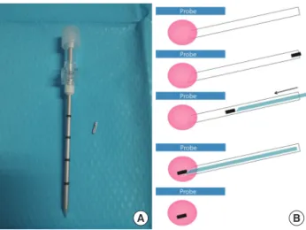
US-guided surgical clip insertion and location confirmation We prepared surgical clips with a disposable clip applier (LigaClip MCA MSM20, Ethicon Endo-Surgery, Somerville, USA; Premium Surgiclip M-9.75, Covidien, Mansfield, USA) for patients scheduled for NAC and who had agreed to surgi- cal clip insertion (Figure 1A). A short skin incision was made using local anesthesia under aseptic conditions. A14/16-gauge coaxial guiding needle (TSK Stericut; TSK Laboratory, Tochi- gi, Japan) was inserted into the center of the malignancy, and the inner stylet was removed under US guidance. One to two
Figure 1. Schematic diagram of the preoperative ultrasonography (US)- guided surgical clip insertion. (A) The coaxial guiding needle with an in- ner stylet and surgical clips. (B) Under US-guidance (blue), the coaxial guiding needle (white) is inserted into the the center of the breast can- cer (pink), and one or two clips (black) are passed through. The inner stylet (light blue) is reinserted for pushing the clip.
B A
surgical clips were passed through the inserted introducer, and the inner stylet was reinserted to complete the clip place- ment (Figure 1B). We confirmed the location of the clips by US immediately after clip insertion. If there were the multiple or large breast cancers, additional clips were placed for lesion extent bracketing as per the radiologist’s judgment. One of three experienced breast radiologists (2–20 years of experi- ence) performed these procedures in consensus. Postproce- dural mammography was performed to confirm objectively the appropriate location of the inserted clips.
Final histopathological results from both the initial biopsy and surgery were reviewed. Final pathological types of breast cancer, tumor (T) stages, and immunochemical markers, in- cluding the estrogen receptor, progesterone receptor, and hu- man epidermal growth factor receptor 2 (HER2) were also evaluated.
Radiologic evaluation and complications after clip insertion Follow-up mammography, US, and MRI were performed within the 10-day period before elective surgery to evaluate treatment response after NAC. After surgical excision, speci- men mammography was also done to evaluate clip retrieval and to assess specimen margin (Figure 2). A pathologist con- firmed the results.
We calculated the time interval from clip insertion to post- procedural mammography, follow-up preoperative mammo- graphy, US and MRI, and BCS. Each interval was expressed by mean±standard deviation (days) and median values. We also recorded the number of inserted clips. Two experienced breast radiologists (I.Y. and SH.C.) retrospectively reviewed the medi- cal records, and all images from the time of clip insertion to BCS were reviewed to confirm the location of clips, clip migra- tion, the presence of complications such as hemorrhage or in- fection, and the effect of clips on treatment assessment. The lo- cation of a clip was categorized as either “within the tumor” or
“outside the tumor.” Clip migration was defined as the clip be- ing located outside the proven malignancy at a distance of more than 1 cm. The signal void or artifact from inserted clips was reviewed for all signals of the breast MRI to evaluate nega- tive effects on the assessment. Finally, we reviewed the total cost of the surgical clips compared to commercial markers.
RESULTS
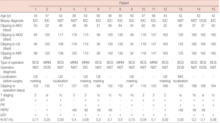
Table 1 shows the summarized results, including immuno- histopathological results and the documented time intervals.
Pathology revealed cases of invasive ductal carcinoma (IDC), ductal carcinoma in situ (DCIS), and invasive carcinoma of no special type. One patient had bilateral cancer consisting of
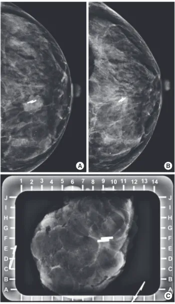
right breast IDC and left breast DCIS. After NAC, the final pathologic types were as follows: 11 residual invasive carcino- mas of no special type, two IDC, and three DCIS. Among these, five lesions in five patients were treated with modified radical mastectomy, while 11 lesions in 10 patients underwent BCS. Image-guided localization or skin marking was per- formed in six patients. There was no difficulty in the pathologi- Figure 2. Mammography of a 40-year-old woman who underwent ul- trasonography-guided surgical clip insertion due to left breast cancer. (A) Postprocedural follow-up mammography was performed after clipping, and showed metal clips in the center of the proven malignant mass. (B) At preoperative final follow-up mammography, the clips were located in the proven malignant mass which had decreased in size. There was no evidence of clip migration or other complications. (C) Specimen mam- mography was performed immediately after surgery, and there were metal clips visualized in the proven malignant lesion without evidence of clip migration. The pathologic result was invasive carcinoma of no spe- cial type, and a clear tumor margin was observed.
A B
C
cal evaluation of a specimen due to inserted surgical clips. The majority of patients presented with T1 tumors (n=7, 43.8%;
T1b with 2, T1c with 5), and 37.5% (n=6) had T2 tumors ex- cept for three DCIS lesions (Tis, 18.8%). The positive rate of estrogen receptor, progesterone receptor, and HER2 status was 81.3% (n=13), 62.5% (n=10), and 25% (n=4), respectively.
The time interval from clip insertion to postprocedural mammography and surgery was 47.1±14.7 days (median, 50.0 days) and 128.6±34.4 days (median, 132.0 days), respectively.
The mean period between clip insertion and preoperative fol- low-up imaging was 122.6±34.8 days (median, 122.5 days) for mammography, 121.3±36.8 days (median, 122.5 days) for US, and 121.6±36.6 days (median, 122.5 days) for MRI. These data are presented in Table 1. The mean number of inserted clips was 2.3±0.7 (median, 2.0; range, 1–4).
There was no mammographic evidence of clip migration during postprocedural follow-up, preoperative final follow- up, and in surgical specimens (Figure 2). Moreover, no com- plication related to the clip insertion was noted during the study period, and no patient complained of heat sensation or pain during the MRI examination. On US, the inserted clips appeared as linear hyperechoic structures with or without posterior shadowing (Figure 3) and on MRI as small signal
voids due to paramagnetic or susceptibility properties of the clips (Figure 4). However, there was no difficulty in evaluating the treatment response to NAC using US and MRI.
Table 1. Summarization of time intervals, operation, and immunohistopathological results for all patients Patient
1 2 3 4 5 6 7 8 9 10 11 12 13 14 15
Age (yr) 45 47 42 38 53 60 56 55 40 37 56 43 42 42 42
Biopsy diagnosis IDC IDC NST NST IDC IDC IDC IDC IDC IDC IDC IDC NST NST DCIS IDC
Clipping to MG1 (days)
50 22 46 49 49 21 56 64 34 62 50 22 65 57 57 50
Clipping to MG2
(days) 94 120 117 119 113 56 130 130 46 118 147 163 125 162 162 160
Clipping to US (days)
94 120 108 119 113 56 130 130 34 118 147 163 125 162 162 160
Clipping to MRI
(days) 98 120 108 120 113 56 130 130 34 118 147 163 125 162 162 160
Type of operation BCS MRM BCS MRM MRM BCS BCS MRM BCS BCS MRM BCS BCS BCS BCS BCS
Operation
diagnosis NST DCIS NST NST IDC NST NST NST NST NST NST NST DCIS NST DCIS NST
Localization
before surgery US
marking - US
localization - US marking US
marking - - US
marking - - US
marking MG
localization - - - Clipping to
operation (days)
103 132 117 127 125 69 132 132 47 119 153 169 133 168 168 164
T staging 2 is 1c 2 2 1c 1c 1c 1b 2 2 2 is 1b is 1c
ER + + + - + + - - + + + + + + + +
PR - - + - + + - - + - + + + + + +
HER2 - + - +(e) -(e) -(e) -(e) - - - - + +(e) -(e) -(e) -
p53 + + + + + + + + + + + + - + + +
Ki-67 (%) 0.11 0.25 0.03 0.4 0.08 0.2 0.1 0.5 0.15 0.04 0.1 0.05 0.05 0.2 0.1 0.08 IDC =invasive ductal carcinoma; NST =invasive carcinoma of no special type; DCIS =ductal carcinoma in situ; MG1 =postprocedural mammography;
MG2=preoperative mammography; US=ultrasonography; MRI=magnetic resonance imaging; BCS=breast-conserving surgery; MRM=modified radical mastec- tomy; MG=mammography; ER=estrogen receptor; PR=progesterone receptor; HER2=human epidermal growth factor receptor 2; (e)=equivocal.
Figure 3. Preoperative ultrasonography (US)-guided surgical clip inser- tion. On US images, the coaxial needle (arrows) is visible as an echo- genic white line and the clips show a linear hyperechoic structure (ar- rowhead) in the center of the proven malignancy.
DISCUSSION
NAC is considered a standard step in the treatment of locally advanced breast cancer in its early stages. NAC is used fre- quently as it leads to better surgical results by conserving the breast while lessening complications and improving cosmetic outcomes [1,5]. The assessment of tumor response to NAC as confirmed on US, mammography, and MRI is important even if pCR is accomplished, which is reported to be 32.9% [3]. In this study, there was no case of pCR after NAC, but most of the enrolled cases showed a partial response to NAC, and some only had the in situ component without invasive foci. More- over, BCS was performed in 10 patients, and among these, six underwent surgery under image-guidance, while the five re- maining patients underwent modified radical mastectomy.
The commercial breast marker is generally placed at a CNB- performed site because radiologists cannot anticipate the exact outcome of NAC. As NAC has become more common, breast marker placement has become routine, especially in cases that may need additional excision based on the pathological find- ings [8-12]. Traditionally, breast markers are left behind for be- nign biopsies; for malignant lesions, markers are excised along with the cancerous lesion. In this way, breast marker usage is considered standard practice in malignant lesions [7].
There have been many types of titanium-based commercial breast markers launched by different companies. Prices range from US $75.00 to US $200 per clip internationally. However, until recently, none of these clips had been cleared for use by the KFDA. Thus, it has been impossible in South Korea to fol-
low guidelines that suggest inserting tissue markers in a proven malignancy prior to NAC. As an alternative, traditionally used, less expensive surgical clips have been proven to be made of safe materials such as titanium, and have been approved by the KFDA. These clips can remain in the patient after surgery without serious complications, except for rare cases of allergic reactions. Moreover, recent studies have also shown that accu- rate tumor bed localization done by placing a surgical clip in the wall of a seroma cavity can assist the planning of radiation therapy following BCS [13,14]. Surgical clips can be easily and safely inserted, and the approximate cost in South Korea is about US $10 per clip. This study was premised upon the hy- pothesis that surgical clips could replace commercial breast markers because of their safety features and low cost.
In our study, all clips inserted as tissue markers were re- moved with the primary breast cancers during surgery. Surgi- cal clips were placed via a commercial coaxial guiding needle used in the CNB of the breast. This procedure was easy to per- form, and clip insertion required less than 5 minutes, as the procedure is similar to that of CNB under US guidance. Since clip insertion is done with US-guided CNB, it is performed with real-time imaging surveillance and regarded as a relatively safe method with few reported complications or adverse events. Therefore, the insertion process itself is not considered an onerous duty by breast radiologists. A similar method of us- ing surgical clips to replace commercial breast markers has been reported by Uematsu [15] and Lee et al. [16], but in that study, the surgical clip was inserted by using an automated gun. Our study is the first to insert surgical clips with a semi- automatic gun via a guiding needle. This method is superior to the automated gun method as there is no need for repeat nee- dle insertion into the mass. Thus, there is no tissue injury due to repeat insertion, and there is less bleeding and a lower prob- ability of tumor cell seeding. If we can perform on-site clipping immediately after CNB for either a benign or malignant lesion, both medical costs and procedure time can be lower than those of the two-step clipping procedure used in our study.
The migration of surgical clips and related complications can be a major limitation of surgical clip insertion. The low tissue resistance of fatty breasts may allow clips to easily migrate;
however, clips are generally inserted into the center of the mass. Thus, the chance of clip migration should be lower be- cause of the higher tissue resistance [17]. Despite the mean time period from clipping to surgery of approximately 4 months (range, 47–169 days), there were no cases of clip mi- gration in our study as confirmed on all imaging modalities, including specimen mammography after surgery. Moreover, there was no case of complications related to clip insertion. The other advantage of clip insertion was that the clips allowed easy Figure 4. Magnetic resonance imaging (MRI) of a 40-year-old woman
who underwent ultrasonography-guided surgical clip insertion due to left breast cancer. (A) There was a 19 mm-sized, fast, washout-enhancing malignancy at initial T1-weighted enhanced MRI with subtraction. (B) At preoperative follow-up MRI after neoadjuvant chemotherapy, a small sig- nal void due to the clips (arrowhead) was observed in the center of the proven malignancy, which was much decreased in size and enhance- ment. However there was no difficulty in evaluating treatment response.
B A
detection of breast cancer on mammography and it was help- ful for surgeons to explain breast cancer to their patients.
Several previous studies have reported that radiopaque markers are useful for tumor localization as well as for the as- sessment of tumor response after NAC, without disturbing ra- diologic multimodality evaluation including MRI [2,6,12,18].
We were able to evaluate tumor response to NAC and confirm the clip location by using multimodality imaging studies; the clips were visualized as a radiopaque metal density on radio- graphy, and as a hyperechoic linear structure with or without posterior shadowing on US. While breast MRI has proved su- perior to mammography and US in assessing tumor response for pCR [5,12], metal clips can cause artifacts on MRI, depend- ing on magnetic susceptibility, clip quality, size, shape, orienta- tion, position, and used MRI parameters [12]. In our study, the surgical clips created a small signal void on MRI; however, the primary malignancy was easily visualized on MRI. Moreover, the clinician can reassure patients by showing them the con- spicuous decrease in exact mass after NAC, along with the in- ternal dense radiopaque clip on mammography.
This study has several limitations. First, it was a retrospective study, and only patients who had agreed to surgical clip inser- tion and had undergone mammography, US, and MRI for scheduled BCS after NAC were selected. Therefore, a selection bias may exist. Second, the number of subjects was limited at only 16, a number too small to provide a reliable overall gener- alization from the study results. Further studies are needed for continued assessment of this procedure. Third, the placement of a surgical clip via a coaxial guide needle is not yet a globally approved method.
We concluded from this small study that surgical clips may replace commercial tissue markers for tumor localization in breast cancer patients undergoing NAC without migration.
Our results have shown that surgical clips are well tolerated and safe for the patient, easily visualized on imaging, do not interfere with treatment response, and are cost-effective.
CONFLICT OF INTEREST
The authors declare that they have no competing interests.
REFERENCES
1. Kaufmann M, von Minckwitz G, Bear HD, Buzdar A, McGale P, Bon- nefoi H, et al. Recommendations from an international expert panel on the use of neoadjuvant (primary) systemic treatment of operable breast cancer: new perspectives 2006. Ann Oncol 2007;18:1927-34.
2. Oh JL, Nguyen G, Whitman GJ, Hunt KK, Yu TK, Woodward WA, et al. Placement of radiopaque clips for tumor localization in patients un- dergoing neoadjuvant chemotherapy and breast conservation therapy.
Cancer 2007;110:2420-7.
3. Abdel-Razeq H, Marei L. Current neoadjuvant treatment options for HER2-positive breast cancer. Biologics 2011;5:87-94.
4. Kim Z, Min SY, Yoon CS, Lee HJ, Lee JS, Youn HJ, et al. The basic facts of Korean breast cancer in 2011: results of a nationwide survey and breast cancer registry database. J Breast Cancer 2014;17:99-106.
5. Lobbes MB, Prevos R, Smidt M, Tjan-Heijnen VC, van Goethem M, Schipper R, et al. The role of magnetic resonance imaging in assessing residual disease and pathologic complete response in breast cancer pa- tients receiving neoadjuvant chemotherapy: a systematic review. Insights Imaging 2013;4:163-75.
6. Dash N, Chafin SH, Johnson RR, Contractor FM. Usefulness of tissue marker clips in patients undergoing neoadjuvant chemotherapy for breast cancer. AJR Am J Roentgenol 1999;173:911-7.
7. Kaufmann M, von Minckwitz G, Mamounas EP, Cameron D, Carey LA, Cristofanilli M, et al. Recommendations from an international consensus conference on the current status and future of neoadjuvant systemic therapy in primary breast cancer. Ann Surg Oncol 2012;19:
1508-16.
8. Margolin FR, Jacobs RP, Denny SR, Schrumpf JD. Clip placement after sonographically guided percutaneous breast biopsy. Breast J 2003;9:
226-30.
9. Kopans DB. Clip placement during sonographically guided breast bi- opsy. AJR Am J Roentgenol 2001;176:1076-7.
10. Guenin MA. Clip placement during sonographically guided large-core breast biopsy for mammographic-sonographic correlation. AJR Am J Roentgenol 2000;175:1053-5.
11. Phillips SW, Gabriel H, Comstock CE, Venta LA. Sonographically guided metallic clip placement after core needle biopsy of the breast.
AJR Am J Roentgenol 2000;175:1353-5.
12. Genson CC, Blane CE, Helvie MA, Waits SA, Chenevert TL. Effects on breast MRI of artifacts caused by metallic tissue marker clips. AJR Am J Roentgenol 2007;188:372-6.
13. Tamai K, Mitsumori M, Fujishiro S, Kokubo M, Ooya N, Nagata Y, et al.
A case of allergic reaction to surgical metal clips inserted for postopera- tive boost irradiation in a patient undergoing breast-conserving thera- py. Breast Cancer 2001;8:90-2.
14. Coles CE, Wilson CB, Cumming J, Benson JR, Forouhi P, Wilkinson JS, et al. Titanium clip placement to allow accurate tumour bed localization following breast conserving surgery: audit on behalf of the IMPORT Trial Management Group. Eur J Surg Oncol 2009;35:578-82.
15. Uematsu T. Commercially available titanium clip placement following a sonographically guided core needle biopsy of the breast. Breast J 2007;
13:624-6.
16. Lee SY, Kook SH, Kwag HJ. The results and usefulness of marker clip placement after ultrasound-guided mammotome excision of breast le- sion. J Korean Radiol Soc 2005;52:207-13.
17. Margolin FR, Kaufman L, Denny SR, Jacobs RP, Schrumpf JD. Metallic marker placement after stereotactic core biopsy of breast calcifications:
comparison of two clips and deployment techniques. AJR Am J Roent- genol 2003;181:1685-90.
18. Baron LF, Baron PL, Ackerman SJ, Durden DD, Pope TL Jr. Sonograph- ically guided clip placement facilitates localization of breast cancer after neoadjuvant chemotherapy. AJR Am J Roentgenol 2000;174:539-40.