Ⅰ. Introduction
Nowadays, dental composite resins are used increasingly by practitioners for esthetic qualities.
As presently posterior composites are suited to be bonded Class Ⅰ and Class Ⅱ cavity preparations.
The mechanism of adhesive bonding is based on acid etching both enamel and dentin of the tooth
* Corresponding author: Jeong-Kil Park Department of Conservative Dentistry
College of Dentistry, Pusan National University, 1-10, Ami-dong, Seo-gu, 602-739, Busan, Korea Tel: 82-51-240-7454
E-mail: [email protected]
The effect of adhesive thickness on microtensile bond strength to the cavity wall
Hwa-Eon Lee, Hyeon-Cheol Kim, Bock Hur, Jeong-Kil Park*
Department of Conservative Dentistry, College of Dentistry, Pusan National University, Busan, Korea
The purposes of this study were to examine the variability of adhesive thickness on the different site of the cavity wall when used total-etch system without filler and simplified self-etch system with filler and to evaluate the relationship between variable adhesive thickness and microtensile bond strength to the cavity wall.
A classⅠcavity in six human molars was prepared to expose all dentinal walls. Three teeth were bonded with a filled adhesive, ClearfilTMSE bond and the other three teeth were bonded with unfilled adhesives, ScotchbondTM Multi Purpose. Morphology and thickness of adhesive layer were examined using fluorescence microscope. Bonding agent thickness was measured at three points along the axial cavity wall, edge of cavity margin ((rriimm)), halfway down each cavity wall ((hhllff)), internal angle of the cavity ((aanngg)). After reproducing the adhesive thickness at rriimm,, hhllff and aanngg,, micro-tensile bond strength were evaluated.
For both bonding agents, adhesive thickness of aanngg was significantly thicker than that of rriimm and h
hllff (P < 0.05). As reproduced the adhesive thickness, microtensile bond strength was increased as adhesive thickness was increased in two bonding agents.
Adhesive thickness of internal angle of the cavity was significantly thicker than that of the cavity margin and the halfway cavity wall for both bonding agents. Microtensile bond strength of the thick adhesive layer at the internal angle of the cavity was higher than that of the thin adhesive layer at the cavity margin and the halfway cavity in the two bonding systems. [J Kor Acad Cons Dent 32(1):9-18, 2007]
Key words: Adhesive thickness, Microtensile bond strength, Cavity wall, Filled adhesive, Unfilled adhesive
- Received 2006.9.13., revised 2006.10.27., accepted 2006.10.30. - ABSTRACT
cavity surface. Although adhesion to phosphoric acid-etched enamel is reliable and long-lasting, adhesion to dentin has been more challenging because of the complex mineral and organic com- ponents of dentin. The bonding mechanism of adhesive resins to dentin proposed by Naka- bayashi1) was described as micromechanical bond- ing due to the impregnation and polymerization of monomers into the exposed collagen of dem- ineralized dentin surface, creating a hybrid layer.
The major role of the adhesive resin is the stabi- lization of the formed hybrid layer and the forma- tion of resin tag in the unplugged dentinal tu- bules2).
In the adhesive dentistry, total-etch technique has led to major improvements. However, the achievement of reliable bond to dental hard tissue without a separate acid etching step represented a major challenge in the past. Recently, self-etch- ing adhesives were introduced. These systems use hydrophilic, acid monomers which are able to demineralize and penetrate enamel and dentin3). Unlike total-etch adhesives, these systems does not completely resolve or remove the smear layer.
Kaaden et al.3) studied that the filled bonding agent Clearfil SE bond resulted in bond strengths higher than those of the unfilled adhesive sys- tems. Perdigao and Lopes4) suggested that self- etching adhesives should provide optimum bond- ing besides a simplification of the bonding proce- dure and a potential decrease in technique sensi- tivity.
Most of the cavity preparations clinically show not only areas of exposed enamel and superficial dentin but also deep dentinal areas. Then vari- able location of exposed dentin such as cavity wall and cavity floor are appeared in the cut cavity.
Thus a layer of cured adhesive of variable thick- ness is present on the cavity surface inevitably5). Adhesive thickness in the cavity wall is variable along the cavity depth by the gravity. In clinical situation, pooling of adhesive was apparent in the internal angle of the cavity and then decreased in thickness toward cavity margin. Peter et al.6) sug- gested that pooling of dentin bonding agents at the internal angle of the cavity arises because of
the difference in viscosity between primers and unfilled adhesives. Perdigao et al.7,8) noticed that air thinning had a tendency to cause pooling of the adhesive into irregularities on the dentin sur- face and at the internal angle of the cavity.
During the application of the adhesive, the manu- facturer recommends that it should be applied in a uniform coating and then excessive air thinning of the adhesive should be avoided9).
Moreover, the variable adhesive thickness on the different area of the cavity would affect on the bond strength. Zheng et al.10) suggested that the effect of the thickness of the adhesive layer on bond strength is material-dependent. In their study, the increase in bond strengths of Clearfil Liner Bond 2V was directly proportional to the thickness of the bonding layer and the bond strengths of Single Bond decreased significantly with increase adhesive resin thickness. They emphasized that care should be taken to avoid excess adhesive resin at line angle in cavities bonded with single bottle system that contain water and ethanol10).
Recently, fillers have been added to single-bottle adhesive systems to reinforce the hybrid layer and increase bond strength. Increased filler load- ing increases viscosity of the bonding system and may reduce its flow11). In general, the variability of film thickness on the different site of the cavity is dependent on the type of bonding agents with or without filler because of the difference of their viscosity and flow. Grossman and Setzer5) showed that the bonding layer was thinnest at the cavity margin and thickest at the internal angle of the cavity for both type of the bonding agents. In addition, they described that layer thickness for filled adhesive, Optibond showed a progressive increase down the cavity wall to the internal angle while unfilled adhesive, Scotchbond Multi- purpose showed great film thickness variability5). Therefore, the variability of the adhesive thick- ness using of the different type of the bonding agents, filled adhesive and unfilled adhesive, should be evaluated on the prepared cavity.
The purposes of this study were to examine the variability of adhesive thickness on the different
site of the cavity wall when used total-etch system without filler and simplified self-etch system with filler and to evaluate the relatio- nship between variable adhesive thickness and microtensile bond strength to the cavity wall.
Ⅱ. Materials and Methods
This study was performed in two parts. One part was to measure adhesive thickness on the cavity wall and the other part was to measure microtensile bond strength.
Part Ⅰ. Adhesive thickness
P
Prreeppaarraattiioonn ooff ssppeecciimmeennss
Six intact, non-carious, non-restored, human molars were used in this study. A class Ⅰ cavity was prepared to expose all dentinal wall (mesiodistal width; 6 ㎜, buccolingual width;
4 ㎜, depth; 6 ㎜). After cavity preparation, three teeth were bonded with a filled adhesive (ClearfilTM SE bond; Kuraray, Medical Inc., Okayama, Japan) and the other three teeth were bonded with unfilled adhesives (ScotchbondTM Multi Purpose; 3M ESPE, St Paul, MN, USA) by Manufacture’s recommendation. Rhodamine B was added in these adhesives. And then a hybrid light-activated resin composite (Z100; 3M ESPE, St. Paul, MN, USA) was filled in six cavities. The
compositions of dentin bonding systems used in the study are described in Table 1.
The bonded teeth were then stored in distilled water at 37℃ for 24 hour prior to sectioning.
Each tooth was sectioned bucco-lingually into a series of 1.0 mm thick slabs using a high-speed precision cut-off machine (Accutom-50; Struers, Ballerup, Denmark) with water coolant. Twelve specimens were prepared in each group (Table 2).
F
Flluuoorreesscceennccee mmiiccrroossccooppyy
These specimens were observed by fluorescence microscope (Axioskop; ZEISS, Oberkochen, Germany). Morphology and thickness of adhesive layer were founded through phase of diffusion with rhodamine B mixed adhesive resin. Bonding agent thickness was measured at three points along the cavity wall, edge of cavity margin ((rriimm)), halfway down each cavity wall ((hhllff)), internal angle of the cavity ((aanngg)).
S
Sttaattiissttiiccaall aannaallyyssiiss
Statistical analysis of the collected data was performed by SPSSTM version 12.0 (SPSS Inc., Chicago, IL, USA). After calculating the means and standard deviations of the adhesive thickness for the specific points in each group, Student’s t- test was used to compare adhesive thickness between two bonding systems. And one-way ANOVA and Scheffe’s test for post-hoc compari-
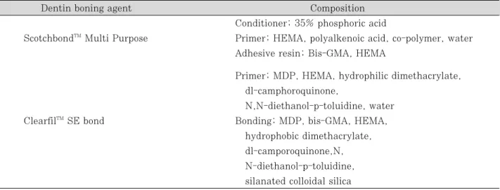
Table 1.Dentin bonding systems used in this study
Dentin boning agent Composition
Conditioner; 35% phosphoric acid
ScotchbondTMMulti Purpose Primer; HEMA, polyalkenoic acid, co-polymer, water Adhesive resin; Bis-GMA, HEMA
Primer; MDP, HEMA, hydrophilic dimethacrylate, dl-camphoroquinone,
N,N-diethanol-p-toluidine, water
ClearfilTMSE bond Bonding; MDP, bis-GMA, HEMA,
hydrophobic dimethacrylate, dl-camporoquinone,N, N-diethanol-p-toluidine, silanated colloidal silica
son was performed to evaluate the difference of adhesive thickness among three points in each group. Statistical significance was defined as P <
0.05.
Part Ⅱ. Microtensile bond strength
P
Prreeppaarraattiioonn ooff ssppeecciimmeennss
Adhesive thickness at each point was repro- duced by multiple coating of adhesive. Adhesive
thickness at the rriimm and the hhllff were same as one coat adhesive layer thickness in both ScotchbondTM Multi Purpose (SM group) and ClearfilTM SE bond (SE group), while adhesive thickness at the aanngg was same as seven coats in SM group and six coats in SE group.
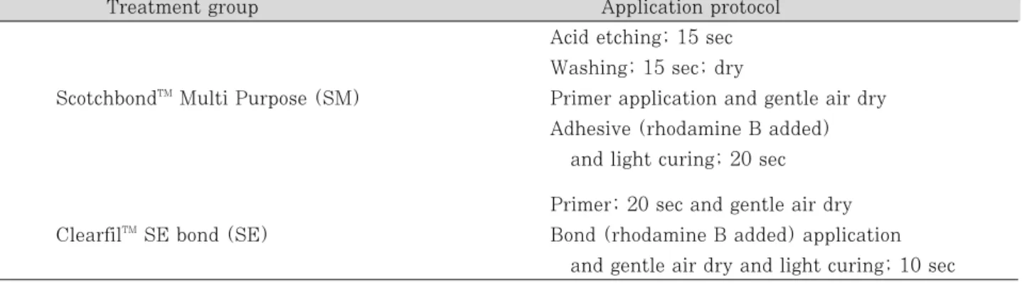
Eight intact, non-carious, non-restored, human molars were used. A class Ⅰ cavity was prepared to expose all dentinal wall. The teeth were sec- tioned longitudinally in mesiodistal direction Table 2.Application protocol for adhesive thickness measurement
Treatment group Application protocol
Acid etching; 15 sec Washing; 15 sec; dry
ScotchbondTMMulti Purpose (SM) Primer application and gentle air dry Adhesive (rhodamine B added)
and light curing; 20 sec Primer; 20 sec and gentle air dry ClearfilTMSE bond (SE) Bond (rhodamine B added) application
and gentle air dry and light curing; 10 sec
Table 3.Application protocol for microtensile bond strength test
Treatment group Application protocol
Acid etching; 15 sec ScotchbondTMMulti Purpose Washing; 15 sec; dry
1 coat (SM 1) Primer application and gentle air dry
Adhesive and light curing; 20 sec
ScotchbondTMMulti Purpose Apply one coat
7 coat (SM 7) Apply consecutive coats without waiting
between application and light curing; 20 sec Application and curing 5 times additionally ClearfilTMSE bond Primer; 20 sec and gentle air dry
1 coat (SE 1) Bond application and gentle air dry
and light curing; 10 sec Apply one coat
ClearfilTMSE bond Apply consecutive coats without waiting between
6 coat (SE 6) application and gentle air dry
and light curing; 10 sec
Application and gentle air dry and curing 4 times additionally
through the prepared cavity using a diamond disc attached low-speed handpiece with air-water cooling. Sectioned sixteen teeth were classified into four groups.
The four groups were etched and bonded in manner described in Table 3. After dentin bond- ing procedure, a hybrid light-activated resin com- posite (Z100; 3M ESPE, St. Paul, MN, USA) was built up free hand in three increments to an approximate height of 6 ㎜. Each increment was 2 ㎜ and light-cured for 20 s.
The teeth were stored in water at 37℃ for 24 hour. The 1 ㎜ × 1 ㎜ sticks were then sectioned mesiodistally and occlusogingivally using a high- speed precision cut-off machine under water coolant. Twenty specimens were prepared in each group.
M
Miiccrrootteennssiillee bboonndd ssttrreennggtthh
The stick was fixed to the test bed using cyano- acrylate adhesive, Zapit (DVA Inc., Corona, CA, USA). The stick was pulled to failure under ten- sion using a Micro Tensile Tester (Bisco inc., Shaumburg, IL, USA) at a crosshead speed of 1 ㎜/min. And then the microtensile bond strength was recorded in mpa.
S
Sttaattiissttiiccaall aannaallyyssiiss
Statistical analysis of the collected data was performed by SPSSTM version 12.0. After calculat-
ing the means and standard deviations of the microtensile bond strength for each group, Student’s t-test was used to compare of micro- tensile bond strength for each group. Statistical significance was defined as P < 0.05.
Ⅲ. Results Part Ⅰ. Adhesive thickness
M
Moorrpphhoollooggyy ooff aaddhheessiivvee llaayyeerr
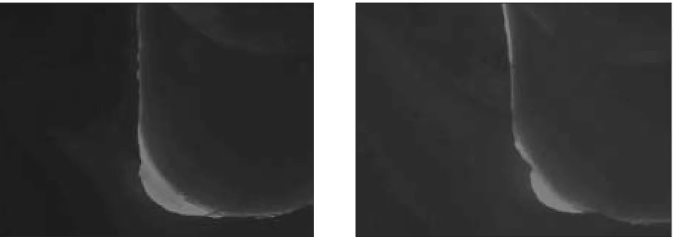
In fluorescence microscopy observation, adhesive layer was pooled only at the internal angle of the cavity for both bonding agents (Figure 1). And for both bonding agents the bonding layer was thinnest at the cavity margin and thickest at the internal angle of the cavity.
ClearfilTM SE bond formed uniform layer over the cavity wall compared to ScotchbondTM Multi Purpose.
A
Addhheessiivvee llaayyeerr tthhiicckknneessss
Table 4 shows the results of adhesive thickness at the specific area in two dentin bonding sys- tems.
In ScotchbondTM Multi Purpose, adhesive thick- ness of aanngg was significantly thicker than that of rim and hhllff (P < 0.05). Adhesive thickness of aanngg was also significantly thicker than that of rim and h
hllff when ClearfilTMSE bond was used (P < 0.05).
Figure 1. Fluorescence microscopic image of ScotchbondTMMulti Purpose (left) and ClearfilTMSE bond (right).
In rr ii mm and hhllff, adhesive layer thickness of Clearfil SE bond was significantly thicker than that of ScotchbondTM Multi Purpose (P < 0.05). In a
a nn gg, however, adhesive layer thickness of ScotchbondTM Multi Purpose was significantly thicker than that of ClearfilTMSE bond (P < 0.05).
Part Ⅱ. Microtensile bond strength
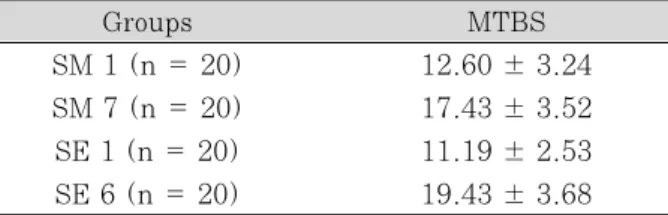
Table 5 and 6 show the results of microtensile bond strength for each group. Microtensile bond strength was highest in the SE 6 group (19.43 ± 3.68).
In ScotchbondTM Multi Purpose, seven coats group was significantly higher microtensile bond strength than one coat group (P < 0.05). In Clea- rfilTM SE bond, six coats group was also signifi- cantly higher microtensile bond strength than one coat group (P < 0.05). For both bonding agents, microtensile bond strength was increased as adhesive thickness was increased.
In one coat groups, microtensile bond strength for both adhesive was not significantly different (P > 0.05). Likewise, in multiple coats groups, microtensile bond strength for both adhesive was not significantly different (P > 0.05).
Ⅳ. Discussion
An ideal dental adhesive would provide high bond strengths. Bond strength is more predictive of a material’s retentive potential12). We observed the bonding agent within the cavity and assessed the film thickness of the adhesive layer along the cavity wall and measured microtensile bond strength at different sites of the cavity wall to better understand the bonding agents as an inter- facial material in clinical situation.
Part Ⅰ. Adhesive thickness
In fluorescence microscopy evaluation, ClearfilTM SE bond formed uniform layer over the cavity wall and the internal angle of the cavity compared to ScotchbondTM Multi Purpose. The highly signifi- cant difference between the coatings of the bond- ing agents on the cavity surface appears to be related to the type of bonding agent with or with- out filler. ClearfilTM SE bond with filler contents of about 10% has increased viscosity and reduced flow3). Film thickness should be even along the entire composite resin-tooth interface to ensure consistent bonding and uniform stress distribution5). The location of failure sites is often Table 4.Adhesive thickness (Mean ± SD, μm)
Location Adhesive thickness Student t-test
SM SE p-value
rriimm (n = 20) 17.91 ± 3.09a 35.77 ± 5.45a < 0.000
h
hllff (n = 20) 18.11 ± 3.20a 36.12 ± 4.98a < 0.000
a
anngg (n = 20) 109.24 ± 39.2b 83.67 ± 6.78b 0.009
ab: Different letter indicates significant differences between the groups in vertical low (P < 0.05).
Table 5.Microtensile bond strength (Mean ± SD, MPa)
Groups MTBS
SM 1 (n = 20) 12.60 ± 3.24 SM 7 (n = 20) 17.43 ± 3.52 SE 1 (n = 20) 11.19 ± 2.53 SE 6 (n = 20) 19.43 ± 3.68
Table 6.Statistical difference by Student t-test Dentin bonding agent p-value
Adhesive SM1 SE1 0.132
thickness SM7 SE6 0.086
p-value < 0.000 < 0.000
interpreted as reflecting the weakest link in the restoration system5). It could be that the high proportion of mixed failures in tensile and shear bond test may be related to variations in bonding agent thickness4,13,14). Therefore, an aspect of uni- form stress distribution, ClearfilTM SE bond would be superior to ScotchbondTMMulti Purpose.
Because of an effect of the gravity, pooling of adhesive was apparent at the internal angle of the cavity and then decreased in thickness toward cavity margin. For both bonding agents, the bonding layer was thinnest at the cavity margin and thickest at the internal of the cavity. This result was agreement with Grossman’s study5). And adhesive layer was pooled only at the inter- nal angle of the cavity for both bonding agents.
Peter et al.6) reported on film thickness reaching 254 ㎛ in the line angle of cavity preparations.
They suggested that the difference in viscosity between primers and adhesives cause the pooling of dentin bonding agents at the internal angle of the cavity in experiments used unfilled adhesives.
And they proposed that air thinning is unable to drive the higher viscosity adhesive through the primed collagen network because of the damming effect of the preparation angle.
Filler should increase viscosity of the adhesives.
It should be that the higher viscosity adhesive, ClearfilTM SE bond is less driven through the primed collagen network than lower viscosity adhesive, ScotchbondTMMulti Purpose. But in this study adhesive layer thickness of unfilled adhe- sive, ScotchbondTM Multi Purpose was significant- ly thicker than that of filled adhesive, ClearfilTM SE bond at the line angle. It seems that the rea- son of this result would be an effect of the gravity and the reduced flow of filled adhesive. Therefore, the higher viscosity, ClearfilTM SE bond would not flow to the bottom well though damming effect of internal angle of the cavity.
At the cavity margin and the halfway cavity wall, on the other hand, adhesive layer thickness of filled adhesive, ClearfilTM SE bond was signifi- cantly thicker than that of unfilled adhesive, ScotchbondTM Multi Purpose. In the case of the filled adhesive, the increased viscosity and the
role of the oxygen inhibition layer is a major cause of thick bonding layers6). Opdam et al.16) reported that thick adhesive layer seems to pre- vent the formation of interfacial gaps between tooth and restoration and act as a superior elastic buffer compared to thinner layers, whereas Hilton and Schwartz17)suggested that thick adhesive lay- er adversely affects bond strengths, increases crack propagation, elevates the thermal co-effi- cient of expansion mismatch with the tooth and decreases the load bearing and wear component of the restoration.
Ultramophological findings were able to demon- strate hydroxyl apatite crystals within the hybrid layer after the use of ClearfilTM SE bond. It was speculated that the bonding mechanism of ClearfilTM SE bond might depend upon interlock- ing with these crystals, possibly resulting in more rigid and compact interface18). Thin hybrid layer and thick adhesive layer may also be advanta- geous3).
Part Ⅱ. Microtensile bond strength
Hybrid layer formation plays an important role in achieving maximum bond strengths between resin and dentin1,19,20). Increased thickness of the adhesive resin film would result in higher bond strengths by improving stress distributions in the bonded assembly10). In this study, microtensile bond strength was increased as adhesive thick- ness was increased in two bonding agents. The increase in the tensile bond strength of both bonding agents with an increase in the thickness of the adhesive layer may be due to the improved stress distributions. The thicker adhesive layer may permit self-alignment of the specimen that corrects for minor deviations in specimens place- ment, thereby, improve stress distributions dur- ing testing, yield higher apparent bond stre- ngths10). In this study, consequently, microtensile bond strength of the thick adhesive layer repro- duced the adhesive thickness of internal angle of the cavity was higher than that of the thin adhe- sive layer reproduced the adhesive thickness at the cavity margin and the halfway cavity in the
two bonding systems. Therefore, on the assump- tion that other conditions are same, microtensile bond strength at the internal angle of the cavity is higher than that at the other sites of cavity wall.
Recently, fillers have been added to adhesive systems to reinforce the hybrid layer and increase bond strength. Some21,22) have advocated the addi- tion of filler to dentin adhesives because the filler might improve the mechanical properties of the material and act as elastic buffer beneath the restorative material. In this study, however, microtensile bond strength between two bonding systems were not significantly different on the specimens reproduced the each sites to the cavity wall. From the results of this study, more rese- arch is needed to know the relationship of adhe- sive thickness and microtensile bond strength in relation to the addition of filler and adhesion strategy.
In many other studies3,13,23), generally bond strengths are higher in superficial than deep dentin. Marshall et al.23) suggested that the nature of the substrate presented for bonding would vary with location.
For this reason, we support that high microten- sile bond strength with thick adhesive layer at the internal angle of the cavity would compensate for the low bond strength due to dentinal struc- ture in the deep dentin when used the two bond- ing systems. Therefore, the phenomenon of the pooling at the internal angle of the cavity would not be problem.
In clinical situation, additionally, simplified two step dentin bonding system, ClearfilTM SE bond has microtensile bond strength comparable with three steps, ScotchbondTM Multi Purpose. Acco- rdingly, it would not be problem that ClearfilTM SE bond is applied to the cavity wall under influence of the gravity. Further studies on the bond strength of adhesive within the prepared cavity are needed considering other variables.
Ⅴ. Conclusions
1. Adhesive layer was pooled only at the internal angle of the cavity for both bonding agents.
ClearfilTM SE bond formed uniform layer over the cavity wall compared to ScotchbondTM Multi Purpose.
2. For both bonding agents, adhesive thickness of internal angle of the cavity was significantly thicker than that of the cavity margin and the halfway cavity wall (P < 0.05).
3. Microtensile bond strength of the thick adhe- sive layer at the internal angle of the cavity was higher than that of the thin adhesive layer at the cavity margin and the halfway cavity in the two bonding systems (P < 0.05).
References
1. Nakabayashi N, Nakamura M, Yasuda N. Hybrid layer as a dentin bonding mechanism. J Esthetic Dent 3(4):
133-138, 1991.
2. Frankenberger R, Kramer N, Petschelt A. Technique sensitivity of dentin bonding: effect of application mis- takes on bond strength and marginal adaptation. Oper Dent 25(4):324-330, 2000.
3. Kaaden C, Powers JM, Friedl KH, Schmalz G. Bond strength of self-adhesives to dental hard tissues. Clin Oral Investig 6(3):155-160, 2002.
4. Perdigao J, Lopes M. Dentin bonding-questions for the new millennium. J Adhes Dent 1(3):191-209, 1999.
5. Grossman ES, Setzer S. Bonding Agents: Adhesive layer thickness and retention to cavity surfaces with time. SADJ 56(6):266-272, 2001.
6. Peter A, Paul SJ, Luthy H, Scharer P. Film thickness of various dentine bonding agents. J Oral Rehabil 24(8):568-573, 1997.
7. Meerbeek BV, Perdigao J, Lambrechts P, Vanherle G.
The clinical performance of adhesives.J Dent 26(1):1- 20, 1998.
8. Perdigao J, Lambrechts P, Meerbeek BV, Braem M, Yildiz E, Yucel T, Vanherle G. The interaction of adhe- sive systems with human dentin. Am J Dent 9(4):167- 173, 1996.
9. Retief DH, Wendt SL, Bradley EL. The effect of adhe- sive thickness on the shear bond strength of Scotchbond 2/Silux to dentin. Am J Dent 2(6):341- 344, 1998.
10. Zheng L, Pereira PNR, Nakajima M, Sano H, Tagami J. Relationship between adhesive thickness and microtensile bond strength. Oper Dent 26(1):97-104, 2001.
11. Gallo JR, Comeaux R, Haines B, Xu X, Burgess JO.
Shear bond strength of four filled dentin bonding sys- tems. Oper Dent 26(1):44-47, 2001.
12. Fortin D, Swift EJ, Denehy GE, Reinhardt JW. Bond strength and microleakage of current dentin adhesives.
Dent Mater 10(4):253-258, 1994.
13. Yoshiyama M, Carvalho R, Sano H, Horner J, Brewer PD, Pashley DH. Interfacial morphology and strength of bonds made to superficial versus deep dentine. Am J Dent 8(6):297-302, 1995.
14. Eick JD, Robinson SJ, Chappell RP, Cobb CM, Spencer P. The dentinal surface: Its influence on dentinal adhesion. PartⅢ. Quintessence Int 24(8):571-582, 1993.
15. Staninec M, Marshall GW, Kawakami M, Low A. Bond strength, interfacial characterization and fracture sur- face anylysis for a new stress-breaking bonding agent.
J Prosthet Dent 74(5):469-475, 1995.
16. Opdam NJM, Roeters FJM, Verdonschot EH.
Adaptation and radiographic evaluation of four adhe- sive systems. J Dent 25(5):391-397, 1997.
17. Hilton TJ, Schwartz RS. The effect of air thinning on dentin adhesive bond strength. Oper Dent 20(4):133- 137, 2001.
18. Perdigao J, Lopes M, Gomes G. Ultramorphology of the hybrid layer - a TEM study of non-decalcified inter- faces. J Dent Res 79:336, 2000.
19. Erickson RL. Surface interactions of dentin adhesive materials. Oper Dent Suppl 5:81-94, 1992.
20. Meerbeek BV, Inokoshi S, Braem M, Lambrechts P, Vanherle G. Morphological aspects of the resin-dentin interdiffusion zone with different dentin adhesive sys- tems. J Dent Res 71(8):1530-1540, 1992.
21. Staninec M, Kawakami M. Adhesion and microleakage tests of a new dentin bonding system. Dent Mater 9(3):204-208, 1993.
22. Fanning DE, Wakefield CW, Robbins JW, Bagley AL.
Effect of a filled adhesive on bond strength of three dentinal bonding agents. Gen Dent 43(3):256-262, 1995.
23. Marshall GW, Marshall SJ, Kinney JH, Balooch M.
The dentin substrate: structure and properites related to bonding. J Dent 25(6):441-458, 1997.
와동벽에서 접착제의 두께가 미세인장 결합강도에 미치는 영향
이화언∙김현철∙허 복∙박정길*
부산대학교 치과대학 보존학교실
이 연구의 목적은 와동벽에서 다른 위치에서의 상아질 접착제의 두께를 평가하고, 이런 다양한 접착제의 두께와 미세 인장 강도 사이의 관계를 평가하기 위한 것이다.
여섯 개의 인간 대구치에 모든 상아질 면이 노출되도록Ⅰ급 와동을 형성하였다. 3개의 치아는 filled adhesive (ClearfilTM SE bond)를 와동 내에 도포하였고, 다른 3개의 치아는 unfilled adhesives (ScotchbondTM Multi Purpose)를 도포하였다. 형광 현미경을 이용하여 접착층의 형태와 두께를 관찰하였다. 접착제의 두께는 수직 와동 벽을 따라 와동 변연, 와동벽 1/2, 와동 내각의 세 지점에서 측정되었다. ScotchbondTM Multi Purpose와 ClearfilTM SE bond가 와동 변연과 와동벽 1/2, 와동 내각에서의 접착제의 두께를 재현하여 미세 인장 결합 강도 를 측정하였다.
이 실험의 결과에서 두 가지 상아질 접착제 모두에서 와동 내각에서의 접착제의 두께가 와동 변연과 와동벽 1/2 위치에서의 두께보다 두꺼웠으며, 와동 내각의 두꺼운 접착제의 미세 인장 결합 강도는 와동 변연과 와동벽 1/2에 서의 얇은 접착제 두께의 미세 인장 결합 강도보다 유의성 있게 높게 나타났다.
주요어: 접착제의 두께, 미세 인장 결합 강도, 와동벽, 필러가 포함된 접착제, 필러가 포함되지 않은 접착제 국문초록