Fluoroscopy-guided Combined (Surgical/Endovascular) Treatment of Dural Arteriovenous Fistula
So Hee Park, Jong-Hoon Kim, Chul-Hoon Chang, Young-Jin Jung
Department of Neurosurgery, College of Medicine, Yeungnam University, Daegu, Korea
For dural arteriovenous fistula (DAVF), when the usual endovascular or neurosurgical approaches are difficult to treat, multi-modal treatment can be helpful. We present a case of a 71-year-old woman with DAVF, who pre- sented with an intracerebral haemorrhage. Digital subtraction angiography revealed a DAVF of the transverse sinus, with cortical venous reflux.
Transvenous and transarterial approaches for coil embolization failed. In the operating room, a small craniotomy was performed, and coil emboliza- tion was done under fluoroscopy. Transcranial venous embolization might be a useful method to occlude DAVF in a case that is difficult to access by usual surgical or endovascular approaches.
J Cerebrovasc Endovasc Neurosurg.
2017 June;19(2):106-110 Received : 21 October 2016 Revised : 25 November 2016 Accepted : 22 March 2017 Correspondence to Young-Jin Jung Department of Neurosurgery, College of Medi- cine, Yeungnam University, 170 Hyeonchung-ro, Nam-gu, Daegu 42415, Korea
Tel : 82-53-620-3790 Fax : 82-53-620-3770 E-mail : [email protected]
ORCID : http://orcid.org/0000-0002-9659-2607
This is an Open Access article distributed under the terms of the Creative Commons Attribution Non- Commercial License (http://creativecommons.org/li- censes/by-nc/3.0) which permits unrestricted non- commercial use, distribution, and reproduction in any medium, provided the original work is properly cited.
Keywords Arteriovenous fistula, Transverse sinuses, Endovascular procedures, Neurosurgical procedures
INTRODUCTION
Dural arteriovenous fistula (DAVF) is an abnormal shunt inside the dura, accounting for 10-15% of intra- cranial arteriovenous malformations.9) DAVF is a rare aetiology of spontaneous intracerebral haemorrhage (ICH),8) but the annual bleeding rate of DAVF is esti- mated at approximately 1.8-20%.7) If the lesion has cortical venous reflux (CVR) or was located at the tentorium, the bleeding risk is higher.1)
DAVF can be treated with endovascular emboliza- tion, surgery or stereotactic radiosurgery alone but sometimes a combined method is necessary. We pres- ent our experience with DAVF with ICH and CVR treated with venous embolization, using a transcranial approach, a combination of surgical and endovascular methods under fluoroscopic guidance.
CASE REPORT
A 71-year-old woman was admitted to our hospital with global aphasia. She had no notable past medical history. Laboratory test showed no evidence of coagulopathy. An initial brain computed tomography (CT) showed an ICH about 15 mL on the left parie- to-occipital area (Fig. 1). Digital subtraction angiog- raphy (DSA) showed a DAVF of the transverse sinus (TS) fed by multiple arteries arising from the left mid- dle meningeal artery and occipital artery. The fistula drained through the right TS, sigmoid sinus (SS), and cortical veins. CVR was seen (Borden classification type III, Cognard classification type IV) and the left TS and SS were not visible (Fig. 2). As the patient was at high risk of a re-bleed because of the CVR and acute haemorrhage, early treatment for DAVF was necessary.
Fig. 1. Axial non-contrast brain computed tomography shows an intracerebral haemorrhage on left parieto-occipital area.
A B
Fig. 2. Digital subtraction angiography, left external carotid artery injection, anteroposterior (A) and lateral (B) views. Angiogram re- vealing an isolated left transverse sinus and dural arteriovenous fistula fed by left middle meningeal artery and occipital artery with ectatic venous drainage.
Under local anaesthesia, a transvenous approach through the superior sagittal sinus and left TS was at- tempted but failed due to tortuous veins. Consequently,
a transarterial embolization via the left middle me- ningeal artery and left occipital artery endeavoured.
However, access through the occipital artery failed.
Through the middle meningeal artery, a microcatheter reached the DAVF but the coil formed a frame out- side the DAVF, so it unsuccessful.
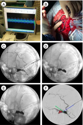
Finally, after moving to the operating room and un- der general anaesthesia, a retromastoid approach, us- ing a small craniotomy (about 2 × 3 cm) was per- formed, around the TS SS junction. We identified the location of the sinus by transcranial Doppler (TCD), and arterial pulsation was observed in the isolated left TS at TCD (Fig. 3A). The sinus was punctured with a 16-gauge angiocath needle. Through the angio- cath needle, a microcatheter was advanced to the si- nus (Fig. 3B).
Once the tip of the microcatheter was located within the sinus, fibered coils were inserted, under fluoro- scopy, in the operating room (Fig. 3C). The coil mass leaned to one side and, therefore, we punctured the sinus at the opposite side with another angiocath nee- dle (Fig. 3D). We deposited additional fibered coils.
A B
C D
E F
Fig. 3. Illustration of coil embolization under C-arm image guid- ing after craniotomy. (A) Arterial pulsation was observed in vein of transverse sinus at transcranial Doppler. (B) An angio- cath needle was placed in the isolated sinus and preparation for coil embolization was performed through the needle. (C) The isolated sinus was punctured with an angiocath needle and coils were inserted. (D) The sinus was punctured at the opposite side with another angiocath needle and coils were inserted. (E) Coil masses were met from both sides. (F) Sketch demonstrated the isolated sinus packed with coils, two punc- tured angiocath needles and feeding arteries (left middle me- ningeal artery and left occipital artery).
Fifty-seven fibered coils were used (Fig. 3E). The left common carotid angiography through the femoral ar- tery showed successful obliteration of the DAVF. To avoid DAVF recurrence, the bone defect was covered with artificial bone made from bone cement, and dura mater tag-up suture was performed.
A week later, CT scans showed haemorrhage reso- lution without increasing size, and the DSA showed no evidence of partial recanalization (Fig. 4). Global aphasia was slightly improved, and the patient under-
went rehabilitation therapy.
DISCUSSION
DAVFs are arteriovenous shunts from a dural arte- rial supply to a dural venous drainage channel.4) Many studies show an association between DAVF and venous thrombosis. Whatever the inciting events, the final common path was arteriovenous shunting and secondary venous hypertension. Nonetheless, this strategy can lead to leptomeningeal retrograde drain- age and predispose these channels to become varicose and potentially rupture.6) When DAVF is accom- panied by CVR, the risk of aggressive symptoms, such as haemorrhage and mortality, is increased; an annual ICH risk of 8.1-19.2% and an annual mortality of 10.4-19.2%.10)
About 20% of patients with a DAVF present with ICH.7) The re-haemorrhage rate is estimated to be up to 35% in the first 2 weeks following an initial hae- morrhage3) and, in other studies, as high as 43% in the first few days.2) When DAVF re-bled, 20% of the patients suffered an unfavourable course including death.3) Thus, DAVF with acute haemorrhage requires prompt treatment.
Treatment of DAVF with CVR is aimed at occlusion of the venous drainage or all arterial supply. In many cases, DAVF is treatable with a single modality.
However, a single approach might sometimes be diffi- cult and, in such instances, treatment typically involves a combination of modalities. Combined approaches, such as venous embolization under transcranial ap- proach, are commonly done4)5) for DAVF that are dif- ficult to treat by the usual transvenous or transarterial approach alone. The advantage of venous emboliza- tion with the transcranial approach is minimising risk and improving outcome. Due to the short distance to the fistula, fistula access is simple, and craniotomy al- lows multiple needle punctures. Furthermore, compared to surgical resection, blood loss is lower. Despite these advantages, there can be a problem with the equipment and patient transfer if the operating room
A B
Fig. 4. DSA, left external carotid artery injection, anteroposterior (A) and lateral (B) views. A week after treatment, follow-up DSA re- vealing complete obliteration of the DAVF on left parieto-occipital area. DSA = digital subtraction angiography; DAVF = dural arterio- venous fistula.
and angio-room are separate.
Our case was difficult to treat by a single modality.
First, we tried an endovascular approach in the an- gio-room. When that failed, we moved to the operat- ing room, did a craniotomy, and punctured the sinus for coil embolization. Embolization performed in the operating room with fluoroscopy may lead to partial occlusion because the resolution of fluoroscopy is worse than the angiogram. In contrast, as the operat- ing room is located on the third floor but the an- gio-room on the first basement level, it is challenging to transfer the patient to the angio-room in general anaesthesia, while leaving multiple devices in the body and brain. As fluoroscopy is highly available, and accessible without moving the patient, we se- lected coil embolization in the operating room using fluoroscopy. In this way, the lesion was completely obliterated.
A growing trend is the hybrid operating room, obvi- ating the need to move the patient, reducing both the risk associated with movement and the time under general anaesthesia. There is an increasing preference
for non-invasive treatment. In determining the treat- ment policy, the transcranial venous approach, in a case like this, might be able to reduce bleeding risk and perioperative risk. In order to reduce the patient's perioperative risk, a hybrid operating room might be helpful in difficult access cases, with the development of instruments and endovascular treatment methods.
CONCLUSION
DAVFs accompanied by ICH and CVR demand ur- gent treatment, due to re-bleeding risk. When DAVF is challenging to treat by a single modality, combined modalities can be beneficial. A venous embolization with transcranial approach has many advantages, in- cluding simple fistula access, allows multiple needle punctures and has low blood loss. Fluoroscopy-guid- ed coil embolization has inherent risks of incomplete obliteration. Hence, to reduce perioperative risk re- lated with combined modalities, along with endovas- cular treatment methods and instrumental develop- ments, a hybrid operating room might be effective.
ACKNOWLEDGMENTS
This work was supported by the 2014 Yeungnam University Research Grant.
Disclosure
The authors report no conflict of interest concerning the materials or methods used in this study or the findings specified in this paper.
REFERENCES
1. Awad IA, Little JR, Akarawi WP, Ahl J. Intracranial du- ral arteriovenous malformations: factors predisposing to an aggressive neurological course. J Neurosurg. 1990 Jun;72(6):839-50.
2. Byun JS, Hwang SN, Park SW, Nam TK. Dural arterio- venous fistula of jugular foramen with subarachnoid hemorrhage : selective transarterial embolization. J Korean Neurosurg Soc. 2009 Mar;45(3):199-202.
3. Duffau H, Lopes M, Janosevic V, Sichez JP, Faillot T, Capelle L, et al. Early rebleeding from intracranial dural arteriovenous fistulas: report of 20 cases and review of the literature. J Neurosurg. 1999 Jan;90(1):78-84.
4. Halbach VV, Higashida RT, Hieshima GB, Mehringer CM, Hardin CW. Transvenous embolization of dural fis- tulas involving the transverse and sigmoid sinuses.
AJNR Am J Neuroradiol. 1989 Mar-Apr;10(2):385-92.
5. Houdart E, Saint-Maurice JP, Chapot R, Ditchfield A, Blanquet A, Lot G, et al. Transcranial approach for ve- nous embolization of dural arteriovenous fistulas. J Neurosurg. 2002 Aug;97(2):280-6.
6. Houser OW, Campbell JK, Campbell RJ, Sundt TM Jr.
Arteriovenous malformation affecting the transverse du- ral venous sinus--an acquired lesion. Mayo Clin Proc.
1979 Oct;54(10):651-61.
7. Iwama T, Hashimoto N, Takagi Y, Tanaka M, Yamamoto S, Nishi S, et al. Hemodynamic and metabolic disturbances in patients with intracranial dural arteriovenous fistulas:
positron emission tomography evaluation before and af- ter treatment. J Neurosurg. 1997 May;86(5):806-11.
8. Jolink WM, van Dijk JM, van Asch CJ, de Kort GA, Algra A, Groen RJ, et al. Outcome after intracranial haemor- rhage from dural arteriovenous fistulae; a systematic re- view and case-series. J Neurol. 2015 Dec;262(12):2678-83.
9. Newton TH, Cronqvist S. Involvement of dural arteries in intracranial arteriovenous malformations. Radiology.
1969 Nov;93(5):1071-8.
10. van Dijk JM, terBrugge KG, Willinsky RA, Wallace MC.
Clinical course of cranial dural arteriovenous fistulas with long-term persistent cortical venous reflux. Stroke.
2002 May;33(5):1233-6.