Copyright © 2019 Korean Neurological Association 583
JCN
Open AccessContinuous Positive Airway Pressure Therapy in a Patient with Pantothenate-Kinase-Associated Neurodegeneration
Dear Editor,
Pantothenate-kinase-associated neurodegeneration (PKAN), also known as Hallervor- den-Spatz Disease, is the most common form of neurodegeneration involving brain iron ac- cumulation. Most patients with classic PKAN are wheelchair-bound by their midteens.1 The first-line treatment for obstructive sleep apnea (OSA), one of the most common sleep disor- ders, is continuous positive airway pressure (CPAP) therapy. Dystonia or dyskinesia hinders the application of CPAP therapy. There have been a few reports on the treatment of OSA by using invasive procedures including intrathecal baclofen or tracheostomy in patients with la- ryngeal dystonia and severe bulbar dysfunction.2 However, evidence is lacking for the useful- ness of CPAP in patients with severe dystonia and OSA. Herein we report a case of PKAN and OSA treated with CPAP, with favorable acceptance and compliance.
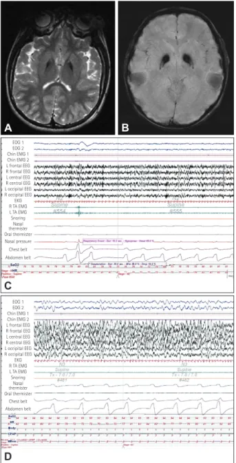
A 12-year-old boy presented at our sleep clinic with the frequent cessation of breathing dur- ing sleep. Recurrent falling with generalized dystonia had started from 2 years of age, and the patient had been wheelchair-bound since the age of 8 years. His younger brother pre- sented similar symptoms. The patient was diagnosed with PKAN caused by gene mutation (R440P and N501I; compound heterozygote) at 9 years of age (Fig. 1A, B). He had a history of deep brain stimulation of the bilateral globus pallidus interna due to severe dystonia. The patient showed progressive retrocollic dysphagia with dystonia, and underwent percutaneous endoscopic gastrostomy. Frequent snoring and cessation of nocturnal breathing was observed during the previous 2 years. His body mass index was 14.6 kg/m2 (weight, 36 kg; height, 157 cm), and his neck circumference was 34 cm. Overnight polysomnography (PSG) was suc- cessfully completed despite severe dystonia, which revealed moderate OSA with an apnea-hy- popnea index (AHI) of 16.2/h. His lowest oxygen level was 69%, and desaturation with the level below 90% accounted for 84.4% of the total sleep time (Fig. 1C). The AHIs in supine and nonsupine positions were 18.0/h and 0/h, respectively. The AHI was markedly higher dur- ing rapid eye movement (REM) sleep (31.4/h) than during non-REM sleep (16.2/h). The pa- tient showed no abnormal limb movements or parasomnia. Following a second PSG session, snoring and OSA completely disappeared at a CPAP of 7 cm H2O (Fig. 1D). After 1 month of CPAP therapy his daytime sleepiness improved and the mean AHI during CPAP therapy decreased to 1.3/h. The patient used CPAP therapy every day; the proportion of days with >4 h/night usage was on 97% of days, and the mean duration was 6.7 h/day.
Our patient presented severe dystonia and dysphagia, possibly associated with the devel- opment of OSA.3 OSA accelerates neurocognitive deterioration and increases mortality rates via various mechanisms such as intermittent hypoxia, sleep fragmentation, and hemodynamic fluctuation.4 Although there is no evidence for the development of sleep-related breathing disorder in PKAN,5 adequate respiratory support is recommended if stridor or laryngeal dys- tonia is observed.6 The present patient received CPAP therapy due to the presence of mod- erate OSA and profound desaturation (down to 69%). His acceptance of and compliance with Hyung Seok Guk
Dae Lim Koo Hyunwoo Nam
Department of Neurology, Boramae Medical Center, Seoul National University College of Medicine, Seoul, Korea
pISSN 1738-6586 / eISSN 2005-5013 / J Clin Neurol 2019;15(4):583-584 / https://doi.org/10.3988/jcn.2019.15.4.583
Received April 30, 2019 Revised June 17, 2019 Accepted June 18, 2019 Correspondence Dae Lim Koo, MD, PhD Department of Neurology, Boramae Medical Center, Seoul National University College of Medicine, 20 Boramae-ro 5-gil,
Dongjak-gu, Seoul 07061, Korea Tel +82-2-870-2473 Fax +82-2-831-0714 E-mail [email protected]
cc This is an Open Access article distributed under the terms of the Creative Commons Attribution Non-Com- mercial License (https://creativecommons.org/licenses/by-nc/4.0) which permits unrestricted non-commercial use, distribution, and reproduction in any medium, provided the original work is properly cited.
LETTER TO THE EDITOR
584 J Clin Neurol 2019;15(4):583-584
CPAP Therapy in a Patient with PKAN
JCN
CPAP usage were excellent, and no complications (e.g., air leak- age or skin irritation) were reported.
Neuronal loss and iron accumulation in the globus pallidus
interna could reduce inhibitory outputs to the pedunculopon- tine nuclei, which are the muscle tone regulators of REM sleep in patients with PKAN.5 In our patient, AHI during REM sleep was twice as high as that during non-REM sleep. Thus, the phe- notype of OSA in patients with PKAN could differ from that in patients without dystonia. This is the first report on the use- fulness of CPAP therapy in a patient with PKAN. Clinicians should diagnose and treat OSA despite the presence severe dystonia or the disabled state of patients. Further studies are needed to elucidate the pathogenesis and efficacy of CPAP therapy in patients with PKAN and OSA.
Author Contributions
Conceptualization: Dae Lim Koo. Data curation: Hyung Seok Guk, Dae Lim Koo. Formal analysis: Dae Lim Koo. Investigation: Hyung Seok Guk, Dae Lim Koo, Hyunwoo Nam. Methodology: Dae Lim Koo. Supervision:
Dae Lim Koo, Hyunwoo Nam. Writing—original draft: Hyung Seok Guk, Dae Lim Koo. Writing—review & editing: Hyung Seok Guk, Dae Lim Koo, Hyunwoo Nam.
ORCID iDs
Hyung Seok Guk https://orcid.org/0000-0003-2839-012X Dae Lim Koo https://orcid.org/0000-0001-6858-6093 Hyunwoo Nam https://orcid.org/0000-0003-3345-7069 Conflicts of Interest
The authors have no potential conflicts of interest to disclose.
REFERENCES
1. Gregory A, Hayflick SJ. Pantothenate kinase-associated neurodegen- eration. In: Adam MP, Ardinger HH, Pagon RA, Wallace SE, Bean LJH, Stephens K, et al, editors. GeneReviews. Seattle: University of Washington, 1993.
2. McCarty SF, Gaebler-Spira D, Harvey RL. Improvement of sleep ap- nea in a patient with cerebral palsy. Am J Phys Med Rehabil 2001;
80:540-542.
3. Marchese-Ragona R, Vianello A, Restivo DA, Pittoni G, Lionello M, Martini A, et al. Sleep-related adductor laryngeal dystonia causing sleep apnea: a sleep-related breathing disorder diagnosed with sleep endoscopy and treated with botulinum toxin. Laryngoscope 2013;
123:1560-1563.
4. Koo DL, Nam H, Thomas RJ, Yun CH. Sleep disturbances as a risk factor for stroke. J Stroke 2018;20:12-32.
5. Fantini ML, Cossu G, Molari A, Cabinio M, Uyanik O, Cilia R, et al.
Sleep in genetically confirmed pantothenate kinase-associated neu- rodegeneration: a video-polysomnographic study. Parkinsons Dis 2010;2010:342834.
6. Hogarth P, Kurian MA, Gregory A, Csányi B, Zagustin T, Kmiec T, et al. Consensus clinical management guideline for pantothenate ki- nase-associated neurodegeneration (PKAN). Mol Genet Metab 2017;
120:278-287.
Fig. 1. Magnetic resonance imaging and polysomnography features of the patient. T2-weighted (A) and T2* susceptibility-weighted im- ages showing a hyperintense center with a dense hypointense rim (eye-of-tiger sign) (B). C: Baseline polysomnography image revealing recurrent episodes of obstructive sleep apnea and the lowest oxygen saturation (down to 69%). D: Complete elimination of snoring and sleep apnea was confirmed at a continuous positive airway pressure of 7 cm H2O by polysomnography. CPAP: continuous positive airway pressure, EEG: electroencephalography, EKG: electrocardiogram, EMG:
electromyography, EOG: electrooculography, HR: heart rate, L: left, R:
right, SaO2: peripheral oxygen saturation, TA: tibialis anterior.
A
C
B
D
EOG 1 EOG 2 Chin EMG 1 Chin EMG 2 L frontal EEG R frontal EEG L central EEG R central EEG L occipital EEG R occipital EEG
EKG R TA EMG L TA EMG Snoring Nasal thermister Oral thermister Nasal pressure Chest belt Abdomen belt
EOG 1 EOG 2 Chin EMG 1 Chin EMG 2 L frontal EEG R frontal EEG L central EEG R central EEG L occipital EEG R occipital EEG
EKG R TA EMG L TA EMG Snoring Nasal thermister Oral thermister
Chest belt Abdomen belt