Comparison of Drug-eluting Coronary Stents, Bare
Coronary Stents and Self-expanding Stents in Angioplasty of Middle Cerebral Artery Stenoses
Jong-Hyeog Lee1, Sung-Min Jo2, Kwang-Deog Jo3, Moon-Kyu Kim4, Sang-Youl Lee4, Seung-Hoon You4
1Department of Radiology, S-Jungang Hospital, Jeju, Republic of Korea
2Department of Neurosurgery, Donghae Dongin Hospital, Donghae, Republic of Korea
Department of 3Neurology, 4Neurosurgery, Gangneung Asan Hospital, College of Medicine, University of Ulsan, Gangneung, Republic of Korea
Objective : The purpose of this study is to investigate the results of treat- ment using stent-angioplasty for symptomatic middle cerebral arterial (MCA) stenosis and comparison of in-stent restenosis between drug-eluting stents (DES), bare metal coronary stents (BMS) and self-expanding stents (SES).
Materials and Methods : From Jan. 2007 to June. 2012, 34 patients (mean age ± standard deviation: 62.9 ± 13.6 years) with MCA stenosis were treated. Inclusion criteria were acute infarction or transient ischemic attacks (TIAs) and angiographically proven symptom related severe stenosis.
Stents used for treatment were DES (n = 8), BMS (n = 13) and SES (n = 13).
National Institutes of Health Stroke Scale (NIHSS) at admission was 2.5 ± 3.1 and mean stenosis rate was 79.0 ± 8.2%. Assessment of clinical and angiographic results was performed retrospectively.
Results : Among 34 patients, periprocedural complications occurred in four cases (11.8%), however, only two cases (6.0%) were symptomatic. All patients were followed clinically (mean follow-up period; 40.7 ± 17.7 months) and 31 were followed angiographically (91.2%. 13.4 ± 8.5 months). There was no occurrence of repeat stroke in all patients; however, mild TIAs re- lated to restenosis occurred in three of 34 patients (8.8%). The mean NIHSS after stent-angioplasty was 1.7 ± 2.9 and 0.8 ± 1.1 at discharge. The modi- fied Rankin score (mRS) at discharge was 0.5 ± 0.9 and 0.3 ± 0.8 at the last clinical follow-up. In-stent restenosis over 50% occurred in five of 31 an- giographically followed cases (16.1%), however, all of these events occurred only in patients who were treated with BMS or SES. Restenosis rate was 0.0% in the DES group and 20.8% in the other group (p = 0.562); it did not differ between BMS and SES (2/11 18.2%, 3/13 23.1%, p = 1.000).
Conclusion : Stent-angioplasty appears to be effective for symptomatic MCA stenosis. As for restenosis, in our study, DES was presumed to be more effective than BMS and SES; meanwhile, the results did not differ between the BMS and SES groups.
J Cerebrovasc Endovasc Neurosurg.
2013 June;15(2):85~95 Received : 25 April 2013 Revised : 4 June 2013 Accepted : 12 June 2013
Correspondence to Seung-Hoon You Department of Neurosurgery, Gangneung Asan Hospital, 415 Bangdong-ri, Sacheon-myeon, Gangneung-si, Gangwon-do, 210-711, Republic of Korea
Tel : 82-33-610-3258 Fax : 82-33-641-8070 E-mail : [email protected]
This is an Open Access article distributed under the terms of the Creative Commons Attribution Non- Commercial License (http://creativecommons.org/li- censes/by-nc/3.0) which permits unrestricted non- commercial use, distribution, and reproduction in any medium, provided the original work is properly cited.
Keywords Intracranial stenosis, Middle cerebral artery, Restenosis, Stent-angioplasty, Drug-eluting stent, Self-expanding stent
INTRODUCTION
Intracranial atherosclerotic stenosis is responsible forapproximately 5% to 10% of all strokes in mixed pa- tient populations, and in the Asian population, this is even the most commonly found vascular lesion.10) Endovascular therapy using primary angioplasty or stenting has been used in treatment of medically re- fractory patients with high-grade intracranial athero- sclerotic stenoses.5)22) However, endovascular manage- ment of intracranial stenosis is associated with a sig- nificant number of potential complications, including acute in-stent thrombosis, thromboembolism, vessel dissection, and even rupture. To date, according to some reports, these complications during angioplasty have varied widely, ranging from 0% to more than 30%.6)7)12)13) Therefore, current treatment decisions are usually based on results of case series for each center.
Prior to introduction of self-expanding stents (SES) for intracranial stent-angioplasty, the only available stents for intracranial stenotic disease were coronary balloon mounted (balloon-expandable) stents. Since that time, many studies of intracranial stent-angioplasty have proceeded in earnest.3)8)11)14)18)27)30)38)
The main trunk of the middle cerebral artery (MCA) represents one of the most common sites of sympto- matic intracranial atherosclerosis.4) However, stent-an- gioplasty for MCA lesions remains challenging be- cause of vascular tortuosity, small vessel diameter, and concern for perforator injury. In addition, MCA lesions are known to be highly prone to restenosis af- ter stent-angioplasty.32)
The authors assessed the treatment results, peri- procedural complications, and angiographic results of stent-angioplasty for symptomatic MCA stenosis, and then attempted to compare the results of restenosis between drug-eluting stents (DES), bare metal coro- nary stents (BMS), and SES.
MATERIALS AND METHODS
Patient characteristics and Stent selection
From January 2007 to June 2012, 35 patients (23 men, 12 women) underwent stent-angioplasty for
MCA stenosis. Patients ranged from 34 to 84 years of age (mean ± standard deviation: 63.3 ± 13.6 years).
We required that candidates for stent-angioplasty have acute or subacute symptomatic MCA territorial infarction (i.e. borderzone infarction) or repeated TIAs, and severe MCA stenotic rate over 70% related to symptoms confirmed with catheter angiography.
Patients with asymptomatic stenosis or mild stenosis under 70% were excluded from endovascular treatment.
Because of actual assessment for clinical and angio- graphic results of stent-angioplasty, an additional in- clusion criterion for this study was successful deploy- ment of a stent on the lesion. Therefore, 34 patients (23 male, 11 female, mean age: 62.9 ± 13.6 years) in whom stents were successfully deployed were in- cluded in this study. Stents used for treatment were DES (n = 8, 23.5%), BMS (n = 13, 38.2%), and SES (n
= 13, 38.2%). The mean National Institutes of Health Stroke Scale (NIHSS) at admission was 2.5 ± 3.1, and mean stenosis rate was 79.0 ± 8.2%. Follow-up (FU) NIHSS was evaluated immediately after the proce- dure, and at discharge. Evaluation of modified Rankin Score (mRS) was performed at discharge and was last performed in the Out-Patient Department. Clinical sta- tus (including NIHSS and mRS) and angiographic re- sults were assessed retrospectively. We then compared the results between DES, BMS, and SES.
Until Apr. 2010, DES or BMS was implanted for in- tracranial stenosis, including MCA stenosis, and, after that time, due to the availability of stents in domestic medical practice, most patients were treated with SES (Wingspan stent, Boston Scientific, Natick, MA).
Pre- and post-operative medications and Endovascular techniques
All patients undergoing stent-angioplasty were giv- en dual antiplatelet therapy for at least three days be- fore the procedure, which consisted of aspirin (100 mg - 300 mg/d orally) and clopidogrel (75 mg/d or- ally), except for one patient, who underwent stent-an- gioplasty as an emergency treatment after a loading dose of dual antiplatelet medication (aspirin 300 mg
A B C D
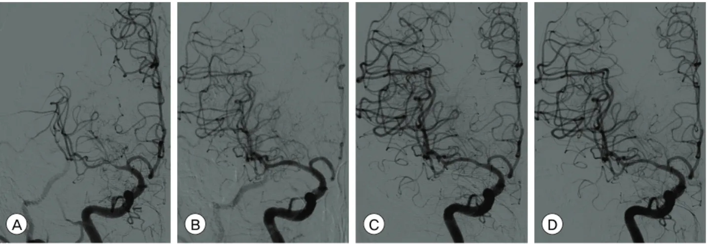
Fig. 1. Catheter angiograms of a 66-year-old male patient who presented with repeated transient ischemic attacks of left arm weakness. (A) Preoperative right internal cerebral artery (ICA) angiogram demonstrates severe stenosis at the right middle cerebral artery (MCA) and flow compromise of the MCA territory. (B) Right MCA was fully recanalized after Wingspan stent (Boston Scientific, Natick, MA, USA) deployment. (C) Follow-up angiogram three months after stent-angioplasty. M1 segment in which stent was implanted is more remodeled than immediately after stent-angioplasty. (D) Follow-up angiogram 13 months after stent-angioplasty.
No change is seen when compared with the three month follow-up angiogram.
and clopidogrel 375 mg). All endovascular procedures were performed under local anesthesia by a neuro- surgeon and a neuroradiologist. A 6 or 7 French guid- ing catheter was inserted into the proximal internal cerebral artery (ICA) after a femoral puncture. A 0.014 inch (0.35 mm) wire was then introduced through the guiding catheter with or without use of a microcatheter. In cases of coronary balloon mounted stents, including DES and BMS, after the wire had passed through the stenotic lesion under the road- map, the stent was delivered to the lesion along the wire and placed across the stenotic lesion by inflation of the balloon. The stent was deployed at the same time when the stenosis was dilated by inflation of the balloon. In cases of self-expanding stents (Wingspan stent), after the wire had passed through the lesion, a Gateway balloon was delivered to the lesion first, and inflated for pre-stent dilatation. A Wingspan stent was delivered to the lesion and placed across the le- sion by unsheathing the stent-delivery catheter system after ballooning-dilatation. This procedure was per- formed under anticoagulation treatment with intra- venous heparin, which was followed by nadroparin calcium (Fraxiparin; 2850 IU anti-Factor Xa, Sanofi Winthrop Industrie, Ambares, FR) dosed at 0.3 ml SC
q 12 hours for at least one day following the procedure. Dual antiplatelet therapy was continued after the procedure and then switched to monotherapy after at least 12 weeks (aspirin 100 mg/d orally).
Measurement of MCA stenosis and restenosis All digital subtraction angiograms were analyzed by a neurosurgeon. The angiographic view that best demonstrated the stenotic lesion was identified dur- ing pre-stenting angiography. This optimal view was used as the working view for stent placement and im- mediate post-stenting angiography. The angle of the chosen working view was recorded and reproduced during FU angiography (Fig. 1). The degree of steno- sis was calculated as the percent stenosis from the catheter angiogram using the following formula:
Percent stenosis (%) = 100 × (1 - S/N), where S is the diameter of the residual lumen at the point of maximum stenosis and N is the width of the dis- ease-free distal MCA (M1 segment) at the point where the walls were approximately parallel. Residual steno- sis was quantified according to the vessel diameter achieved immediately after stent-angioplasty, and restenosis was calculated by comparing the lesion di- ameter of the follow up angiogram and that of the
Comparison variables All patients treated (n=34)
Patients followed by
angiography (n=31) P value
Age 62.9 ± 13.6 63.6 ± 13.6 0.837
Reference diameter (mm) 2.5 ± 0.2 2.5 ± 0.3 0.959
Preop. stenosis rate (%) 79.0 ± 8.2 78.9 ± 8.1 0.955
Preop. stenosis rate (%) 6.7 ± 2.4 6.8 ± 2.4 0.885
DES 8 (23.5%) 7 (22.6%)
Used stents BMS 13 (38.2%) 11 (35.5%) 0.954
SES 13 (38.2%) 13 (41.9%)
Postop. residual stenosis rate (%) 4.0 ± 9.2 4.3 ± 9.6 0.922
at admission 2.5 ± 3.1 2.5 ± 3.2 0.865
NIHSS Postop 1.7 ± 2.9 1.8 ± 3.0 0.865
Discharge 0.8 ± 1.1 0.8 ± 1.1 0.881
mRS Discharge 0.5 ± 0.9 0.6 ± 0.9 0.933
At last FU 0.3 ± 0.8 0.3 ± 0.8 0.996
Clinical FU period (months) 40.7 ± 17.7 40.3 ± 18.1 0.928
Preop= preoperative; Postop= postoperative; DES= drug-eluting stents; BMS= bare metal stents;
SES= self-expanding stents; NIHSS= National Institutes of Health Stroke Scale; mRS= modified Rankin scale; FU= follow up
Table 1. Comparisons of all patients who had undergone stent-angioplasty and the patients who have been conducted an- giographic follow-up either
postoperative angiogram immediately after treatment.
Restenosis was categorized into two groups: insignif- icant to mild restenosis (0-49%), and moderate to se- vere restenosis (50-100%).
Statistical analysis
Between-group comparisons were made according to the types of stents used. Age, sex, past history of medical disease (hypertension, diabetes mellitus, and hypercholesterolemia), angiographic data, including diameter of the disease-free distal MCA (reference di- ameter), length of stenosis, stenosis rate (preoperatively, postoperatively, and at FU angiography), and clinical status such as NIHSS and mRS were analyzed. We compared continuous variables using Student's t-tests and categorical variables using Pearson's Chi-Square tests. Comparisons were made according to con- fidence intervals of 95%. All statistical tests were two-sided and all analyses were performed using stat- istical software (SPSS for Windows, 15.0 standard ver- sion, IBM, NY). Values are expressed as mean ± standard deviation. A probability value less than 0.05 was considered statistically significant.
RESULTS
Of 35 patients, intracranial stents were successfully deployed at the first trial in 34 patients. In one pa- tient, we attempted to perform stent-angioplasty us- ing a Wingspan stent; stent delivery failed after suc- cessful pre-stent balloon angioplasty due to a severely tortuous ICA course. Therefore, we assessed the treat- ment results of stent-angioplasty with these 34 pa- tients who were treated successfully. Overall results are shown in Table 1. NIHSS on the day after angio- plasty showed improvement in most patients (2.5 ± 3.1 at admission vs. 1.7 ± 2.9 after the procedure, p = 0.014) and then continued to show improvement (0.8
± 1.1, at discharge, p = 0.001) and so did mRS (0.5 ± 0.9 at discharge to 0.3 ± 0.8 at last FU, p = 0.003).
Procedure-related complications occurred in four cas- es (11.8%); however, they were symptomatic in only two cases (5.8%). One was a case of MCA rupture af- ter inflation of ballooning deployment of the coronary stent, and the other, a case of thromboembolism, ap- peared to be related to the procedure with a coronary stent not even detected angiographically during the
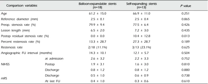
Comparison variables Balloon-expandable stents
(n=18) Self-expanding stents
(n=13) P value
Age 61.2 ± 15.0 66.9 ± 11.0 0.251
Reference diameter (mm) 2.5 ± 0.1 2.5 ± 0.4 0.865
Preop. stenosis rate (%) 79.9 ± 9.4 77.5 ± 6.4 0.426
Lesion length (mm) 6.5 ± 2.0 7.2 ± 3.0 0.435
Postop residual stenosis rate (%) 0.0 ± 0.0 10.4 ± 12.8 0.013
Percent restenosis rate (%) 13.3 ± 28.7 27.3 ± 28.7 0.189
Restenosis rate 2/18 (11.1%) 3/13 (23.1%) 0.625
Angiographic FU interval (months) 14.3 ± 10.1 12.1 ± 5.7 0.504
at admission 2.6 ± 3.2 2.2 ± 3.3 0.752
NIHSS Postop 1.9 ± 3.1 1.6 ± 3.0 0.810
Discharge 0.8 ± 1.2 0.8 ± 1.2 0.880
mRS Discharge 0.5 ± 1.0 0.6 ± 0.9 0.738
At last FU 0.4 ± 1.0 0.3 ± 0.6 0.610
Table 2. Comparisons of patients treated with coronary balloon-expandable stents (including drug-eluting stents and bare metal stents) vs. self-expanding stents.
procedure. Although the former was healed com- pletely with the repetition of balloon inflation and de- flation after stent deployment, a small amount of SAH and multiple scattered infarctions were devel- oped in the MCA territory. And, in the latter, the ag- gravated hemiparesis showed improvement to a near- ly previous state six months after the procedure. Two asymptomatic complications described above were proximal vessel (i.e. cervical segment of ICA) dis- sections caused by guiding catheters. In one case, it was healed spontaneously after the procedure, and the other was treated with another stent deployment without clinical sequela.
There were no cases of repeated infarction during the FU periods; however, mild TIAs occurred in three patients within one year after stent-angioplasty (7.2 ± 5.5 months). All of these patients' symptoms were mild but identical to preoperative ones, and proved to be related to severe in-stent restenosis over 70% on FU angiography. We attempted to retreat these pa- tients with a drug-eluting balloon (DEB) in two of three patients, and with another stent in the other pa- tient, because the stenotic lesion was located beside the center of the previously implanted stent. All of these patients have been in a symptom-free state for
at least seven months after retreatment (40 and 14 months after retreatment with DEB, seven months with another stent, respectively).
Angiographic findings
The mean percent stenosis before the procedure was 79.0 ± 8.2%, and the reference diameter (vessel diame- ter just distal to lesion) was 2.5 ± 0.2 mm, and the length of the lesion was 6.7 ± 2.4 mm. The residual stenosis was 4.0 ± 9.2% immediately after stent deployment. FU angiograms were obtained in 31 of 34 patients (91.2%) at a mean of 13.4 ± 13.4 months after the procedure. There were no differences in the group characteristics between whole patients who had undergone stent-angioplasty and the patients who have been conducted angiographic FU either (Table 1).
The mean percent of restenosis was 19.2 ± 29.1% on FU angiograms. Each of the documented restenosis occurred as "in-stent" restenosis, as a sequela of thick- ening of the intima into the stent strut, not as a result of stent recoil or stent kinking. On the other hand, the overall rate of moderate-to-severe restenosis (≥ 50%) was 16.1% (five of 31 cases). Two cases were patients treated with coronary balloon-expandable stents, and the other three patients were treated with the SES (2/18, 11.1% vs. 4/13, 23.1%, p = 0.625). Therefore, we
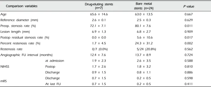
Comparison variables Drug-eluting stents (n=7)
Bare metal
stents (n=24) P value
Age 65.6 ± 14.6 63.0 ± 13.5 0.667
Reference diameter (mm) 2.6 ± 0.1 2.5 ± 0.3 0.629
Preop. stenosis rate (%) 72.1 ± 7.1 80.1 ± 7.6 0.011
Lesion length (mm) 6.9 ± 1.3 6.8 ± 2.7 0.909
Postop residual stenosis rate (%) 0.0 ± 0.0 5.6 ± 10.6 0.017
Percent restenosis rate (%) 1.7 ± 4.5 24.3 ± 31.2 0.002
Restenosis rate 0/7 (0.0%) 5/24 (20.8%) 0.562
Angiographic FU interval (months) 12.4 ± 7.6 13.7 ± 8.9 0.724
at admission 1.9 ± 2.3 2.6 ± 3.5 0.588
NIHSS Postop 1.7 ± 2.6 1.8 ± 3.2 0.810
Discharge 0.9 ± 1.5 0.8 ± 1.1 0.886
mRS Discharge 0.7 ± 1.5 0.2 ± 0.5 0.598
At last FU 0.7 ± 1.5 0.2 ± 0.5 0.411
Table 3. Comparisons of patients treated with drug-eluting stents vs. bare metal stents including coronary balloon-expandable stents and self-expanding stents.
compared the results between the groups of patients who were treated with coronary balloon-expandable stents and SES (Table 2). In this comparison, we found that only one variable differed i.e. post- operative residual stenosis rate, even though the dif- ference of restenosis rate itself did not reach statistical significance. There were no cases of restenosis in pa- tients treated with DES. Thus, we compared patients treated with DES and the others (Table 3). Some varia- bles differed between these two groups. Postoperative residual stenosis rate and percent restenosis rate dif- fered significantly. We then compared the BMS and SES, because both stents have the same characteristics in terms of the bare metal stent, but not the same de- sign, such as balloon-expandable stents vs. self-ex- panding stents, which should be deployed after sub- optimal balloon angioplasty (Table 4). In this compar- ison, the only significant difference was postoperative residual stenosis. However, in comparison of this be- tween DES vs. BMS, it was nearly the same, but the percent restenosis rate was different, although it did not reach statistical significance (DES vs. BMS, 1.7 ± 4.5 vs. 20.7 ± 35.1, p = 0.105) (Table 5). Meanwhile, in cases of SES, both were significantly different from
DES (Postoperative residual stenosis: 10.4 ± 12.8, p = 0.013, Percent restenosis rate: 27.3 ± 28.7%, p = 0.008) (Table 5). No association was observed between de- velopment of restenosis and past history of medical disease, including hypertension (p = 0.394), diabetes mellitus (p = 1.000), and hypercholesterolemia (p = 1.000).
DISCUSSION
The success rate of stent-angioplasty was 97.1% in this study, and the procedural complication rate was 5.7% in the case of symptomatic patients. No in- farction or hemorrhage was observed over one-year follow-up periods. The reported success rate and pro- cedural risk of intracranial angioplasty varied accord- ing to the reports.9)36) Yoon et al.36) reported risk of disabling stroke or death of 6% (two of 32 patients), and mortality rate of 3% (one of 32 patients). Marks et al.20) reported a rate of periprocedural death and stroke of 8.3%, and, in a Taiwan study, Jiang et al.15) reported a complication rate up to 11.8%. In a sys- temic review in 2009, the success rate of stent angio- plasty for intracranial arterial stenosis was reported from 71.4% to 100%, and the periprocedural minor or
Comparison variables Coronary bare metal stents (n=11) Self-expanding stents (n=13) P value
Age 58.4 ± 15.2 66.9 ± 11.0 0.124
Reference diameter (mm) 2.5 ± 0.8 2.5 ± 0.4 0.981
Preop. stenosis rate (%) 84.8 ± 7.1 77.5 ± 6.4 0.014
Lesion length (mm) 6.3 ± 2.3 7.2 ± 3.0 0.401
Postop residual stenosis rate (%) 0.0 ± 0.0 10.4 ± 12.8 0.013
Percent restenosis rate (%) 20.7 ± 35.1 27.3 ± 28.7 0.615
Restenosis rate 2/11 (18.2%) 3/13 (23.1%) 1.000
Angiographic FU interval (months) 15.5 ± 11.7 12.2 ± 5.7 0.372
at admission 3.1 ± 3.7 2.2 ± 3.3 0.556
NIHSS Postop 2.0 ± 3.6 1.6 ± 3.0 0.778
Discharge 0.8 ± 1.0 0.8 ± 1.2 0.913
mRS Discharge 0.4 ± 0.5 0.3 ± 0.9 0.407
at last FU 0.2 ± 0.4 0.2 ± 0.6 0.820
Table 4. Comparisons of patients treated with coronary bare metal stents vs. self-expanding stents.
Comparison variables
Coronary drug-eluting stents (n=11)
Coronary bare metal stents
(n=11) P value
Self- expanding stents
(n=13) P value
Age 65.6 ± 14.6 58.4 ± 15.2 0.336 66.9 ± 11.0 0.819
Reference diameter (mm) 2.6 ± 0.1 2.5 ± 0.8 0.345 2.5 ± 0.4 0.659
Preop. stenosis rate (%) 72.1 ± 7.1 84.8 ± 7.1 0.002 77.5 ± 6.4 0.105
Lesion length (mm) 6.9 ± 1.3 6.3 ± 2.3 0.460 7.2 ± 3.0 0.796
Postop residual stenosis rate (%) 0.0 ± 5.6 0.0 ± 0.0 - 10.4 ± 12.8 0.013
Percent restenosis rate (%) 1.7 ± 4.5 20.7 ± 35.1 0.105 27.3 ± 28.7 0.008
Restenosis rate 0/7 (0.0%) 2/11 (18.2%) 0.359 3/13 (23.1%) 0.521
Angiographic FU interval (months) 12.4 ± 7.6 15.5 ± 11.7 0.540 12.2 ± 5.7 0.947
at admission 0.9 ± 2.3 3.1 ± 3.7 0.445 2.2 ± 3.3 0.797
NIHSS Postop 1.7 ± 2.6 2.0 ± 3.6 0.858 1.6 ± 3.0 0.942
Discharge 0.9 ± 1.5 0.8 ± 1.0 0.947 0.8 ± 1.2 0.885
mRS Discharge 0.7 ± 1.5 0.4 ± 0.5 0.478 0.3 ± 0.9 0.852
At last FU 0.7 ± 1.5 0.2 ± 0.4 0.390 0.2 ± 0.6 0.313
Table 5. Comparisons of patients treated with coronary drug-eluting stents vs. coronary bare metal stents and self-expanding stents.
major stroke and death rates ranged from 0% to 50%
with a median of 7.7%.13) Meanwhile, Zhang et al.37) reported feasible results of stent-angioplasty for MCA stenosis. Although the size of the study was relatively smaller than that of the previous reports, in compar- ison, the author's result appears to show an equiv- alent or slightly better clinical outcome.
As for the restenosis, in the Stenting of Symptomatic Atherosclerotic Lesions in the Vertebral or Intracranial Arteries (SSYLVIA) Study, the rate of successful stent placement was 95%, however, restenosis rate of ≥ 50%
at six months was 32.4%.31) Recently, the incidence of in-stent restenosis and retreatment of this condition were intensively investigated from data of the US Wingspan Registry, and the rate of restenosis was re- ported as 29.8%, and an additional 4.5% of stent thromboses occurred during an average follow up pe- riod of 5.9 months.19) The rate of recurrent stroke in the subgroup with in-stent restenosis was 6.8% and the rate of TIA was 13.8%.19) In this study, overall restenosis over 50% was 16.1% (five of 31 cases) and only 9.7% (3/31) for symptomatic cases at a mean an-
A B C
Fig. 2. Catheter angiograms of a 68-year-old female patient who presented with MCA borderzone infarct. (A) Preoperative left ICA angiogram demonstrates severe stenosis at M1 segment and definite flow compromise caused by the stenosis. (B) Postoperative an- giogram after stent-angioplasty with a drug-eluting coronary stent (Endeavor, Medtronic Vascular, Santa Rosa, CA) shows no re- sidual stenosis and fully recovered MCA flow. (C) One year follow-up angiogram shows no restenosis compared with immediate postoperative angiogram.
giographic follow up period of 13.4 months.
However, due to the diverse composition of the kinds of stents, these figures should not be taken at face value. All cases of restenosis were bare metal stent, including coronary BMS and SES, and in case of SES (Wingspan stent), restenosis rate was 23.1%. This re- sult is not much better than that of previous reports.
However, on the contrary, restenosis did not occur in cases with DES, although the number of cases was very small (Fig. 2). DES has been known to be effec- tive for reducing the risk of in-stent restenosis due to its controlled local release of antiproliferative agents, as compared with BMS in coronary stenting.23)28)29) Off-label use of drug-eluting coronary stents for treat- ment of intracranial atherosclerotic lesions has re- cently been reported.1)6)27) Park et al.24) reported prom- ising results on drug-eluting stents for intracranial stenosis. The DES appears to be a good option for prevention of in-stent restenosis, however, technical difficulty due to stent stiffness remains a serious problem in generalizing and drawing definitive con- clusions regarding the safety and effectiveness for in-
tracranial lesions, especially distal ICA or MCA le- sions, although restenosis itself was solved in part with these stents.16)33)
Findings of this study revealed a tendency of a sim- ilar rate of restenosis in cases of coronary BMS and SES (18.2% at BMS, 23.1% at SES, p = 1.000) (Table 4).
The only meaningful difference was immediate post- operative residual stenosis rate. Percentage of residual stenosis in cases of balloon expandable stents such as DES and BMS was nearly zero, however, in case of SES, it was over 10%. This might be due to the pre- stent balloon angioplasty without simultaneous stent- ing of the Gateway Wingspan system. Technically, it places restrictions on sufficient dilatation of the le- sions because of concern regarding serious arterial in- jury, such as dissection and even rupture. However, because of the similar results of the coronary BMS and SES, it might not be a risk factor. On the other hand, Shin et al.26) pointed out the speed of inflation as a risk factor for in-stent restenosis in Wingspan stenting. Although this explanation could have im- portant implications for Wingspan stenting, it may
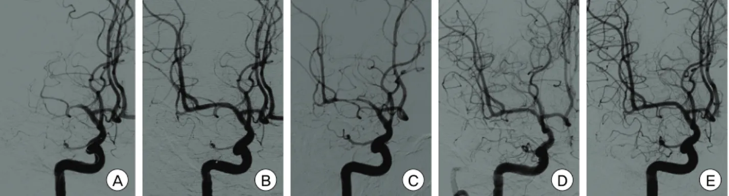
A B C D E
Fig. 3. Catheter angiograms of a 47-year-old male patient who presented with right MCA borderzone infarct. (A) Preoperative right ICA angiogram demonstrates MCA occlusion. (B) Postoperative angiogram after stent-angioplasty with a bare metal coronary stent (Coroflex, B. Braun, DE) shows no residual stenosis and fully recovered MCA flow. (C) Three month follow-up angiogram shows se- vere in-stent restenosis up to 80% with mild flow compromise. (D) Follow-up angiogram immediately after retreatment (balloon an- gioplasty) with a drug-eluting balloon (SeQuent® Please, B.Braun, DE) shows fully recovered MCA flow compromise, but insignif- icant residual stenosis remained. (E) One year follow-up angiogram after retreatment demonstrates a fully dilated M1 segment with- out any residual stenosis or restenosis.
not be applicable in the same way in cases of stenting with balloon-expandable coronary stents, because their use required significantly less stenting time (ap- proximately 20 to 30 seconds in the author's series).
Further studies should be required in order to clarify the association between in-stent restenosis and rele- vant risk factors, including technical aspects men- tioned above, and other medical history.
In cases of restenosis, we attempted to administer retreatment only in symptomatic patients (three of five cases). In one case, retreatment was administered using another Wingspan system, because the stenotic lesion was located beside the center of the previously implanted stent. We treated the other two patients with the DEB (Fig. 3). All of these patients have been in a symptom-free state for at least seven months since retreatment (40 and 14 months after retreatment with a drug-eluting balloon, seven months with an- other stent, respectively). Several reported options, such as balloon angioplasty, another stent-angioplasty, etc, can be adopted for retreatment of restenosis.
Currently, balloon angioplasty has been adopted in the majority of cases.12) Recently, the combination of mechanical balloon dilatation and local drug applica- tion was confirmed to be very efficient for treatment
of coronary in-stent restenosis, and Vajda et al.34) re- ported successful treatment results of a DEB for the patients with in-stent restenosis after stent-angioplasty for intracranial stenotic diseases. In addition, they in- troduced another combination of DEB followed by the SES, instead of a current bare balloon with a bare stent, such as a Gateway balloon and Wingspan stent.35) Although conduct of further studies is need- ed, the DEB appears to be a promising alternative op- tion for in-stent restenosis.
CONCLUSIONS
In this study, stent-angioplasty appeared to be safe and effective for MCA stenotic disease, however, a limitation of the current study was that the number of patients included was relatively small. It should prompt further studies on a larger scale.
The rate of in-stent restenosis was 16.1% as a whole, however, DES showed better results for prevention of restenosis, compared with BMS or SES. In this study, although the postoperative residual stenosis was one of the most noticeable differences between coronary balloon-expandable stents and SES, it appears to have little direct relationship with in-stent restenosis.
REFERENCES
1. Abou-Chebl A, Bashir Q, Yadav JS. Drug-eluting stents for the treatment of intracranial atherosclerosis: initial experience and midterm angiographic follow-up. Stroke.
2005 Dec;36(12):e165-8.
2. Albuquerque FC, Levy EI, Turk AS, Niemann DB, Aagaard-Kienitz B, Pride GL Jr, et al. Angiographic pat- terns of Wingspan in-stent restenosis. Neurosurgery.
2008 Jul;63(1):23-7.
3. Al Hasan M, Murugan R. Stenting versus aggressive medical therapy for intracranial arterial stenosis: more harm than good. Crit Care. 2012 May;16(3):310.
4. Arenillas JF, Molina CA, Montaner J, Abilleira S, González-Sánchez MA, Alvarez-Sabín J. Progression and clinical recurrence of symptomatic middle cerebral artery stenosis: a long-term follow-up transcranial Doppler ul- trasound study. Stroke. 2001 Dec;32(12):2898-904.
5. Bose A, Hartmann M, Henkes H, Liu HM, Teng MM, Szikora I, et al. A novel, self-expanding, nitinol stent in medically refractory intracranial atherosclerotic stenoses:
the Wingspan study. Stroke. 2007 May;38(5):1531-7.
6. Boulos AS, Agner C, Deshaies EM. Preliminary evidence supporting the safety of drug-eluting stents in neuro- vascular disease. Neurol Res. 2005;27 Suppl 1:S95-102.
7. Chimowitz MI, Lynn MJ, Derdeyn CP, Turan TN, Fiorella D, Lane BF, et al. Stenting versus aggressive medical therapy for intracranial arterial stenosis. N Engl J Med. 2011 Sep;365(11):993-1003.
8. Derdeyn CP, Fiorella D, Lynn MJ, Barnwell SL, Zaidat OO, Meyers PM, et al. Impact of operator and site ex- perience on outcomes after angioplasty and stenting in the SAMMPRIS trial. J Neurointerv Surg. 2012 Sep 12;0:1-6.
9. De Rochemont Rdu M, Turowski B, Buchkremer M, Sitzer M, Zanella FE, Berkefeld J. Recurrent symptomatic high-grade intracranial stenoses: safety and efficacy of undersized stents-initial experience. Radiology. 2004 Apr;231(1):45-9.
10. De Silva DA, Woon FP, Lee MP, Chen CP, Chang HM, Wong MC. South Asian patients with ischemic stroke:
intracranial large arteries are the predominant site of disease. Stroke. 2007 Sep;38(9):2592-4.
11. Fiorella D, Levy EI, Turk AS, Albuquerque FC, Niemann DB, Aagaard-Kienitz B, et al. US multicenter experience with the wingspan stent system for the treatment of in- tracranial atheromatous disease: periprocedural results.
Stroke. 2007 Mar;38(3):881-7.
12. Fiorella D, Levy EI, Turk AS, Albuquerque FC, Pride GL Jr, WooHH, et al. Target lesion revascularization af- ter wingspan: assessment of safety and durability.
Stroke. 2009 Jan;40(1):106-10.
13. Gröschel K, Schnaudigel S, Pilgram SM, Wasser K, Kastrup A. A systematic review on outcome after stent- ing for intracranial atherosclerosis. Stroke. 2009 May;40 (5):e340-7.
14. Henkes H, Miloslavski E, Lowens S, Reinartz J, Liebig T, Kühne D. Treatment of intracranial atherosclerotic stenoses with balloon dilatation and self-expanding stent
deployment (Wingspan). Neuroradiology. 2005 Mar;47(3):
222-8.
15. Jiang WJ, Du B, Leung TW, Xu XT, Jin M, Dong KH.
Symptomatic Intracranial Stenosis: cerebrovascular com- plications from elective stent placement. Radiology. 2007 Apr;243(1):188-97.
16. Kim JS, You SH, Kim SR, Kim SD, Kim YW, Park IK, et al. Usefulness of drug-eluting stents in angioplasty and stenting of the vertebral artery origin: comparison with bare stents. Korean J Cerebrovasc Surg.
2005;7(2):135-42.
17. Lee J, Kwon S, Lee JH, Suh DC, Kim JS. Percutaneous transluminal angioplasty for symptomatic middle cere- bral artery stenosis: long-term follow-up. Cerebrovasc Dis. 2003;15(1-2):90-7.
18. Lee JH, Yun JK, Kim DW, Kang SD. Clinical and angio- graphic outcomes of Wingspan stent placement for treat- ment of symptomatic intracranial stenosis: single center experience with 19 cases. J Cerebrovasc Endovasc Neurosurg. 2012 Sep;14(3):157-63.
19. Levy EI, Turk AS, Albuquerque FC, Niemann DB, Aagard-Kienitz B, Pride L, et al. Wingspan in- stent restenosis and thrombosis: incidence, clinical pre- sentation, and management. Neurosurgery. 2007 Sep;61(3):644-50; discussion 650-1.
20. Marks MP, Marcellus ML, Do HM, Schraedley-Desmond PK, Steinberg GK, Tong DC, et al. Intracranial angio- plasty without stenting for symptomatic atherosclerotic stenosis: long-term follow-up. AJNR Am J Neuroradiol.
2005 Mar;26(3):525-30.
21. Marks MP, Marcellus M, Norbash AM, Steinberg GK, Tong D, Albers GW. Outcome of angioplasty for athe- rosclerotic intracranial stenosis. Stroke. 1999 May;30(5) :1065-9.
22. Marks MP, Wojak JC, Al-Ali F, Jayaraman M, Marcellus ML, Connors JJ, et al. Angioplasty for symptomatic in- tracranial stenosis: clinical outcome. Stroke. 2006 Apr;37(4):1016-20.
23. Morice MC, Serruys PW, Sousa JE, Fajadet J, Ban Hayashi E, Perin M, et al. A randomized comparison of a sirolimus-eluting stent with a standard stent for coro- nary revascularization. N Engl J Med. 2002 Jun;346(23):
1773-80.
24. Park S, Lee DG, Chung WJ, Lee DH, Suh DC.
Long-term outcomes of drug-eluting stents in sympto- matic intracranial stenosis. Neurointervention. 2013 Feb;8(1):9-14.
25. Scheller B, Hehrlein C, Bocksch W, Rutsch W, Haghi D, Dietz U, et al. Two year follow-up after treatment of coronary in-stent restenosis with a paclitaxel-coated bal- loon catheter. Clin Res Cardiol. 2008 Oct;97(10):773-81.
26. Shin YS, Kim BM, Suh SH, Jeon P, Kim DJ, Kim DI, et al. Wingspan stenting for intracranial atherosclerotic stenosis: clinical outcomes and risk factors for in-stent restenosis. Neurosurgery. 2013 Apr;72(4):596-604; dis- cussion 604.
27. Steinfort B, Ng PP, Faulder K, Harrington T, Grinnell V, Sorby W, et al. Midterm outcomes of paclitaxel-eluting stents for the treatment of intracranial posterior circu- lation stenoses. J Neurosurg. 2007 Feb;106(2):222-5.
28. Stettler C, Wandel S, Allemann S, Kastrati A, Morice MC, Schömig A, et al. Outcomes associated with drug-eluting and bare-metal stents: a collaborative net- work meta-analysis. Lancet. 2007 Sep;370(9591):937-48.
29. Stone GW, Ellis SG, Cox DA, Hermiller J, O'Shaughnessy C, Mann JT, et al. A polymer-based, pa- clitaxel-eluting stent in patients with coronary artery disease. N Engl J Med. 2004 Jan;350(3):221-31.
30. Tarlov N, Jahan R, Saver JL, Sayre JW, Ali LK, Kim D, et al. Treatment of high risk symptomatic intracranial atherosclerosis with balloon mounted coronary stents and Wingspan stents: single center experience over a 10 year period. J Neurointerv Surg. 2012 Jan;4(1):34-9.
31. The SSYLVIA Study Investigators. Stenting of sympto- matic atherosclerotic lesions in the vertebral or intra- cranial arteries (SSYLVIA): study results. Stroke. 2004 Jun;35(6):1388-92.
32. Turk AS, Levy EI, Albuquerque FC, Pride GL Jr, Woo H, Welch BG, et al. Influence of patient age and steno- sis location on wingspan in-stent restenosis. AJNR Am J Neuroradiol. 2008 Jan;29(1):23-7.
33. Vajda Z, Aguilar M, Göhringer T, Horváth-Rizea D, Bäzner H, Henkes H. Treatment of intracranial athero
sclerotic disease with a balloon-expandable paclitaxel eluting stent: procedural safety, efficacy and mid-term patency. Clin Neuroradiol. 2012 Sep;22(3):227-33.
34. Vajda Z, Güthe T, Perez MA, Heuschmid A, Schmid E, Bäzner H, et al. Neurovascular in-stent stenoses: treat- ment with conventional and drug-eluting balloons. AJNR Am J Neuroradiol. 2011 Nov-Dec;32(10):1942-7.
35. Vajda Z, Güthe T, Perez MA, Kurre W, Schmid E, Bäzner H, et al. Prevention of intracranial in-stent reste- noses: predilatation with a drug eluting balloon, fol- lowed by the deployment of a self-expanding stent.
Cardiovasc Intervent Radiol. 2013 Apr;36(2):346-52.
36. Yoon W, Seo JJ, Cho KH, Kim MK, Kim BC, Park MS, et al. Symptomatic middle cerebral artery stenosis treat- ed with intracranial angioplasty: experience in 32 patients. Radiology. 2005 Nov;237(2):620-6.
37. Zhang L, Huang Q, Zhang Y, Deng B, Liu J, Hong B, et al. A single-center study of Wingspan stents for symptomatic atherosclerotic stenosis of the middle cere- bral artery. J Clin Neurosci. 2013 Mar;20(3):362-6.
38. Zhou Y, Yang QW, Xiong HY. Angioplasty with stent- ing for intracranial atherosclerosis: a systematic review. J Int Med Res. 2012 Feb;40(1):18-27.