224
Introduction
Pulmonary embolism (PE) is a relatively common cardiovas- cular emergency, and is associated with a high mortality.1) In this respect, early diagnosis of PE is clinically important. Be- cause of the variable and nonspecific clinical presentations of PE, a diagnostic testing like computed tomographic pulmonary an- giography (CTPA) is necessary to diagnose and determine the extent of disease. However, we sometimes experience a patient with a significant discrepancy between clinical findings and CTPA results. Here, we describe a patient with PE, in whom CTPA did not suggest the therapeutic role of surgical embolec- tomy. Notwithstanding, based on the clinical and echocardio- graphic judgment, the patient was referred to surgery, resulting in a progressive, hemodynamic improvement postoperatively.
Case
A 42-year-old man presented to the emergency department with recently aggravated severe dyspnea. The patient had been treated for Crohn’s disease. He experienced acute PE 2 years ago and thereafter had been on warfarin, with a target INR of
2.0 to 3.0. The last follow-up CTPA that had been conducted 7 weeks before revealed a decreased extent of eccentric filling defects in descending braches of both pulmonary arteries, which was improved compared to the initial exam (Fig. 1A). Con- comitant baseline echocardiography revealed mildly to mod- erately depressed right ventricular (RV) systolic function, mild tricuspid regurgitation (TR) and pulmonary hypertension with estimated systolic pulmonary arterial pressure of 62 mm Hg. The patient was alert and well oriented. His blood pressure was 81/70 mm Hg, pulse 89 bpm, respiration rate 40 breaths per minute, and body temperature 36.0°C with oxygen satu- ration of 81% on room air. Serum level of cardiac enzymes was within normal ranges, but BNP was elevated at 396 pg/mL, and D-dimer was also elevated at 1,940 ng/mL. Serum level of hsCRP was within normal range. PT INR was 5.19. Initial arterial blood gas analysis showed combined metabolic acido- sis and respiratory alkalosis (pH 7.237, pCO2 12.8 mm Hg, HCO3 9.6 mmol/L) and PaO2 of 126 mm Hg with supple- mental oxygen at 10 liters/min via a partial rebreathing mask.
Shortly after arrival at emergency department, he collapsed.
pISSN 1975-4612/ eISSN 2005-9655 Copyright © 2011 Korean Society of Echocardiography www.kse-jcu.org http://dx.doi.org/10.4250/jcu.2011.19.4.224
CASE REPORT J Cardiovasc Ultrasound 2011;19(4):224-227
Importance of Clinical and
Echocardiographic Hemodynamic
Assessment in Chronic Pulmonary Embolism
Won-Seok Choe, MD, Do-Yoon Kang, MD, Jung-Han Yoon, MD, Min-Ho Lee, MD,
Myung-Jin Cha, MD, Hyung-Kwan Kim, MD, Yong-Jin Kim, MD, Goo-Yeong Cho, MD and Dae-Won Sohn, MD
Division of Cardiology, Department of Internal Medicine, Seoul National University College of Medicine, Seoul National University Hospital, Seoul, Korea
We describe a 42-year-old man presenting to the emergency department with cardiogenic shock. He had a prior history of acute pulmonary embolism (PE), and had been on anticoagulation for 2 years. Although computed tomographic pulmonary angiogra- phy performed at the emergency department showed no change in the extent of PE and did not support a role of surgical treat- ment, pulmonary embolectomy was recommended by attending physician based on clinical and echocardiographic hemodynam- ic findings like unstable vital sign and markedly enlarged right ventricle with severely depressed systolic function. Surgery confirmed the presence of fresh thrombi. After surgery, hemodynamic status was progressively improved, but the patient died due to pneumonia and pulmonary hemorrhage.
KEY WORDS: Pulmonary embolism · Computed tomography · Echocardiography.
• Received: September 14, 2011 • Revised: November 3, 2011 • Accepted: November 30, 2011
• Address for Correspondence: Hyung-Kwan Kim, Division of Cardiology, Department of Internal Medicine, Cardiovascular Center, Seoul National University College of Medicine, 101 Daehak-ro, Jongno-gu, Seoul 110-744, Korea Tel: +82-2-2072-0243, Fax: +82-2-762-9662, E-mail: [email protected]
• This is an Open Access article distributed under the terms of the Creative Commons Attribution Non-Commercial License (http://creativecommons.org/licenses/by-nc/3.0) which permits unrestricted non-commercial use, distribution, and reproduction in any medium, provided the original work is properly cited.
online © ML Comm
Importance of Echo in Pulmonary Embolism Assessment | Won-Seok Choe, et al.
225 Mechanical ventilation support for respiratory failure was ini-
tiated. Due to high clinical suspicion of acute PE, CTPA with venous phase imaging of lower extremities was conducted.
But CTPA did not demonstrate any interval change in the ex- tent of pre-existing chronic PE (Fig. 1B), and CT venography also did not demonstrate a source of pulmonary embolism.
There was no lung parenchymal lesion which could explain the patient’s acute respiratory failure. However, echocardiogra- phy revealed an increased RV size with severely depressed RV systolic function (Fig. 2A) in relation to D-shaped left ventri- cle (arrows in Fig. 2B), indicative of hemodynamically signifi- cant pulmonary hypertension and RV failure. Moderate TR was found with maximal velocity of TR jet of 3.58 m/sec, cor- responding to an estimated systolic pulmonary arterial pres- sure of 71 mm Hg with an assumption of RA pressure of 20 mm Hg (Fig. 2C). Because of refractory respiratory failure un- responsive to maximum medical management, he was subse- quently connected to a venovenous extracorporeal membrane oxygenation system in medical intensive care unit. Although CTPA did not support the role of surgical intervention, pul- monary embolectomy under cardiopulmonary bypass was per- formed on the third hospital day with a high probability of hemodynamically significant acute PE, based on clinical and echocardiographic findings. In the operating room, both pul- monary arteries were extensively inspected, and a large amount of organized thrombi with fresh thrombi were found in right and left pulmonary arteries extending to the segmental artery levels. The thrombi were removed successfully (Fig. 3) with- out unexpected events.
His hemodynamic parameters had progressively improved over the following 2 days, with a significant reduction in sys- tolic pulmonary artery pressure to 40 mm Hg on invasive mon- itoring. The serial echocardiography also revealed improve- ments in RV systolic dysfunction (Fig. 2D) and the degree of interventricular septal flattening (Fig. 2E). However, pulmo- nary hemorrhage and ventilator associated pneumonia devel- oped and he finally passed away on the 43rd postoperative day.
Discussion
Since the first introduction of multi-detector CT, CTPA has become the method of choice for the diagnosis of suspected acute PE in everyday clinical practice.2) In hemodynamically unstable patients with acute PE, CTPA is strongly recommend- ed because of its high sensitivity for detecting ‘fresh’ emboli in
“large” pulmonary artery branches.3) However, there are two points that we should keep in mind in this setting. First, the predictive value of CTPA depends on the pretest probability of PE. Stein et al.3) have reported that in patients with a high pretest probability of PE, as assessed by the Wells score,4) a negative CTPA result had a low (60%) negative predictive value for PE, highlighting that clinical finding should be con- sidered a high priority over CTPA findings in therapeutic de- cision-making. Second, in the setting of acute thromboembol- ic event on top of chronic PE, CTPA can underestimate the distribution of pulmonary emboli in the pulmonary branches.
This is especially true when the emboli are linearly located along the pulmonary arterial wall or at the subsegmental ar- tery level despite use of multi-detector CTPA.5) In addition,
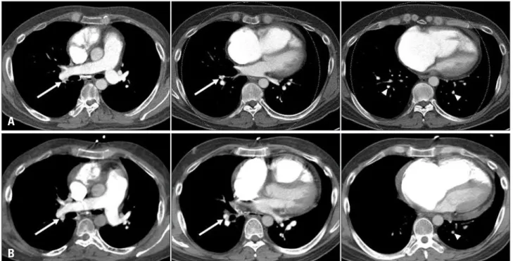
Fig. 1. A: Baseline computed tomographic pulmonary angiography (CTPA) performed 7 weeks before the index visit to emergency department (ED).
Transverse sections show chronic pulmonary embolism (PE) with eccentric filling defects (white arrows) in the right descending pulmonary artery and segmental artery of the right lower lobe. Multiple focal filling defects (arrowheads) are also seen in subsegmental arteries of the right lower and left lower lobes. B: CTPA performed immediately after the ED visit. No definite interval change is seen in the extent of pre-existing chronic PE.
B A
Journal of Cardiovascular Ultrasound 19 | December 2011
226
we suggest based on this case that in-situ thrombosis occur- ring slowly on chronic thromboemboli is difficult to detect with CTPA. As shown in Fig. 3, fresh thrombi were clearly demonstrated on operative findings, although CTPA could not suggest their presence. In this situation, clinical presenta- tion of the patient must be discrepant from anatomic burden of pulmonary emboli. In this particular condition, echocardiog-
raphy is able to guide therapeutic decision-making because it can reflect hemodynamic status of the patient, not simple ana- tomical thrombi burden.
Echocardiography is frequently performed in PE patients, because it permits reliable, qualitative, hemodynamic assess- ment of RV size, RV systolic function, and pulmonary arterial pressure.6)7) Prognostic information can be obtained with the help of echocardiography.8)9) Furthermore, in a critically ill pa- tient with worsening symptoms who cannot undergo CTPA without delay, bedside echocardiography contributes to early diagnosis and treatment of PE. Kucher et al.10) have suggested that echocardiographic signs of RV pressure overload and dys- function in a hemodynamically unstable patient with high pre- test probability of PE may justify invasive treatment for PE.
In this case, worsened hemodynamic parameters on serial echocardiography helped us to apply an invasive approach for recurrent PE. Since the patient was on a venovenous extracor- poreal membrane oxygenation system, intravenous thrombol- ysis was not considered an initial treatment option. Therefore, surgical embolectomy was performed, and an immediate he- modynamic improvement was achieved. This clinical decision was not supported by CTPA. As described above, CTPA can underestimate the burden of pulmonary thrombi, especially those that is distributed along the pulmonary arterial wall, at
Fig. 3. Surgical specimen of pulmonary embolectomy. Whitish-yellow organized thromboemboli with fresh red thrombi are clearly shown.
RPA: right pulmonary artery, RUL: right upper lobe, RML: right middle lobe, RLL: right lower lobe, LPA: left pulmonary artery, LUL: left upper lobe, LLL: left lower lobe.
Fig. 2. A: Modified apical four-chamber view of the transthoracic echocardiography shows an enlarged right ventricular (RV) cavity with depressed RV systolic function. B: Parasternal short-axis view demonstrates an enlarged RV with flattening of the interventricular septum deviated to the left ventricle (white arrows). C: Tricuspid regurgitant jet with a maximum velocity of 3.58 m/sec. RA pressure was assumed to be 20 mmHg based on inferior vena cava response to respiration, resulting in an estimated pulmonary arterial systolic pressure of 71 mm Hg. D: Follow-up echocardiography performed on the 6th postoperative day shows improved RV systolic function and (E) improved degree of interventricular septal flattening (arrow).
D A
E B
C
Importance of Echo in Pulmonary Embolism Assessment | Won-Seok Choe, et al.
227 the subsegmental artery level, and in-situ thrombosis on top
of chronic thrombi. Therefore, we believe that the present case is important and instructive in that hemodynamic as- sessment with echocardiography should be conducted to more accurately guide therapeutic strategy in clinically sus- pected PE patients.
References
1. Kucher N, Rossi E, De Rosa M, Goldhaber SZ. Massive pulmonary embolism. Circulation 2006;113:577-82.
2. Schoepf UJ, Goldhaber SZ, Costello P. Spiral computed tomography for acute pulmonary embolism. Circulation 2004;109:2160-7.
3. Stein PD, Fowler SE, Goodman LR, Gottschalk A, Hales CA, Hull RD, Leeper KV Jr, Popovich J Jr, Quinn DA, Sos TA, Sostman HD, Tapson VF, Wakefield TW, Weg JG, Woodard PK; PIOPED II Investigators. Multidetector computed tomography for acute pulmonary embolism. N Engl J Med 2006;354:2317-27.
4. Wells PS, Anderson DR, Rodger M, Ginsberg JS, Kearon C, Gent M, Turpie AG, Bormanis J, Weitz J, Chamberlain M, Bowie D, Barnes D, Hirsh J. Derivation of a simple clinical model to categorize pa-
tients probability of pulmonary embolism: increasing the models utility with the SimpliRED D-dimer. Thromb Haemost 2000;83:416-20.
5. Henzler T, Barraza JM Jr, Nance JW Jr, Costello P, Krissak R, Fink C, Schoepf UJ. CT imaging of acute pulmonary embolism. J Cardiovasc Comput Tomogr 2011;5:3-11.
6. Come PC. Echocardiographic evaluation of pulmonary embolism and its response to therapeutic interventions. Chest 1992;101:151S-62S.
7. Lee JH, Park JH. Role of Echocardiography in Patients with Acute Pul- monary Thromboembolism. J Cardiovasc Ultrasound 2008;16:9-16.
8. Casazza F, Bongarzoni A, Capozi A, Agostoni O. Regional right ven- tricular dysfunction in acute pulmonary embolism and right ventricular in- farction. Eur J Echocardiogr 2005;6:11-4.
9. Kurzyna M, Torbicki A, Pruszczyk P, Burakowska B, Fijałkowska A, Kober J, Oniszh K, Kuca P, Tomkowski W, Burakowski J, Wawrzyńska L. Disturbed right ventricular ejection pattern as a new Dop- pler echocardiographic sign of acute pulmonary embolism. Am J Cardiol 2002;90:507-11.
10. Kucher N, Luder CM, Dörnhöfer T, Windecker S, Meier B, Hess OM. Novel management strategy for patients with suspected pulmonary em- bolism. Eur Heart J 2003;24:366-76.