THE HAND
Nodular Melanoma on the Tip of the Thumb
Su Hyun Choi, Hong Bae Jeon, Ja Hea Gu
Department of Plastic Surgery, Dankook University Hospital, Cheonan, Chungnam, Korea
Received: September 21, 2016 Revised: [1] November 7 2016 [2] November 16 2016 Accepted: November 23, 2016 Correspondence to: Ja Hea Gu epartment of Plastic Surgery, Dankook University Hospital, 201 Manghyang-ro, Dongnam-gu, Cheonan 31116, Korea TEL: +82-41-550-3873
FAX: +82-41-554-6477 E-mail: [email protected]
This is an Open Access article distributed under the terms of the Creative Commons Attribution Non-Commercial License (http://creativecommons.org/licenses/bync/
3.0/) which permits unrestricted noncommercial use, distribution, and reproduction in any medium, provided the original work is properly cited.
Nodular type malignant melanoma is uncommon in fingers. In previous publications, treatment, diagnosis and case reports of subungal melanoma is often, however fingertip lesion was not focused. A 64-year-old woman who had a non-healing red and dark colored nodular mass with ulceration over the finger tip in the right thumb visited our clinics. Biopsy results was malignant melanoma then we performed amputation surgery of distal phalanx. Lymph node biopsy and resection margin was negative for melanoma. Chemotherapy was administered immediately. After 5 months, pulmonary nodular lesion was found and diagnosed as metastatic malignant melanoma by the wedge resec- tion surgery. The patient is treated for additional chemotherapy consistently and disease free for 2 years. Nodular type melanoma of the finger is uncom- mon and it could be presented as ulceration and amelanotic nodular mass.
Therefore we recommend biopsy to diagnose correctly if there are chronic non healing lesions on the fingers.
Keywords: Melanoma, Nodular melanoma, Chronic ulcer, Finger tip J Korean Soc Surg Hand 2016;21(4):238-242.
http://doi.org/10.12790/jkssh.2016.21.4.238
INTRODUCTION
The occurrence of malignant melanoma on fingers is rare;
however melanoma of the finger is a highly malignant tumor with rapid disease progression1. Acral malignant melanoma has a low incidence, accounting for 5% of total malignant melanomas1. Pathological symptoms are char- acterized by acral splash melanoma, early radioactive growth, shallow invasion and less likelihood of metasta- sis. Early treatment normally results in a favorable prog- nosis. In the late stage, the tumor exhibits vertical growth, a high rate of lymph node metastasis and poor prognosis.
However, early clinical symptoms of these patients are mild and easily overlooked. In addition, patients can eas- ily access their wounds, then they themselves treat their minor wounds, and the primary physician can misdiag- nose the condition as it is a nonspecific small lesion and it could be misunderstood as a minor traumatic lesion or pyogenic granuloma.
In melanomas, both clinical presentation and derma- toscopical features are extremely variable2. Melanomas have dermatoscopical features indicating their melano- cytic origin, such as pigment, aggregated brown or black globules, and site specific features on palms and soles.
and dermatoscopically relatively featureless, and in such cases even experienced dermatologists may have dif- ficulty recognizing the melanoma. In previous publica- tions, treatment, diagnosis and case reports of subungual melanoma were frequently reported; however, there was no focus on the fingertip lesion3.
We report a case of a nodular type malignant mela- noma in a 64-year-old woman, whose major complaints were skin ulceration and nodular lesion. The aim of this case report is to emphasize that in the differential diag- nosis of skin lesions that take a longer time to heal, or in cases of an amelanotic mass of unknown origin, it is necessary for clinicians to take malignant melanoma into consideration as a potential diagnosis.
CASE REPORT
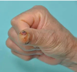
A 64-year-old woman visited our clinic with a complaint of a non-healing ulcer lesion over the ulnar side of the tip of the right thumb for 3 weeks. It appeared to have started as a nodule which had developed 9 months ago. The nod- ule gradually increased in size, and it caused pain and swelling with bullae on her right thumb. She tried to re- move the bullae herself, but the lesion did not resolve. On physical examination, the lesion was 5mm in diameter and its surface was red and dark in color and ulcerated (Fig. 1). There were no signs of infection. Based on the clinical appearance, a diagnosis of pyogenic granuloma
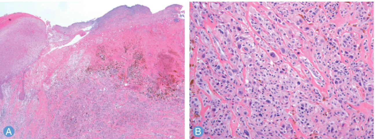
There was a dark colored nodular lesion inside the ulceration (Fig. 2). Histological examination revealed a malignant melanoma. Three pathologists reviewed the slides to confirm the diagnosis because of the presence of rare melanocytes (Fig. 3). Tumor depth was 5.5 mm and tumor cells with ulceration were positive for melanocytic markers. One week later, magnetic resonance imaging showed that postoperative scar tissue was detected on the ulnar side of the tip of the right thumb and there was no lesion infiltrating into the bone marrow space. We performed amputation surgery on interphalangeal joint level by institutional tumor board’s recommendation
Fig. 1. Preoperative view. The lesion was 5 mm in diameter and its surface was red and dark color, ulcerated.
Fig. 2. Intraoperative finding. (A) Nodular lesion was found inside the small ulceration (B) Diameter of the nodule was about 5.5 mm.
A B
considering patient size and lesion location (Fig. 4). In addition, axillary sentinel lymph node biopsy was per- formed. Frozen section reported that there was absence of melanoma in the lymph node and amputated product.
Positron emission tomography–computed tomography (PET-CT) was done to evaluate of distant metastasis.
The result demonstrated small sized pulmonary nodular opacity in right lower lobe posteior basal segment and the size was too small to characterize. A few small sized
lymph nodes with fat hilum along right axillary area without significant fluorodeoxyglucose uptake seemed to be reactive lymphadenitis. Chest CT scan found no evidence of metastasis with linear subsegmental atelec- tasis in left lower lobe. According to these results, this le- sion was staged on II-c.
Chemotherapy was administered immediately after the diagnosis of melanoma. However, contrast-enhanced computed tomography of the chest showed a nodular lesion in the right middle lobe, a finding that suggested the presence of metastases after 5 months. The nodular lesion was diagnosed as metastatic malignant melanoma by wedge resection surgery. The clinical findings of the patient and appearance of the masses were consistent with the diagnosis of nodular melanoma. The patient is being treated with chemotherapy consistently and no re- currence was detected for 2 years.
DISCUSSION
Acral lentiginous melanoma is the least common type among the 4 types of melanoma. However, it is a com- mon type of melanoma among Asians and dark-skinned individuals2. It is known to affect the glabrous skin, and it has a predilection for palms, soles and subungual areas.
Furukawa et al.3 and Kato et al.4 reported 15 and 34 cases of thumb melanoma and all these cases were located on the subungual or proximal site. Kuchelmeister et al.5 Fig. 3. Microphotograph of the tumor. (A) Low-power magnification reveals dermal infiltration of tumor cells with abundant
brownish pigment and skin ulceration (H&E stain, ×40). (B) Higher magnification of tumor cells demonstrates marked nuclear pleomorphism with macronucleoli (H&E stain, ×200).
A B
Fig. 4. Postoperative finding. Amputation was done at interpha- langeal joint level. (A) Volar side view of amputated thumb. (B) Dorsal view of stump.
A
B
casians and they showed that these acral type of mela- nomas were located on the palmar, plantar, subungual, and dorsal sites of the hands and feet. Nine percent of the nodular type of melanoma was detected, then authors suggested nodular melanoma could be occur on acral site. However, the lesion was limited on the palmar, plan- tar, subungal and dorsal site of the extremities. Finger tip melanoma is very rare, and it has not been focused or reported previously.
In this case, the patient overlooked her lesion and tried to remove it herself. The nodular lesion was increased in size and it caused painful swelling and ulceration. Based on the clinical appearance, a diagnosis of pyogenic gran- uloma was suspected and excisional biopsy was planned.
However, during the operation, the authors found that the nodule was dark colored, and hence, malignant mela- noma was suspected. As in a previous report, the patient was already in an advanced stage1. In order to make an early diagnosis, provide early treatment, and improve the survival period, a biopsy should be performed in all cases of suspected malignant melanoma of the finger that present with expanded areas of black patches, darkening of the patches, and appearance of uneven borders. The treatment of principal in digital melanoma for this pa- tient was amputation at one joint proximal to the mela- noma along with sentinel node biopsy. Authors followed
In conclusion, melanoma of the finger tip is uncom- mon and it could present with a nevus, ulceration, an amelanotic nodular mass, and pain. Therefore, we rec- ommend biopsy for correct diagnosis, if there is a chronic non-healing lesion on the fingers.
REFERENCES
1. Yang Z, Xie L, Huang Y, et al. Clinical features of malignant melanoma of the finger and therapeutic efficacies of dif- ferent treatments. Oncol Lett. 2011;2:811-5.
2. Situm M, Buljan M, Kolic M, Vucic M. Melanoma: clinical, dermatoscopical, and histopathological morphological characteristics. Acta Dermatovenerol Croat. 2014;22:1-12.
3. Furukawa H, Tsutsumida A, Yamamoto Y, et al. Melanoma of thumb: retrospective study for amputation levels, surgi- cal margin and reconstruction. J Plast Reconstr Aesthet Surg. 2007;60:24-31.
4. Kato T, Suetake T, Sugiyama Y, Tabata N, Tagami H. Epi- demiology and prognosis of subungual melanoma in 34 Japanese patients. Br J Dermatol. 1996;134:383-7.
5. Kuchelmeister C, Schaumburg-Lever G, Garbe C. Acral cutaneous melanoma in caucasians: clinical features, his- topathology and prognosis in 112 patients. Br J Dermatol.
2000;143:275-80.
6. Kozlow JH, Rees RS. Surgical management of primary dis- ease. Clin Plast Surg. 2010;37:65-71.
무지첨부에 발생한 결절성 흑색종
최수현·전홍배·구자혜
단국대학교 병원 성형외과
결절형 흑색종은 손가락에 드물게 발생한다. 이전에 발표된 문헌에서도 손톱하 결절성 흑색종은 진단이나 치료 등이 증 례 보고된 바 있으나 손가락 첨부에 발생한 경우는 없었다. 64세 여자 환자가 오른쪽 무지첨부에 장기간 낫지 않는 궤양 을 동반한 어두운 붉은색의 결절성 종양을 주소로 본원을 내원하였다. 조직 검사 결과 악성 흑색 종으로 확진되어 말단 지골의 절단 수술을 시행하였다. 림프절 생검 및 절제부위에서 추가로 흑색종이 발견되지는 않았으나 항암치료가 수술 후 바로 시행되었다. 그러나 5개월 후, 폐 결절 병변이 발견되었고 폐 쐐기 절제 수술에 의해 전이성 악성 흑색 종으로 진단되어 추가 화학 요법을 시행받고 2년간 재발하지 않고 있다. 손가락의 결절형 흑색 종은 드물고 궤양이나 흑색이 아닌 결절로 나타날 수 있다. 따라서 저자들은 손가락에 만성 비 치유 병변이 있는 경우는 반드시 조직 검사를 시행해야 한다고 생각한다.
색인단어: 흑색종, 결절형 흑색종, 만성 궤양, 수지 첨부
접수일 2016년 9월 21일 수정일 1차: 2016년 11월 7일, 2차: 2016년 11월 16일 게재확정일 2016년 11월 23일
교신저자 구자혜
충청남도 천안시 동남구 망향로 201 (31116) 단국대학교 병원 성형외과
TEL 041-550-3873 FAX 041-554-6477 E-mail [email protected]