ISSN 2234-3806 • eISSN 2234-3814
https://doi.org/10.3343/alm.2020.40.3.253
Isolation of Small Extracellular Vesicles From Human Serum Using a Combination of Ultracentrifugation With Polymer-Based Precipitation
Kyung Ju Ryu , Ph.D.1,2, Ji Young Lee , B.S.2, Chaehwa Park , Ph.D.1,2, Duck Cho , M.D., Ph.D.3, and Seok Jin Kim , M.D., Ph.D.1,4
1Department of Health Sciences and Technology, Samsung Advanced Institute for Health Sciences and Technology, Sungkyunkwan University, Seoul, Korea;
2Samsung Biomedical Research Institute, Samsung Medical Center, Seoul, Korea; 3Department of Laboratory Medicine and Genetics, Samsung Medical Center, Sungkyunkwan University School of Medicine, Seoul, Korea; 4Division of Hematology and Oncology, Department of Medicine, Samsung Medical Center, Sungkyunkwan University School of Medicine, Seoul, Korea
Methods for reproducibly isolating and enriching small extracellular vesicles (EVs) from blood are essential for clinical utilization of small EVs in cancer patients. We combined ul- tracentrifugation (UC) with polymer-based precipitation (ExoQuick [EQ] or Total Exosome Isolation [TEI] kit) to isolate small EVs (diameter, 30–150 nm) from the serum of breast cancer patients. We compared the performance of four cycles of UC (UC4x) with that of two cycles of UC followed by enrichment using the EQ (UC2x→EQ) or TEI (UC2x→TEI) kits. The mean concentration of small EVs isolated from 1 mL of serum using UC2x→EQ (139.0±29.1 μg) and UC2x→TEI (140.4±5.0 μg) did not differ from that obtained using UC4x (141.8±26.9 μg). The mean number of EV particles obtained using UC4x was 29.2±
9.9×10⁹ per mL of serum, whereas UC2x→EQ and UC2x→TEI yielded higher numbers of EVs (50.7±17.0×10⁹ and 59.3±20.6×10⁹, respectively). Concentrations of EV micro- RNAs, including miR-21 and miR-155, did not differ between the three methods. In con- clusion, performing UC prior to the use of polymer-based precipitation kits could be feasi- ble for isolating small EVs from human serum in large sample-based translational researches.
Key Words: Extracellular vesicle, Precipitation, Ultracentrifugation, Isolation, Performance
Received: May 31, 2019
Revision received: September 25, 2019 Accepted: November 14, 2019 Corresponding author:
Seok Jin Kim, M.D., Ph.D.
Division of Hematology-Oncology, Department of Medicine, Samsung Medical Center, Sungkyunkwan University School of Medicine, 81 Irwon-ro, Gangnam-gu, Seoul 06351, Korea
Tel: +82 2-3410-1766 Fax: +82 2-3410-1754 E-mail: [email protected]
© Korean Society for Laboratory Medicine This is an Open Access article distributed under the terms of the Creative Commons Attribution Non-Commercial License (http://creativecom- mons.org/licenses/by-nc/4.0) which permits unrestricted non-commercial use, distribution, and reproduction in any medium, provided the original work is properly cited.
Extracellular vesicles (EVs), small membrane-bound structures secreted by various types of cells, are involved in cell-to-cell com- munication by transferring cargos such as nucleic acids [1, 2].
The delivery of nucleic acids, including microRNAs (miRNAs), through EVs to target cells may promote tumor growth and me- tastasis [3]. The term ‘exosome’ is used to describe a popula- tion of small EVs (30–150 nm) that can be discriminated by size from microvesicles (100–1,000 nm) [4, 5]. However, the Mini- mal Information for Studies of Extracellular Vesicles 2018 study recommended the use of ‘small EVs’ rather than ‘exosomes’ for describing EVs sized <100 nm [6]. For clinical utilization of
small EVs in human disorders, it is essential to reproducibly iso- late and enrich small EVs from blood and body fluids [7, 8].
Currently, several methodologies exist for the isolation and analysis of small EVs including ultracentrifugation (UC), size-ex- clusion chromatography (SEC), immunoaffinity capture-based methods, and polymer-based precipitation kits [9-11]. Although UC has long been considered the gold standard for isolating small EVs, there is an unmet need for methods that can com- pletely separate small EVs from other nonvesicular entities such as apoptotic bodies.
Combinations of techniques have been evaluated in a bid to
2017-03-16 https://crossmark-cdn.crossref.org/widget/v2.0/logos/CROSSMARK_Color_square.svg
254 www.annlabmed.org https://doi.org/10.3343/alm.2020.40.3.253 enhance the isolation efficiency and enrichment of small EVs.
Recent studies have shown that coupling one cycle of UC with SEC provided better results than UC or SEC alone [12, 13], al- though serum protein contamination remained a problem when using this approach [13]. However, as studies utilizing human samples for clinical purposes usually require a large number of patients, polymer-based precipitation kits are used to isolate EVs from human samples because they do not require specialized equipment and have a large and scalable sample capacity. De- spite this, polymer-based precipitation has limited value because it results in larger amounts of non-EV contaminants, such as proteins and polymeric materials, than other methods [9]. To overcome this disadvantage, attempts have been made to use UC followed by enrichment using a polymer-based precipitation kit to isolate EVs from bovine milk; this combined method was reported to provide a better yield than polymer-based precipita- tion alone [14]. However, there are limited data on the efficiency of coupling UC with a polymer-based precipitation technique for isolating EVs from the blood of cancer patients.
We used an approach combining UC and enrichment using one of the two commercially available polymer-based precipita- tion kits (ExoQuick [EQ], System Biosciences, Mountain View, CA, USA; or Total Exosome Isolation [TEI], Invitrogen Life Tech- nologies, Carlsbad, CA, USA) to compare the isolation efficiency of UC alone (four cycles of UC [UC4x]) with that of UC followed by enrichment using EQ or TEI kits (two cycles of UC followed by the enrichment of small EVs using EQ [UC2x→EQ] or TEI [UC2x→TEI], respectively; Fig. 1A). The isolated small EVs were characterized according to the recommendations for EV research and definition [11].
Archived serum samples from breast cancer patients (N=30) that had been stored at -80°C were used (Institutional Review Board of Samsung Medical Center, File No. SMC 2017-12-068, study period (2017–2019); the nine biosamples were provided by Samsung Medical Center BioBank, 2018-0004). As a prepa- ration step, we first performed a brief round of low-speed cen- trifugation to eliminate cellular debris and large particles by se- quentially centrifuging 1 mL of serum at 2,000×g at 4°C for 10 minutes and 10,000×g at 4°C for 30 minutes. The supernatant was then filtered through a 0.22-μm filter and ultracentrifuged at 110,000×g for 120 minutes at 4°C.
For UC4x, the samples underwent three further cycles of UC (110,000×g) at 4°C for 70 minutes, and the pellet was discarded at each step (Fig. 1A). For UC2x→EQ and UC2x→TEI, the sam- ples underwent two cycles of UC (110,000×g) at 4°C for 120 and 70 minutes, respectively. As all tests were conducted on the same
sample, each group essentially comprised three samples con- taining 1 mL of serum. The resulting pellet was reconstituted in 0.5 mL of phosphate-buffered saline (PBS; Gibco, NY, USA) and added to 0.1 mL of EQ solution or TEI according to the manu- facturer’s protocols. The samples were incubated for 30 min- utes at 4°C and centrifuged twice at 1,500×g for 30 and 5 min- utes, respectively. The final pellet was reconstituted in 100 μL of PBS (Fig. 1A).
The particle number of the isolated small EVs was quantified using the nanoparticle tracking analysis (NTA), and the protein concentration was assessed by the bicinchoninic acid (BCA) method using the Pierce BCA Protein Assay kit (Thermo Fisher Scientific, Rockford, IL, USA). The small EVs were characterized by immunoblotting for the presence of EV markers (Western blot- ting) after the isolated EVs were lysed in RIPA buffer (0.5% so- dium deoxycholate, 1% Nonidet P-40, 150 mM NaCl, 50 mM Tris [pH 7.5], 0.1% sodium dodecyl sulfate [SDS], and 1 mM phenylmethylsulfonyl fluoride). Ten micrograms of each protein sample was electrophoresed on a 4–12% SDS polyacrylamide gel and incubated overnight at 4°C with the following antibodies:
anti-CD63 (sc-5275), anti-CD81 (sc-7637), anti-Alix (sc-53540), anti-TSG101 (sc-7964) (Santa Cruz Biotechnology, Santa Cruz, CA, USA), and anti-albumin (Cell signaling Technologies, Dan- vers, MA, USA). The morphology of the isolated small EVs was analyzed by transmission electron microscopy (TEM).
Because the nucleic acid cargo carried by small EVs is impor- tant for their potential role in cancer patients, we extracted the RNA from the small EVs to evaluate the concentration of EV cargo miRNAs. As miR-21, miR-101, miR-155, miR-223, and miR- 451a have been reported to be selectively enriched in EVs from patients with breast cancer, we measured their concentrations in small EVs [15]. Small EV cargo miRNA was analyzed after to- tal RNA was extracted from the isolated small EVs using the miRNeasy Micro Kit (Qiagen, Valencia, CA, USA), according to the manufacturer’s instructions. RNA concentration was mea- sured using a NanoDrop ND-100 Spectrophotometer (Nano- Drop Technologies, Wilmington, DE, USA). The RNA was re- verse-transcribed using the TaqMan microRNA Reverse Tran- scription Kit (Thermo Fisher Scientific) reagent containing spe- cific miRNA primers (Thermo Fisher Scientific) for five miRNAs (miR-21-5p, miR-101-3p, miR-155-5p, miR-223-3p, and miR- 451a), and real-time PCR (ABI PRISM 7900HT, Applied Biosys- tems, Foster City, USA) was performed in triplicate using the cDNA from each sample with 2× TaqMan Universal Master Mix II (with no AmpErase Uracil N-Glycosylase, UNG) and a 20 × TaqMan miRNA expression assay (Thermo Fisher Scientific).
The differences between the three methods were analyzed by ANOVA using SPSS (version 23.0; IBM SPSS Inc., Armonk, NY, USA). P <0.05 was considered significant, and two-sided tests were used in all calculations. Graphs were plotted using Graph- Pad Prism 5.0 (GraphPad Software, Inc., San Diego, CA, USA).
Analysis of supernatants after the first round of UC revealed protein bands, including albumin and immunoglobulin heavy and light chains, which diminished after the final step of each method (EQ, TEI, and U4, Fig. 1B). Silver-stained gels showed that the final pellets resuspended in PBS after the final step of the three methods (UC2x→EQ, UC2x→TEI, and UC4x) contained
extremely low concentrations of albumin compared with the se- rum sample, although several bands reflecting contaminating proteins were still observed (Fig. 1C). Western blotting for albu- min and CD63 using 10 μg of protein from each sample also showed that the ratio of albumin to CD63 was significantly lower in the end-product of the three methods than in the serum sam- ple (Fig. 1D). These findings indicate that UC coupled to EQ or TEI might achieve an enrichment of small EVs comparable to that obtained using UC alone, as non-EV-related proteins, such as albumin and immunoglobulins, could be eliminated. The mean concentration of small EVs isolated from 1 mL of serum by UC2x Fig. 1. Comparison of EV isolation methods. (A) Study overview: three methods for isolating EVs from serum samples of breast cancer pa- tients. (B) Silver-stained polyacrylamide gel of supernatants from the first to the final EV enrichment steps. (C) Silver-stained polyacrylamide gel of small EVs enriched after the final step. (D) Western blotting analysis for CD63 and albumin in serum and small EVs isolated using the three methods (UC4x: four cycles of UC; UC2x→EQ: two cycles of UC followed by enrichment using the EQ kit; UC2x→TEI: two cycles of UC followed by enrichment using the TEI kit). Ten micrograms of protein were used from each sample.
Abbreviations: EV, extracellular vesicle; UC, ultracentrifugation; EQ, ExoQuick; TEI, Total Exosome Isolation kit; PBS, phosphate-buffered saline; IgGH, immu- noglobulin G heavy chain; IgGL, immunoglobulin G light chain.
A
B C D
256 www.annlabmed.org https://doi.org/10.3343/alm.2020.40.3.253
→EQ and UC2x→TEI (139.0±29.1 μg and 140.4±5.0 μg, re- spectively) was not significantly different from that obtained us- ing UC4x (141.8±26.9 μg, Fig. 2A).
The concentration of small EVs we obtained was higher than that normally observed in other samples. This could be associ- ated with the type of underlying disorders, as the concentration of small EVs isolated using UC4x from 1 mL of serum from heal- thy people and lymphoma patients in our previous pilot experi- ments was approximately 30 μg and 70 μg, respectively (data not shown).
TEM imaging analysis showed that the small EVs isolated us-
ing the three methods were all rounded or cup-shaped and within the expected size range (Fig. 2B). The small EVs isolated using the three methods were then characterized by Western blotting for CD63, CD81, Alix, and TSG101. Although CD63 expression was slightly lower in the small EVs isolated using UC2x→EQ, the expression of the other EV markers, including Alix, CD81, and TSG101, did not differ significantly between the three methods (Fig. 2C).
The number and size of small EVs isolated by the three meth- ods were analyzed using a Nanosight NS300 instrument (Mal- vern Instruments, Worcestershire, UK) with NTA (Fig. 2D). The Fig. 2. Characterization of extracellular vesicle. (A) Protein concentrations in small EVs isolated using the three methods. (B) Transmission electron microscopy images of small EVs; size bar=100 nm. (C) Western blotting analysis for Alix, CD63, TSG101, and CD81 using 10 µg of small EVs isolated using the three methods (UC4x: four cycles of UC; UC2x→EQ: two cycles of UC followed by enrichment using the EQ kit; UC2x→TEI: two cycles of UC followed by enrichment using the TEI kit). (D) Size distribution of small EVs and comparison of three methods using nanoparticle tracking analysis. (E-G) Comparison of mean number of EV particles, mean size, and mode size among the three methods. The data represent the mean±SD of three independent experiments.
Abbreviations: see Fig. 1.
A B C
D
E F G
https://doi.org/10.3343/alm.2020.40.3.253 www.annlabmed.org 257
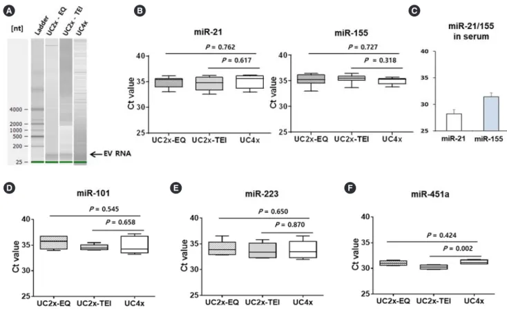
Fig. 3. Analysis of miRNA cargo in EV. (A) The quality of EV RNA was evaluated using an Agilent2100 bioanalyzer with an RNA 6000 Pico kit. (B) miR-21 and miR-155 concentrations in the small EVs did not differ among the three methods. (C) Serum concentrations of miR-21 and miR-155. (D-F) miR-101, miR-223, and miR-451a concentrations did not differ among the three methods. The data represent the mean±SD of three independent experiments. Boxes and whiskers indicate the minimum–maximum, and the lines represent the median of three independent experiments (B, D, E, and F).
Abbreviations: miRNA, microRNA; EV, extracellular vesicle; UC, ultracentrifugation; EQ, ExoQuick; TEI, Total Exosome Isolation kit; UC4x, four cycles of UC;
UC2x-EQ, two cycles of UC followed by enrichment using the EQ kit; UC2x-TEI, two cycles of UC followed by enrichment using the TEI kit.
15 267
Fig. 3. Analysis of miRNA cargo in EV. (A) The quality of EV RNA was evaluated using an 268
Agilent2100 bioanalyzer with an RNA 6000 Pico kit. (B) miR-21 and miR-155 269
concentrations in the small EVs did not differ among the three methods. (C) Serum 270
concentrations of miR-21 and miR-155. (D – F) miR-101, miR-223, and miR-451a 271
concentrations did not differ among the three methods. The data represent the mean ± SD of 272
three independent experiments. Boxes and whiskers indicate the minimum–maximum, and 273
the lines represent the median of three independent experiments (B, D, E, and F).
274
Abbreviations: miRNA, microRNA; EV, extracellular vesicle; UC, ultracentrifugation; EQ, 275
ExoQuick; TEI, Total Exosome Isolation kit; UC4x, four cycles of UC; UC2x-EQ, two 276
cycles of UC followed by enrichment using the EQ kit; UC2x-TEI, two cycles of UC 277
followed by enrichment using the TEI kit.
278 279
A B C
15 267
Fig. 3. Analysis of miRNA cargo in EV. (A) The quality of EV RNA was evaluated using an 268
Agilent2100 bioanalyzer with an RNA 6000 Pico kit. (B) miR-21 and miR-155 269
concentrations in the small EVs did not differ among the three methods. (C) Serum 270
concentrations of miR-21 and miR-155. (D – F) miR-101, miR-223, and miR-451a 271
concentrations did not differ among the three methods. The data represent the mean ± SD of 272
three independent experiments. Boxes and whiskers indicate the minimum–maximum, and 273
the lines represent the median of three independent experiments (B, D, E, and F).
274
Abbreviations: miRNA, microRNA; EV, extracellular vesicle; UC, ultracentrifugation; EQ, 275
ExoQuick; TEI, Total Exosome Isolation kit; UC4x, four cycles of UC; UC2x-EQ, two 276
cycles of UC followed by enrichment using the EQ kit; UC2x-TEI, two cycles of UC 277
followed by enrichment using the TEI kit.
278 279
D E F
mean number of EV particles was 29.2±9.9×10⁹ per mL of se- rum for UC4x, whereas UC2x→EQ and UC2x→TEI yielded a higher number of EV particles; however, there was no significant difference between UC2x→EQ (50.7±17.0×10⁹ per mL of se- rum) and UC2x→TEI (59.3±20.6×10⁹ per mL of serum, Fig.
2E). The mean diameters (UC2x→EQ: 131.3±17.4 nm, UC2x→
TEI: 127.4±11.8 nm, and UC4x: 130.5±23.1 nm) and mode diameters of the small EVs isolated using the three methods did not differ significantly (Fig. 2F, G).
Taken together, these results indicate that 1 mL of serum could be the minimum volume required for our new method, similar to the UC method. As we had no internal control for EV miRNA, we extracted RNA from equal amounts of serum and determined miRNA concentrations. The concentrations of miR-21 and miR- 155 in the small EVs isolated did not differ significantly among the three methods (Fig. 3A, B); however, their concentrations differed from those in serum samples (Fig. 3C). The difference
between serum concentrations of miRNA and EV cargo miRNA might be associated with the different roles of circulating free miRNAs and exosomal miRNAs. The concentrations of miR-101, miR-223, and miR-451a also did not differ among the three me- thods (Fig. 3D-F).
In conclusion, our results suggest that performing UC prior to using a polymer-based precipitation kit could be a feasible and convenient method for isolating small EVs from human serum in terms of yield and quality of EV cargos such as miRNAs. This approach may be useful for large sample-based translational re- searches, and further studies are warranted to validate its reli- ability for clinical application.
DATA AVAILABILITY
Please contact the corresponding author for access to the un- derlying data.
258 www.annlabmed.org https://doi.org/10.3343/alm.2020.40.3.253
ACKNOWLEDGEMENTS
We thank all our colleagues for participating in the research and express our gratitude to the 20-20 project of Samsung Medical Center and the National Research Foundation of Korea (NRF- 2017R1A2B4005136).
AUTHOR CONTRIBUTIONS
Conception and design: SJK. Acquisition of data and experiments:
KJR, JYL. Analysis and interpretation of data: KJR, CP, DC, SJK.
Drafting of manuscript: KJR, SJK. All authors read and approved the final manuscript.
CONFLICTS OF INTEREST
No potential conflicts of interest relevant to this article are re- ported.
RESEARCH FUNDING
This study was supported by a grant from the Basic Science Research Program through the National Research Foundation of Korea (NRF), which is funded by the Ministry of Education, Science, and Technology (NRF-2017R1A2B4005136).
ORCID
Kyung Ju Ryu https://orcid.org/0000-0003-0986-1475 Ji Young Lee https://orcid.org/0000-0003-4396-5091 Chaehwa Park https://orcid.org/0000-0001-7624-304X Duck Cho https://orcid.org/0000-0001-6861-3282 Seok Jin Kim https://orcid.org/0000-0002-2776-4401
REFERENCES
1. Sharma A, Khatun Z, Shiras A. Tumor exosomes: cellular postmen of cancer diagnosis and personalized therapy. Nanomedicine (Lond) 2016;
11:421-37.
2. EL Andaloussi S, Mäger I, Breakefield XO, Wood MJ. Extracellular vesi- cles: biology and emerging therapeutic opportunities. Nat Rev Drug Discov 2013;12:347-57.
3. Tkach M and Théry C. Communication by extracellular vesicles: where we are and where we need to go. Cell 2016;164:1226-32.
4. Cocucci E and Meldolesi J. Ectosomes and exosomes: shedding the confusion between extracellular vesicles. Trends Cell Biol 2015;25:364- 72.
5. Raposo G and Stoorvogel W. Extracellular vesicles: exosomes, microves- icles, and friends. J Cell Biol 2013;200:373-83.
6. Théry C, Witwer KW, Aikawa E, Alcaraz MJ, Anderson JD, Andriantsito- haina R, et al. Minimal information for studies of extracellular vesicles 2018 (MISEV2018): a position statement of the International Society for Extracellular Vesicles and update of the MISEV2014 guidelines. J Extra- cell Vesicles 2018;7:1535750.
7. Skog J, Würdinger T, van Rijn S, Meijer DH, Gainche L, Sena-Esteves M, et al. Glioblastoma microvesicles transport RNA and proteins that pro- mote tumour growth and provide diagnostic biomarkers. Nat Cell Biol 2008;10:1470-6.
8. Melo SA, Luecke LB, Kahlert C, Fernandez AF, Gammon ST, Kaye J, et al. Glypican-1 identifies cancer exosomes and detects early pancreatic cancer. Nature 2015;523:177-82.
9. Li P, Kaslan M, Lee SH, Yao J, Gao Z. Progress in exosome isolation techniques. Theranostics 2017;7:789-804.
10. Lobb RJ, Becker M, Wen SW, Wong CS, Wiegmans AP, Leimgruber A, et al. Optimized exosome isolation protocol for cell culture supernatant and human plasma. J Extracell Vesicles 2015;4:27031.
11. Lötvall J, Hill AF, Hochberg F, Buzás EI, Di Vizio D, Gardiner C, et al.
Minimal experimental requirements for definition of extracellular vesi- cles and their functions: a position statement from the International So- ciety for Extracellular Vesicles. J Extracell Vesicles 2014;3:26913.
12. Koh YQ, Almughlliq FB, Vaswani K, Peiris HN, Mitchell MD. Exosome enrichment by ultracentrifugation and size exclusion chromatography.
Front Biosci (Landmark Ed) 2018;23:865-74.
13. An M, Wu J, Zhu J, Lubman DM. Comparison of an optimized ultracen- trifugation method versus size-exclusion chromatography for isolation of exosomes from human serum. J Proteome Res 2018;17:3599-605.
14. Yamada T, Inoshima Y, Matsuda T, Ishiguro N. Comparison of methods for isolating exosomes from bovine milk. J Vet Med Sci 2012;74:1523-5.
15. Melo SA, Sugimoto H, O’Connell JT, Kato N, Villanueva A, Vidal A, et al.
Cancer exosomes perform cell-independent microRNA biogenesis and promote tumorigenesis. Cancer Cell 2014;26:707-21.