서 론
악성 종양 환자에서 림프절의 전이 유무를 확인하는 것은 초기에 정확한 병기 및 예후와 관련된 정보를 알 수 있고 수술 후 보조치
료의 필요성을 평가해서 조기에 치료를 할 수 있도록 해주기 때 문에 중요하다.1) 림프절 전이를 평가하기 위해 1992년 Morton 등2) 이 생체 염료를 이용하여 악성흑색종(malignant melanoma, MM) 에서 감시림프절의 정확성과 유용성을 보고하였고, 이후 많은 발 전이 이루어져 현재는 생체 염료 이외에도 방사성 동위원소, 감 마 탐색자 등을 사용한다.3,4) 1994년에는 Giuliano 등5)에 의해 유 방암에서 감시림프절 지도화가 널리 사용되게 되었고, 다른 여러 악성 종양에서도 림프절의 전이에 대한 많은 정보들이 축적됨에 따라 다양한 종양에서 감시림프절 생검에 대한 연구가 활발하게 진행되고 있다. 병기에 따라 일부 논란은 있지만 최근 연구들에
Copyright © 2019 by The Korean Orthopaedic Association
“This is an Open Access article distributed under the terms of the Creative Commons Attribution Non-Commercial License (http://creativecommons.org/licenses/by-nc/4.0/) which permits unrestricted non-commercial use, distribution, and reproduction in any medium, provided the original work is properly cited.”
The Journal of the Korean Orthopaedic Association Volume 54 Number 6 2019 Received December 18, 2018 Revised February 26, 2019
Accepted February 27, 2019
Correspondence to: Jae Woo Park, M.D.
Department of Orthopedic Surgery, Yeungnam University Medical Center, 170 Hyeonchung-ro, Nam-gu, Daegu 42415, Korea
TEL: +82-53-620-3640 FAX: +82-53-628-4020 E-mail: [email protected] ORCID: https://orcid.org/0000-0002-4346-8848
근골격계 악성 종양 환자의 림프절 전이 발견을 위한 양전자 방출 컴퓨터 단층 촬영기(Positron Emission
Tomography/Computed Tomography)의 유용성
신덕섭 • 나호동 • 박재우
영남대학교병원 정형외과
The Efficacy of Detecting a Sentinel Lymph Node through Positron Emission To mography/Computed Tomography
Duk-Seop Shin, M.D., Ph.D., Ho Dong Na, M.D., and Jae Woo Park, M.D.
Department of Orthopedic Surgery, Yeungnam University Medical Center, Daegu, Korea
Purpose: Lymph node metastasis is a very important prognostic factor for all skin cancers and some sarcomas. A sentinel lymph node (SLN) biopsy is the most useful technique for identifying SLNs. Recently, a new generation of diagnostic tools, such as single photon emission computed tomography/computed tomography (SPECT/CT) and positron emission tomography/CT (PET/CT) enabled the detection of SLNs.
This study compared the efficacy of PET/CT for detecting lymph node metastases with a SLN biopsy in a single medical center.
Materials and Methods: From 2008 to 2018, 72 skin cancers of sarcoma patients diagnosed with some lymph node involvement in a whole body PET/CT reading were assessed. Patients suspected of lymph node metastasis were sent to biopsy and those suspected to be reactive lesions were observed. The analysis was performed retrospectively using the medical records, clinical information, PET/CT readings, and pathology results.
Results: The age of patients ranged from 14 to 88 years and the mean follow-up period was 2.4 years. Twenty-two patients were suspected of a lymph node metastasis and confirmed. The sensitivity, specificity, positive predictive value and negative predictive value of PET/CT images in sarcoma and non-sarcoma tumors were increased significantly when the expert’s findings were considered together.
Conclusion: PET/CT is effective in detecting lymph node metastases.
Key words: skin neoplasm, sarcoma, sentinel lymph node, positron emission tomography/computed tomography
따르면 MM의 경우에는 감시림프절 생검술이 초기 종양 평가에 중요하다.6) MM을 제외한 피부 및 피부 부속기 악성 종양 중 림 프절로 전이될 가능성이 있는 종양으로는 편평상피세포암(squa- mous cell carcinoma, SCC), Merkel 세포암(Merkel cell carcinoma), 색소상피편평세포암(pigmented epitheloid melanocytoma) 등이 있 고,7) 각각 4.5%-28%,8-10) 16%-42%,11-13) 14%-46%14,15)에서 림프절 전이가 발견된다고 보고하였다. 최근, Matthey-Giè 등7)은 림프절 전이 유무가 강력한 생존 예측인자라는 가정하에 비흑색종 피부 암에서 감시림프절 생검술을 표준 절차로 시행하는 것이 좋다고 주장하였으나 대규모 임상 시험이 부족해서 림프절 전이에 대한 표준 검사가 아직 확립되지 않은 상태이다. 비록 감시림프절 절 제술이 근치적 림프절 절제술보다 부작용을 많이 감소시켰지만 여전히 창상 감염, 환부 혈종, 림프부종, 혈전 정맥염, 심부 정맥 혈전증 등이 발생할 수 있고,16) 수술 시간이 길어진다는 단점이 있기 때문에 일괄적으로 모든 피부의 악성 종양에서 감시림프절 생검술을 시행하기는 어려울 것으로 생각된다.
한편, 골 및 연부조직 육종 등에서는 림프절 전이 빈도도 낮고 이에 대한 연구가 많지 않다. Fong 등17)에 따르면 골 및 연부조직 육종의 약 2.6%에서 림프절 전이가 발견될 정도로 빈도가 낮고, 드물지만 상피성육종, 횡문근육종, 혈관육종 등에서 림프절 전이 가 있는 증례들이 보고되었다. 최근까지 일부 근골격계 악성 종 양에서 림프절 전이를 평가하는 데 있어서 감시림프절 생검술의 효용성과 중요성이 대두되고 있지만 림프절 전이의 이환율이 낮 은 육종에서 표준으로 시행하기에 이점이 있는지는 아직 불명확 하다.18)
이 같은 이유로 인해 감시림프절 생검술을 모든 정형외과 영역 의 악성 종양 환자에서 일괄적으로 시행하기에는 어려움이 있을 것으로 생각되어 감시림프절 생검술 대신 양전자 방출 컴퓨터 단 층 촬영기(positron emission tomography/computed tomography, PET/CT)가 정형외과 영역의 악성 종양 환자에서 림프절 전이를 발견하는 능력을 평가해 보고 임상적으로 유용할 것인지를 평가 해 보고자 한다.
대상 및 방법
2008년부터 2018년까지 영남대학교병원에서 골 및 연부조직 육 종, 피부 및 피부 부속기 악성 종양을 진단을 받은 후 PET/CT를 촬영한 환자 251명을 선별하였다. 이 중 다발성 전이암으로 판명 되거나 림프종 등으로 판명된 178명과 추적 관찰에 실패한 1명 은 제외하고, 72명의 환자를 대상으로 하였다. 이번 연구는 의무 기록, PET/CT 영상 자료와 판독 소견, 병리 소견 등을 바탕으로 후향적 분석을 시행하였다. 골 및 연부조직 악성 종양을 진단 받 은 모든 환자에서 PET/CT를 촬영하였으며, PET/CT는 핵의학 전 문의가 직접 판독하였다. PET/CT와 임상 소견을 종합적으로 평
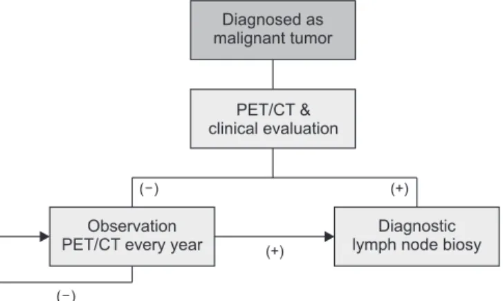
가해서 감시림프절로 생각되는 부위에 전이가 의심될 경우 감시 림프절 생검을 시행하였고, 그렇지 않은 경우는 감시림프절 생검 을 시행하지 않았다. 림프절 생검은 진단적 목적으로 병기 설정 을 위해 시행하였다. 초기 PET/CT상에서 림프절 전이가 의심되 지 않았다 하더라도 임상적으로 평가를 해서 림프절이 촉지되는 등의 의심 소견이 있는 경우에는 생검을 시행하였다. 처음에 림 프절의 전이가 없었던 환자군은 추시 관찰을 하면서 PET/CT를 촬영하여 이전에 없던 림프절(fluorine-18 fluoro-deoxy-glucose, FDG) 섭취 증가가 발견되면 생검을 통하여 전이 여부를 확인하 였다(Fig. 1). 원발 종양의 절제는 근골격계 종양학 전문의에 의 해 변연이 충분하도록 절제하였고, 신속 동결 조직절편검사상 경 계에 종양 침범이 없도록 하였다. 감시림프절 생검술은 원발 종 양의 절제와는 독립적으로 각 부위별 전문의에 의해 시행되었고, 수술 시간도 독립적으로 측정하였다. 감시림프절의 조직검사는 해부 병리학 전문의 1인이 평가하였고, PET/CT의 결과를 조직검 사 결과 및 임상경과와 비교하여 정확도를 평가하였다. PET/CT 영상에서 FDG 섭취 증가 여부를 확인하고, 핵의학 전문의와 근 골격계 종양학 전문의가 임상정보 등을 통하여 림프절 전이 유무 를 평가하였다. 초기 PET/CT에서 림프절 전이가 발견되지 않은 환자군을 추시 관찰하던 도중 림프절 전이가 발생한 경우 초기 병기 평가에서 감시림프절 생검을 하였다면 현미경적 전이를 확 인할 수 있었을 것이라는 가정하에 PET/CT가 림프절 전이를 발 견해 내지 못한 경우로 평가하였다. 림프절 생검을 시행한 환자 에서는 경과관찰하여 감시림프절 생검으로 인한 부작용을 확인 하였다.
결 과
전체 72명의 환자 중 남녀 비는 48명:24명이었고, 평균 연령은
Figure 1. Concepts of the study. All patients diagnosed with malignant tumor underwent positron emission tomography/computed tomography (PET/CT) and lymph node metastases were evaluated through a sentinel lymph node biopsy and clinical course.
57.8세로 확인되었다. 평균 추시 기간은 2.4년이었으며, 전체 환 자 중 22명에서 림프 전이가 발생하였다(Table 1). 12명은 감시림 프절 생검을 통해 림프절 전이를 확진하였고, 10명은 임상경과와 CT, 초음파 등의 영상 검사를 통해 림프절 전이를 확인하였다.
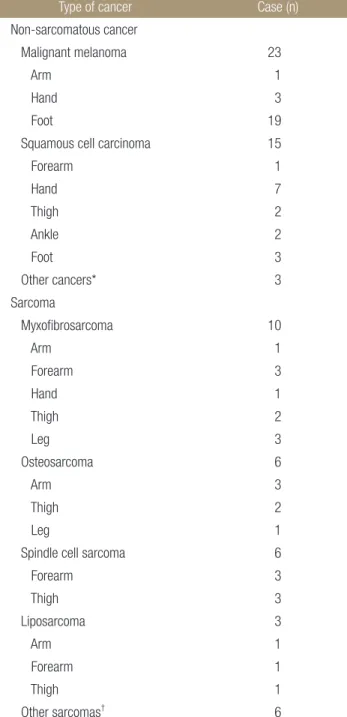
피부 및 피부 부속기 악성 종양 중에서는 MM이 23명으로 가 장 많았고, SCC는 15명이었다. 한선선암종(eccrine porocarcino- ma), 악성한선극세포종(malignant hidroacanthoma), 사마귀양암종 (verrucous carcinoma)은 각각 1명씩 있었다(Table 2). 육종 중에서 는 점액섬유육종이 10명으로 가장 많았고, 골육종과 방추세포육 종이 각각 6명이었다. 지방 육종은 3명, 악성말초신경초종양은 2 명이었고, 활막육종, 평활근육종, 상피성육종은 각 1명이었다. 그 외 2명이 있었다(Table 2).
초기 PET/CT상 34명에서 감시림프절로 생각되는 부위에 FDG 섭취 증가가 관찰되었다. 피부 및 피부 부속기암 중에서는 MM이 9명으로 가장 많았고, SCC가 4명, 한선선암종이 1명이었다. 육종 중에서는 점액섬유육종이 6명으로 가장 많았고, 골육종이 5명, 지 방육종이 3명이었다. 활막육종, 방추세포육종, 악성말초신경초종 양, 상피성육종이 각각 1명씩 있었고, 기타 미분류 육종 2명이었 다. 초기 PET/CT상에서 원발 종양 이외에 FDG 섭취 증가가 발견 되지 않았던 환자는 38명이었고, 이 중에서 31명은 최종 추시 관 찰까지 림프절 전이가 발견되지 않았다. 초기 PET/CT상에서 원 발 종양 이외에 FDG 섭취 증가가 발견되지 않았으나 추시 관찰 중에 림프절 전이가 새로 발견된 7명과 초기 PET/CT상에서 감시 림프절로 생각되는 부위에 FDG 섭취 증가가 보였던 34명에 대해 임상적으로 분석을 시행하였다(Table 3).
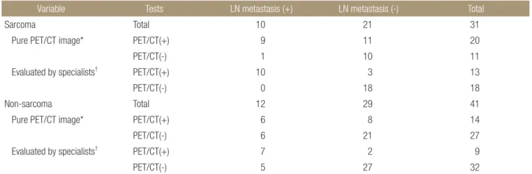
우선 림프절 전이 유무를 감시림프절 생검과 임상경과 및 CT, 자기공명영상(magnetic resonance imaging) 등 다른 영상 검사를 통해 평가한 경우를 확인하여 보았다. 육종 중에서 최종적으로 림프절에 전이가 있는 것으로 평가된 경우는 10명이었고, 림프절 에 전이가 없는 것으로 평가된 경우는 21명이었다. PET/CT 결과 와 이를 임상의사와 핵의학 및 영상의사가 판독한 결과는 Table 4 와 같다. 비육종성 악성 종양은 림프절에 전이가 있는 것으로 평 가된 경우는 12명, 없는 것으로 평가된 경우는 29명이었다. 마찬 가지로 PET/CT 결과와 이를 전문의가 판독한 결과는 Table 4와
같다. 이를 바탕으로 PET/CT 영상 소견의 민감도와 특이도, 양 성예측도, 음성예측도를 평가해보면 육종은 각각 90.0%, 47.6%, 45.0%, 90.9%이고, 비육종성 악성 종양의 경우는 각각 50.0%, 72.4%, 42.9%, 77.8%이다(Table 5). 이에 전문의의 소견을 추가하 면 육종과 비육종성 악성 종양 모두에서 민감도와 특이도, 양성 예측도, 음성예측도가 상승하였다(Table 5).
Table 1. Demographic Data
Variable Data
Sex (male:female) 48:24
Age (yr) 57.8 (14–88)
Follow-up period (yr) 2.4 Increased LN uptake in initial PET/CT 36
LN metastasis (+) 22
Values are presented as number only, median (range), or mean only. LN, lymph node; PET/CT, positron emission tomography/computed tomography.
Table 2. Types of Non-Sarcomatous Cancer and Sarcomatous Cancer
Type of cancer Case (n)
Non-sarcomatous cancer
Malignant melanoma 23
Arm 1
Hand 3
Foot 19
Squamous cell carcinoma 15
Forearm 1
Hand 7
Thigh 2
Ankle 2
Foot 3
Other cancers* 3
Sarcoma
Myxofibrosarcoma 10
Arm 1
Forearm 3
Hand 1
Thigh 2
Leg 3
Osteosarcoma 6
Arm 3
Thigh 2
Leg 1
Spindle cell sarcoma 6
Forearm 3
Thigh 3
Liposarcoma 3
Arm 1
Forearm 1
Thigh 1
Other sarcomas† 6
*Other cancers included verrucous carcinoma, eccrine porocarcinoma and malignant hidroacanthoma. †Other sarcomas included malignant peripheral nerve sheath tumor, synovial sarcoma, epitheloid sarcoma and unclassified sarcoma.
Table 3. Patients Who Were Positive in at Least One Test in PET/CT and SLNB Case No.SexAge (yr)HistologyLocationInitial PET/CTPET/CT with specialist’s advicesFinal LN metastasisDiagnostic method for final LN metastasisSLNB timeSide effect of SLNB 1F79MMLt. toe--+Clinically/other imaging study-N/A 2*F74MMLt. sole--+SLNB5 h 25 minWound problem, hematoma 3*M72MMLt. sole--+SLNB2 h 10 minLymphedema 4F88MMRt. toe+++Clinically/other imaging study-N/A 5*M73MMRt. heel--+SLNB2 h 50 minLymphedema 6M53MMLt. heel--+Clinically/other imaging study-N/A 7*M60MMRt. finger+++SLNB1 h 30 min- 8F75MMLt. sole+--Clinically-N/A 9*M67MMRt. toe+++SLNB3 h 50 minLymphocele 10F55MMLt. heel+--Clinically/other imaging study-N/A 11F52MMRt. heel+--Clinically/other imaging study-N/A 12M27MMLt. foot+--Clinically/other imaging study-N/A 13*M58SCCRt. finger++-FNANull- 14*M71SCCLt. forearm-++SLNB1 h 15 minWound problem 15*M48SCCRt. heel+++SLNB2 h 55 min- 16M85SCCLt. thigh+++Clinically/other imaging study-N/A 17*M80MyxofibrosarcomaRt. leg-++SLNB1 h 5 min- 18M24LiposarcomaRt. forearm+--Clinically/other imaging study-N/A 19M65MyxofibrosarcomaRt. thigh+--Clinically/other imaging study-N/A 20M60MMLt. foot+--Clinically/other imaging study-N/A 21F74MyxofibrosarcomaRt. forearm+--Clinically/other imaging study-N/A 22M79MyxofibrosarcomaLt. hand+++Clinically-N/A 23M31OsteosarcomaRt. distal humerus+++Clinically/other imaging study-N/A 24F17OsteosarcomaRt. distal femur+--Clinically/other imaging study-N/A 25F21Malignant triton tumorLt. leg+++Clinically-N/A 26*F75Synovial sarcomaRt. ankle+++SLNB1 h 45 min- 27*F70SCCLt. ankle++-SLNB 30 min- 28M17OsteosarcomaRt. distal femur+++Clinically-N/A 29M75MyxofibrosarcomaRt. forearm+--Clinically/other imaging study-N/A 30*M15OsteosarcomaLt. humerus++-SLNBNull-
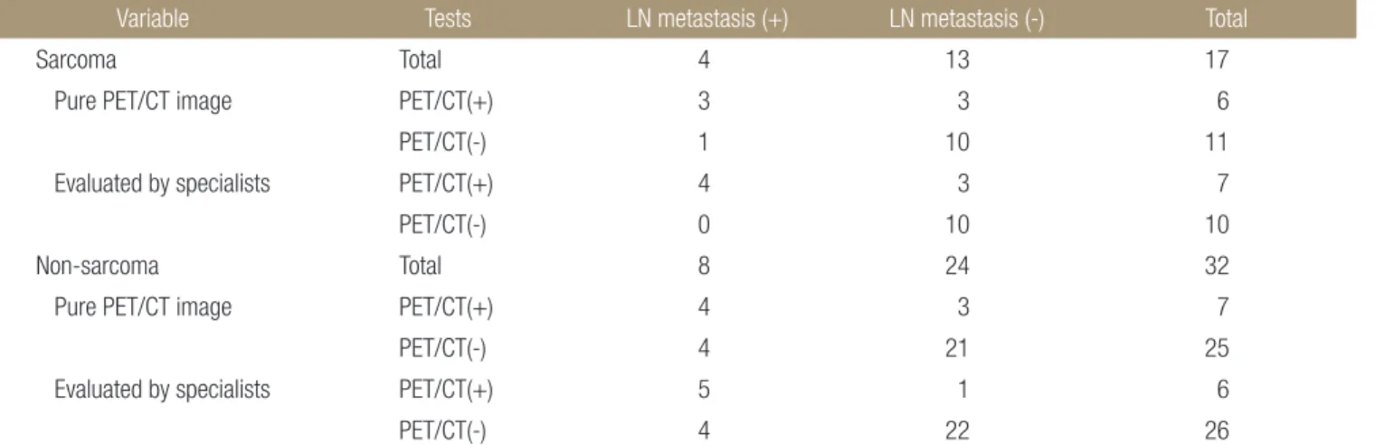
다음으로는 림프절 전이가 생검 이외의 다른 방법을 통해 있 다고 평가된 경우를 제외하고 최종적으로 병리검사를 통해 림프 절 전이 유무를 확인한 18명과 최종 추시까지 전혀 전이 및 재발 이 발견되지 않은 환자를 대상으로 평가하였다(Table 6). 전문의 의 소견을 참고로 한 경우, 위의 경우와 마찬가지로 육종과 비육 종성 악성 종양 모두에서 정확도가 상승하는 것을 확인할 수 있 었다(Table 7).
감시림프절 생검을 통하여 현미경적으로 림프절 전이 여부를 확인한 18명 중에서 2예는 세침흡인술을 통하여 확인하였다. 그 리고 감시림프절 생검술을 시행한 환자 중 13명은 독립적으로 시 간을 측정하였고, 평균 약 2.5시간이 소요되었다. 감시림프절 생 검술을 하고 나서 총 6건의 합병증이 발생하였고, 림프부종이 3 건, 국소 염증이나 부종 등 수술 부위 상처와 관련된 문제가 2건 있었으며 림프류가 1건 있었다(Table 3).
고 찰
악성 종양 환자에서 림프절의 전이 유무를 확인하는 것은 초기에 정확한 병기 및 예후와 관련된 정보를 알 수 있을 뿐 아니라 수술 후 보조치료의 필요성을 평가하고 이와 관련된 치료를 조기에 시 행할 수 있게 하기 때문에 중요하다. 림프절 전이를 평가하는 방 법 중에 감시림프절 절제술은 최근 유방암, MM 등의 종양에서 효용성이 인정되어 초기 종양 평가 시에 널리 사용되고 있다.
특히 MM은 임상적으로 림프절 전이가 없을 것으로 여겨지더 라도 실제로 림프절 전이가 발견되는 빈도가 많게는 24%까지 보 고되고 있어,2,19-21) 림프절 전이를 확인하는 방법에 관해 연구가 이미 많이 진행되었으며, 감시림프절 생검술이 권장되고 있다. 흑 색종 이외의 피부 및 피부 부속기 암의 경우는 대규모 임상 시험 이 없어서 아직 정립되어 있지는 않다. 최근에는 감시림프절 생 검술이 일부 악성 종양에서 초기 진단 시에 표준 절차로 권장될 정도로 정확도가 높고, 불필요한 림프절 곽청술로 인해 발생하 는 합병증을 최소화할 수 있어서,21-23) 감시림프절 생검술을 다른 악성 종양으로도 적용 범위를 넓힐 수 있는가에 대한 연구가 활 발히 진행되고 있다. 비록 감시림프절 절제술이 림프절을 진단해 내는 데 정확도가 높고, 림프절 곽청술보다 합병증을 감소시켰지 만 여전히 림프부종, 상처 감염이나 회복 지연 등의 위험이 있으 며, 림프절 전이가 흔하지 않은 종양의 경우에는 일반적으로 시 행하기에는 절차가 번거롭고, 수술 시간이 더 소요된다. Wright- son 등16)에 따르면 감시림프절 생검술을 시행한 약 4.6%에서 합병 증이 발생하였다고 보고하였다. 혈종이 2.3%로 가장 많았고, 상처 벌어짐 및 상처 감염 등의 상처 연관 합병증이 1.3%로 뒤를 이었 으며, 그 외에도 림프부종, 심부 정맥 혈전증 등이 있었다. 본 연 구에서는 감시림프절 생검술을 하고 나서 총 6건의 합병증이 발 생하였고, 림프부종이 3건, 국소 염증이나 부종 등 수술 부위 상 Table 3. Continued Case No.SexAge (yr)HistologyLocationInitial PET/CTPET/CT with specialist’s advicesFinal LN metastasisDiagnostic method for final LN metastasisSLNB timeSide effect of SLNB 31F54MyxofibrosarcomaRt. leg+--Clinically-N/A 32*F20LiposarcomaLt. thigh++-SLNB2 h 25 min- 33M63Spindle cell sarcomaLt. forearm+--Clinically-N/A 34M81LiposarcomaRt. humerus+++Clinically-N/A 35M44Unclassified sarcomaLt. thigh+--Clinically-N/A 36F82Unclassified sarcomaLt. medial thigh+++Clinically-N/A 37*M74MyxofibrosarcomaRt. forearm++-SLNBNull- 38*M18OsteosarcomaLt. shoulder+++SLNBNull- 39*M37Eccrine porocarcinomaRt. sole+--FNANull 40*M47Epitheloid sarcomaRt. forearm+++SLNB2 hr- 41*M67MMRt. heel+++SLNB3 h 55 minLymphedema All 31 patients who had no lymph node (LN) metastasis in the positron emission tomography/computed tomography (PET/CT) and the sentinel LN biopsy are excluded from this Table. Patients with at least one positive result in PET/CT or sentinel LN biopsy (SLNB) were summarized. *Biopsies were performed to confirm the presence of metastases. F, female; M, male; MM, malignant melanoma; SCC, squamous cell carcinoma; Lt., left; Rt., right; FNA, fine needle aspiration; N/A, not applicable.
처와 관련된 문제가 2건 있었으며 림프류가 1건 있었다. 또 감시 림프절 생검술은 평균 2.5시간이 더 소요되었다. 따라서 본 연구 에서는 종양의 림프절 전이를 평가하는 데 절차가 비교적 간편하 고, 환자 마취가 따로 필요없으며, 합병증 위험도 훨씬 적은 PET/
CT가 감시림프절 생검술을 대체할 수 있을지를 평가해 보았다.
Wagner 등24)은 임상적인 정보를 완전히 배제하고 PET/CT가 림프절 전이 발견에 대한 정확도를 평가하였다. PET/CT상에서 조금이라도 림프절에 전이가 의심될 경우를 양성으로 평가하였 을 때 민감도 16.7%, 특이도 94.4%, 양성예측도 42.9%, 음성예측
도 81.7%로 평가되었고, PET/CT의 결과를 핵의학 전문의가 분 석하였을 때는 민감도 11.1%, 특이도 100%, 양성예측도 100%, 음 성예측도 81.6%로 나타났다. Rinne 등25)은 임상정보 및 이학적 정 보를 바탕으로 PET/CT가 림프절의 전이를 발견하는 데 대한 정 확도를 평가하였고, 민감도가 97.1%, 특이도가 100%로 나타났다.
El-Maraghi와 Kielar26)에 따르면, PET/CT가 MM에서 감시림프절 생검술을 대체하기에는 아직 근거가 부족하다고 하였다. 하지만 이번 연구결과에서 임상정보를 참고로 할 경우 PET/CT의 민감 도와 특이도가 상승하는 것을 확인할 수 있다. 본 연구에서 비육 종성 악성 종양을 임상정보 없이 PET/CT 영상만으로 평가했을 때보다 임상정보를 참고로 하고 핵의학 및 근골격계 종양 전문의 의 소견을 참고로 하면, 민감도와 특이도가 상승하는 것을 확인 할 수 있었다(Table 5, 6). 또, 전문의의 소견을 참고로 할 경우 양 성예측도와 음성예측도가 각각 83.3%, 84.6%로 평가되었다. 이는 비육종성 악성 종양에서 PET/CT상에서 림프절 전이가 있는 경 우에 실제로 있을 확률과 PET/CT상에서 림프절 전이가 없는 경 우에 실제로 전이가 없을 확률이 상당히 높음을 나타내며, 악성 종양 진단 시에 일괄적으로 침습적인 검사방법인 감시림프절 생 검을 하기에 앞서 PET/CT를 먼저 촬영하여 감시림프절 생검이 필요한 환자들을 선별해내는 검사로 유용할 수 있음을 시사한다.
근골격계 육종의 경우 림프절의 전이 빈도가 낮고, 국소 침범 이나 혈행 전이가 더 많기 때문에 이에 대한 연구가 많이 시행되 지 않았다.17,18) Weingrad와 Rosenberg27)는 1978년에, Mazeron과 Suit28)는 1987년에 육종 전이에 대한 논문을 요약하였는데, 각각 47건과 122건의 연구결과를 종합해 보면 림프절 전이의 전체 빈 도가 9.1%, 10.8%로 나타났고,17) 육종에서 림프절을 통한 전이 빈 도가 낮기 때문에 모든 환자에서의 감시림프절 절제술은 의미가 Table 4. Sarcoma and Non-Sarcoma Cases
Variable Tests LN metastasis (+) LN metastasis (-) Total
Sarcoma Total 10 21 31
Pure PET/CT image* PET/CT(+) 9 11 20
PET/CT(-) 1 10 11
Evaluated by specialists† PET/CT(+) 10 3 13
PET/CT(-) 0 18 18
Non-sarcoma Total 12 29 41
Pure PET/CT image* PET/CT(+) 6 8 14
PET/CT(-) 6 21 27
Evaluated by specialists† PET/CT(+) 7 2 9
PET/CT(-) 5 27 32
Lymph node (LN) metastases were confirmed by biopsy, clinical information, and imaging findings. *LN metastases were first evaluated by the pure positron emission tomography/computed tomography (PET/CT) image and a sentinel LN biopsy was performed to evaluate the LN metastasis.
†Additionally, the oncologic specialists including the doctors of nuclear medicine, orthopedic science and imaging medicine evaluated the presence of the LN metastasis using PET/CT. The cases corresponding to each were classified.
Table 5. Sensitivity, Specificity, PPV, and NPV of Sarcoma and Non- Sarcoma Cases
Variable Pure PET/CT
image (%)
Evaluated by specialists (%) Sarcoma
Sensitivity 90.0 100.0 Specificity 47.6 85.7
PPV 45.0 76.9
NPV 90.9 100.0
Non-sarcoma
Sensitivity 50.0 58.3 Specificity 72.4 93.1
PPV 42.9 77.8
NPV 77.8 84.4
Lymph node metastases were confirmed by biopsy, clinical information, and imaging findings. PPV, positive predictive value; NPV, negative predictive value; PET/CT, positron emission tomography/computed tomography.
없다고 평가하였다.27) 하지만 Blazer 등18)에 따르면, 일부 고등급 (high grade) 육종의 경우 국소 림프절 전이 경향을 보이는 경우가 있고, 이 경우에는 림프절 전이를 확인할 필요가 있다고 제시하 고 있다. Ross 등29)은 상피성육종이 국소 림프절 전이가 44%까지 발견된다고 보고하였고, Blazer 등18)은 림프절 전이가 생존율 예 측에 독립적인 예후 인자로 나타났다고 보고하였다. 따라서 상피 성육종의 경우 림프절의 전이에 대한 평가를 해야 하고 이를 통 해 조기에 전신 치료를 받을 수 있도록 해야 한다고 Halling 등30) 이 보고하였다. 활막육종의 경우 2%-17%까지 림프절 전이가 발 견된다고 보고하였고,17,27,28) 최근 보고서에 따르면 종양의 완전 절 제 뿐만 아니라 림프절 전이의 평가가 중요하다고 주장하였다.
이 외에도 투명세포종도 림프절 전이를 평가하는 것이 의미가 있 을 것으로 평가하였다.18) 육종에서 림프절 전이를 평가하기 위하 여 Blazer 등18)은 감시림프절 생검술을 추천하였고, 횡문근육종 의 경우에는 아직 감시림프절 생검술의 이득이 명확하게 입증되 지 않았다고 보고하였다. 육종의 하위분류별로 감시림프절 생검 의 정확도에 대해서는 그 증례가 많지 않아서 아직 통계적으로는 평가된 바 없고, 단지 림프절 전이에 대한 평가가 필요할 것으로 생각되는 육종에서는 감시림프절 생검술을 추천하는 정도이다.
이에 대해 본 연구에서는 육종에서 림프절 전이를 평가하는 데 PET/CT의 능력을 추가로 평가해 보았다. 본 연구에서는 림프절 에 전이가 발견된 육종은 골육종 3예, 점액섬유육종 2예, 상피육 종 1예, 악성말초신경초종양 1예, 활막육종 1예, 지방육종 1예가 있었다. 육종의 경우도 마찬가지로, 전문의의 소견을 참고로 하면 PET/CT가 림프절 전이를 발견해내는 데 민감도와 특이도가 모 두 상승하였다. 육종의 경우는 병리학적 방법으로 림프절 전이를 확인을 한 경우만을 평가했을 때 양성예측도는 57.1%로 평가되 었고, 음성예측도는 100%로 확인되었다. 따라서 육종에서는 선별 검사로서의 PET/CT는 부족한 것으로 생각되나 PET/CT상에서 림프절에 전이가 발견되지 않은 경우는 실제로도 전이가 없을 확 률이 매우 높으므로 PET/CT를 감시림프절 생검을 하기에 앞서 서 시행함으로써 불필요한 감시림프절 생검술을 시행하지 않을 수 있도록 해 줄 것이다. 하지만 육종의 경우는 육종의 종류에 따 라 성질이 다르므로 더 정확한 평가를 위해서는 증례를 보충하여 각 육종의 종류에 따른 림프절 전이를 발견해내는 PET/CT의 정 확도를 평가해 볼 필요가 있을 것으로 여겨진다.
이번 연구는 후향적인 연구로 초기 PET/CT에서 림프절의 전 이가 없는 경우라도 추적 PET/CT 촬영이나 다른 영상 검사 혹은 임상경과를 통해 추후에 림프절 전이가 의심되는 경우는 수술적 으로 림프절을 절제하였고, 조직검사에서 림프절에 전이가 확인 Table 6. Sarcoma and Non-Sarcoma Cases
Variable Tests LN metastasis (+) LN metastasis (-) Total
Sarcoma Total 4 13 17
Pure PET/CT image PET/CT(+) 3 3 6
PET/CT(-) 1 10 11
Evaluated by specialists PET/CT(+) 4 3 7
PET/CT(-) 0 10 10
Non-sarcoma Total 8 24 32
Pure PET/CT image PET/CT(+) 4 3 7
PET/CT(-) 4 21 25
Evaluated by specialists PET/CT(+) 5 1 6
PET/CT(-) 4 22 26
Cases of confirmed lymph node (LN) metastases by non-biopsy method were excluded. PET/CT, positron emission tomography/computed tomography.
Table 7. Sensitivity, Specificity, PPV, and NPV of Sarcoma and Non- Sarcoma Cases
Variable Pure PET/CT
image (%)
Evaluated by specialists (%) Sarcoma
Sensitivity 75.0 100.0
Specificity 76.9 76.9
PPV 50.0 57.1
NPV 90.9 100.0
Non-sarcoma
Sensitivity 50.0 55.6
Specificity 87.5 95.7
PPV 57.1 83.3
NPV 84.0 84.6
Lymph node metastases were confirmed by biopsy only. PPV, positive predictive value; NPV, negative predictive value; PET/CT, positron emission tomography/computed tomography.
된 경우는 초기 PET/CT가 림프절의 전이를 발견해 내는 데 실패 한 것으로 평가하였는데 이로 인하여 PET/CT의 민감도가 감시 림프절 절제술에 비해서 저평가되었을 가능성이 있다. 또한 현실 적으로 모든 환자에서 감시림프절 생검을 수행하는 것이 불가능 하여 이를 보완하기 위해 임상경과, 추적 PET/CT, 다른 영상 소 견들을 바탕으로 림프절의 전이 유무를 평가하는 방법을 사용하 였는데 이것이 이 논문의 한계점으로 여겨진다. 이를 보완하기 위하여 림프절의 전이가 병리소견으로 확인된 경우만을 모아서 추가로 한번 더 평가하였다(Table 6, 7).
이번 연구를 통해 PET/CT가 임상정보와 전문가의 소견을 참 고로 할 경우 검사의 정확도가 상승하며, 양성예측도와 음성예측 도 모두 상승하는 것을 확인할 수 있었다. 비육종성 종양과 육종 모두에서 침습적인 검사인 감시림프절 생검을 하기 전에 시행함 으로써 감시림프절 생검이 실제로 필요한지를 판단하는 방법으 로 유용할 수 있을 것으로 생각된다. 비육종성 종양의 경우는 양 성예측도가 높으므로 PET/CT를 찍어서 감시림프절 생검술을 시 행할 환자들을 선별하는 검사로도 활용이 가능할 것으로 생각되 며, 육종의 경우는 양성예측도가 낮아서 선별검사로는 충분치 않 으나 PET/CT상에서 림프절 전이가 없는 경우는 굳이 불필요한 감시림프절 생검을 하지 않아도 될 것으로 여겨진다. 이를 통해 림프절의 전이가 없는 환자에서 불필요한 감시림프절 생검술을 하지 않을 수 있어 감시림프절 생검술의 부작용을 피할 수 있을 것으로 생각된다.
결 론
이번 연구 결과 아직 PET/CT가 단독으로는 정형외과 영역의 악 성 종양 환자의 림프절 전이를 진단하는 데 완전히 감시림프절 생검술을 대체하기에는 부족할 것으로 생각된다. 하지만 전문가 의 평가가 함께 이루어진다면, PET/CT의 민감도와 특이도, 양성 예측도, 음성예측도가 모두 상승하는 것을 확인할 수 있었다. 추 후 더 많은 증례를 추가하고, 종양별로 나누어 분석을 하면 PET/
CT가 육종 및 비육종성 악성 종양의 림프절 전이를 평가하는 데 좋은 검사법이 될 수 있을 것으로 생각한다. 임상적으로는 감시 림프절 생검을 모든 환자에서 일괄적으로 시행하기보다 PET/CT 를 먼저 촬영하여 감시림프절 절제술이 필요한지를 평가하는 검 사로써 유용할 수 있을 것으로 생각된다.
CONFLICTS OF INTEREST
The authors have nothing to disclose.
REFERENCES
1. Kawada K, Taketo MM. Significance and mechanism of lymph node metastasis in cancer progression. Cancer Res.
2011;71:1214-8.
2. Morton DL, Wen DR, Wong JH, et al. Technical details of intraoperative lymphatic mapping for early stage melanoma.
Arch Surg. 1992;127:392-9.
3. Gershenwald JE, Tseng CH, Thompson W, et al. Improved sentinel lymph node localization in patients with primary melanoma with the use of radiolabeled colloid. Surgery.
1998;124:203-10.
4. Alex JC, Weaver DL, Fairbank JT, Rankin BS, Krag DN.
Gamma-probe-guided lymph node localization in malignant melanoma. Surg Oncol. 1993;2:303-8.
5. Giuliano AE, Kirgan DM, Guenther JM, Morton DL. Lym- phatic mapping and sentinel lymphadenectomy for breast cancer. Ann Surg. 1994;220:391-8; discussion 398-401.
6. Wong SL, Balch CM, Hurley P, et al. Sentinel lymph node biopsy for melanoma: American Society of Clinical Oncol- ogy and Society of Surgical Oncology joint clinical practice guideline. Ann Surg Oncol. 2012;19:3313-24.
7. Matthey-Giè ML, Boubaker A, Letovanec I, Demartines N, Matter M. Sentinel lymph node biopsy in nonmelanoma skin cancer patients. J Skin Cancer. 2013;2013:267474.
8. Ross AS, Schmults CD. Sentinel lymph node biopsy in cuta- neous squamous cell carcinoma: a systematic review of the English literature. Dermatol Surg. 2006;32:1309-21.
9. Liu YY, Rozen WM, Rahdon R. Sentinel lymph node biopsy for squamous cell carcinoma of the extremities: case report and review of the literature. Anticancer Res. 2011;31:1443-6.
10. Renzi C, Caggiati A, Mannooranparampil TJ, et al. Sentinel lymph node biopsy for high risk cutaneous squamous cell carcinoma: case series and review of the literature. Eur J Surg Oncol. 2007;33:364-9.
11. Allen PJ, Bowne WB, Jaques DP, Brennan MF, Busam K, Coit DG. Merkel cell carcinoma: prognosis and treatment of pa- tients from a single institution. J Clin Oncol. 2005;23:2300-9.
12. Ames SE, Krag DN, Brady MS. Radiolocalization of the sen- tinel lymph node in Merkel cell carcinoma: a clinical analysis of seven cases. J Surg Oncol. 1998;67:251-4.
13. Gupta SG, Wang LC, Peñas PF, Gellenthin M, Lee SJ, Nghiem P. Sentinel lymph node biopsy for evaluation and treatment of patients with Merkel cell carcinoma: the Dana-Farber ex-
perience and meta-analysis of the literature. Arch Dermatol.
2006;142:685-90.
14. Zembowicz A, Carney JA, Mihm MC. Pigmented epithelioid melanocytoma: a low-grade melanocytic tumor with meta- static potential indistinguishable from animal-type melano- ma and epithelioid blue nevus. Am J Surg Pathol. 2004;28:31- 40.
15. Magro CM, Crowson AN, Mihm MC Jr, Gupta K, Walker MJ, Solomon G. The dermal-based borderline melano- cytic tumor: a categorical approach. J Am Acad Dermatol.
2010;62:469-79.
16. Wrightson WR, Wong SL, Edwards MJ, et al. Complications associated with sentinel lymph node biopsy for melanoma.
Ann Surg Oncol. 2003;10:676-80.
17. Fong Y, Coit DG, Woodruff JM, Brennan MF. Lymph node metastasis from soft tissue sarcoma in adults. Analysis of data from a prospective database of 1772 sarcoma patients. Ann Surg. 1993;217:72-7.
18. Blazer DG 3rd, Sabel MS, Sondak VK. Is there a role for sentinel lymph node biopsy in the management of sarcoma?
Surg Oncol. 2003;12:201-6.
19. Nowecki ZI, Rutkowski P, Nasierowska-Guttmejer A, Ruka W. Sentinel lymph node biopsy in melanoma patients with clinically negative regional lymph nodes: one institution's ex- perience. Melanoma Res. 2003;13:35-43.
20. Thompson JF, McCarthy WH, Bosch CM, et al. Sentinel lymph node status as an indicator of the presence of meta- static melanoma in regional lymph nodes. Melanoma Res.
1995;5:255-60.
21. Doting MH, Hoekstra HJ, Plukker JT, et al. Is sentinel node biopsy beneficial in melanoma patients? A report on
200 patients with cutaneous melanoma. Eur J Surg Oncol.
2002;28:673-8.
22. McMasters KM, Reintgen DS, Ross MI, et al. Sentinel lymph node biopsy for melanoma: controversy despite widespread agreement. J Clin Oncol. 2001;19:2851-5.
23. Rutkowski P, Nowecki ZI, Nasierowska-Guttmejer A, Ruka W. Lymph node status and survival in cutaneous malignant melanoma--sentinel lymph node biopsy impact. Eur J Surg Oncol. 2003;29:611-8.
24. Wagner JD, Schauwecker D, Davidson D, et al. Prospective study of fluorodeoxyglucose-positron emission tomography imaging of lymph node basins in melanoma patients under- going sentinel node biopsy. J Clin Oncol. 1999;17:1508-15.
25. Rinne D, Baum RP, Hör G, Kaufmann R. Primary staging and follow-up of high risk melanoma patients with whole- body 18F-fluorodeoxyglucose positron emission tomogra- phy: results of a prospective study of 100 patients. Cancer.
1998;82:1664-71.
26. El-Maraghi RH, Kielar AZ. PET vs sentinel lymph node bi- opsy for staging melanoma: a patient intervention, compari- son, outcome analysis. J Am Coll Radiol. 2008;5:924-31.
27. Weingrad DN, Rosenberg SA. Early lymphatic spread of os- teogenic and soft-tissue sarcomas. Surgery. 1978;84:231-40.
28. Mazeron JJ, Suit HD. Lymph nodes as sites of metastases from sarcomas of soft tissue. Cancer. 1987;60:1800-8.
29. Ross HM, Lewis JJ, Woodruff JM, Brennan MF. Epithelioid sarcoma: clinical behavior and prognostic factors of survival.
Ann Surg Oncol. 1997;4:491-5.
30. Halling AC, Wollan PC, Pritchard DJ, Vlasak R, Nascimento AG. Epithelioid sarcoma: a clinicopathologic review of 55 cases. Mayo Clin Proc. 1996;71:636-42.
근골격계 악성 종양 환자의 림프절 전이 발견을 위한 양전자 방출 컴퓨터 단층 촬영기(Positron Emission
Tomography/Computed Tomography)의 유용성
신덕섭 • 나호동 • 박재우
영남대학교병원 정형외과
목적: 악성 종양 환자에서 림프절 전이를 발견하는 것은 초기에 정확한 병기 및 예후와 관련된 정보를 알 수 있고 수술 후 보조치료 (adjuvant therapy)의 필요성을 평가해서 조기에 치료를 할 수 있도록 해주기 때문에 중요하다. 본 연구를 통해 정형외과 영역의 악 성 종양 환자를 평가함에 있어 양전자 방출 컴퓨터 단층 촬영기(positron emission tomography/computed tomography, PET/CT)가 sentinel lymph node biopsy와 비교하여 유용한지를 알아보고자 한다.
대상 및 방법: 2008년부터 2018년까지 악성 근골격계 종양으로 진단받고 PET/CT를 촬영한 환자 251명 중 72명의 환자를 선별하 여 후향적 분석을 시행하였다. 림프절 전이가 의심되는 환자군은 조직검사를 시행하였다. 분석은 의무기록, 임상정보, PET/CT 영상 및 판독 소견, 병리 결과를 통해 시행하였으며, 임상경과 및 병리검사 결과를 PET/CT 결과와 비교하여 PET/CT의 정확도를 평가하 였다.
결과: 환자의 나이는 14세에서 88세까지 분포하였고, 평균 추시 기간은 2.4년이었으며, 22명에서 림프절의 전이가 확인되었다. 육 종 및 비육종 종양에서 PET/CT 영상의 민감도, 특이도, 양성예측도, 음성예측도는 전문가의 소견을 함께 고려할 경우 상당히 상승하 는 것을 확인하였다.
결론: 본 연구를 통하여 전문가의 소견을 함께 고려한다면, 림프절의 전이를 발견하는 데 PET/CT의 유용성은 증가할 것으로 생각된 다.
색인단어: 피부암, 육종, 감시림프절, 양전자 방출 컴퓨터 단층 촬영기
접수일 2018년 12월 18일 수정일 2019년 2월 26일 게재확정일 2019년 2월 27일 책임저자 박재우
42415, 대구시 남구 현충로 170, 영남대학교병원 정형외과
TEL 053-620-3640, FAX 053-628-4020, E-mail [email protected], ORCID https://orcid.org/0000-0002-4346-8848
Copyright © 2019 by The Korean Orthopaedic Association
“This is an Open Access article distributed under the terms of the Creative Commons Attribution Non-Commercial License (http://creativecommons.org/licenses/by-nc/4.0/) which permits unrestricted non-commercial use, distribution, and reproduction in any medium, provided the original work is properly cited.”
대한정형외과학회지:제 54권 제 6호 2019