INTRODUCTION
Digital tomosynthesis generates slice images using conven- tional X-ray and offers several advantages over conventional projection radiography. For example, tomographic imaging al- lows for the determination of depth and improves the conspi- cuity of structures by removing the visual untidiness associated with the overlying anatomy. It also provides better contrast of local structures by limiting the overall image dynamic range to that of a single slice (1-3). Earlier conventional geometric to-
mography found limited use of this method because of image blurring, excessive radiation dose, and a long image-acquisition time. However, digital tomosynthesis overcomes these limita- tions by enabling the reconstruction of the numerous slices ob- tained from the patient based on the low-dose acquisition of data from a single imaging session (1, 4).
Tomosynthesis also allows for an arbitrary number of in-fo- cus planes to be generated retrospectively from a sequence of projection radiographs acquired during a single motion of the X-ray tube. By adding and shifting these projection radio-
J Korean Soc Radiol 2011;65(5):495-503
Received July 31, 2011; Accepted August 26, 2011 Corresponding author: Seung Yon Baek, MD Department of Radiology and Medical Research Institute, School of Medicine, Ewha Womans University, 911-1 Mok-dong, Yangcheon-gu, Seoul 158-710, Korea.
Tel. 82-2-2650-5173 Fax. 82-2-2650-5302 E-mail: [email protected]
Copyrights © 2011 The Korean Society of Radiology
Purpose: To compare digital tomosynthesis with endoscopic retrograde cholangio- pancreatography (ERCP) for the evaluation of common bile duct (CBD) stones as a complementary diagnostic tool.
Materials and Methods: Ninety six consecutive patients clinically suspected of hav- ing CBD stones underwent ERCP and digital tomosynthesis over 22 months, from De- cember, 2008 to May, 2010. Fourteen patients were excluded. Therefore 82 patients were included in this study. The images were retrospectively reviewed to compare the results with the final analysis based on the consensus of two abdominal radiologists.
An evaluation of the presence of CBD stones was followed by a determination of the margins for the stones, scored with a five-point conspicuity scale.
Results: Among the 82 patients, 54 collectively had 89 CBD stones and 28 had no stones. The sensitivity and specificity for the detection of CBD stones were 91.0%
and 80.6% for ERCP, 92.1% and 93.5% for digital tomosynthesis, respectively. The average score was 3.29 for ERCP and 3.89 for digital tomosynthesis in 77 similar detected stones. Digital tomosynthesis demonstrated significantly better conspicui- ty than ERCP (p = 0.001).
Conclusion: Digital tomosynthesis is an effective and complementary diagnostic method for the evaluation of CBD stones.
Index terms Choledocholithiasis
Tomography, X-Ray Computed Cholangiopancreatography, Endoscopic Retrograde
Comparative Study between Digital Tomosynthesis and Endoscopic Retrograde Cholangiopancreatography for the Evaluation of
Common Bile Duct Stones: Focus on Detection and Stone Conspicuity
1총담관결석 평가에서 디지털단층영상합성법과 내시경역행담췌관조영술의 비교 연구1
Ji-Mi Huh, MD
1, Seung Yon Baek, MD
1, Yunmi Hwang, MD
1, Jeong Kyong Lee, MD
1, Yookyung Kim, MD
1, Sun Young Yi, MD
2Departments of 1Radiology, 2Internal Medicine and Medical Research Institute, School of Medicine, Ewha Womans University, Seoul, Korea
men, 40 women; mean age: 62.1 years; age range; 16-92 years), clinically suspected of having CBD stones, and underwent ERCP as well as digital tomosynthesis concurrently over 22 months, from December, 2008 to May, 2010. Among the 96 patients, 14 were excluded because of massive pneumobilia (n
= 9), poor quality of the obtained images due to incomplete study (n = 3), multiple (> 10) stones (n = 2). Consequently, 82 patients were included in this study.
Imaging Technique
Digital tomosynthesis with a digital FPD R/F X-ray system (Sonialvision Safire, Shimadzu, Japan) was acquired by con- tinuous fluoroscopic exposure from top to bottom with a 40°
range and the target as the center of the CBD. Images collect- ed with 36 frames and a 768 × 768 matrix were reconstructed to a 2 mm thickness interval with filtered back projection. To focus on the CBD, the target point was made at a 120 mm depth from the skin with a 70 mm buffer in all directions and continuous X-ray exposure for 2.5 seconds (Fig. 1). ERCP and digital tomosynthesis were performed with the patient under conscious sedation. Every patient underwent an endoscopical stone removal procedure during ERCP.
Image Analysis
The images were retrospectively reviewed to compare the results with the final analysis based on the consensus of two abdominal radiologists. An evaluation of the presence of CBD stones was followed by a determination of the margins of the stones, as scored by a five-point conspicuity scale. The five-point scale was defined by degree of conspicuity of CBD stones as the range of clear margin (1: 0-¼ of circumference, 2: ¼-½ of circumference, 3: ½-¾ of circumference, 4: ¾-entire of circumference, 5: entire circumference). We regarded the results of the stone-removal procedure as the gold standard for stone detection.
Statistical Analysis
Statistical analyses were performed with using SPSS (re- lease 17.0.2; SPSS Inc., Chicago, IL, USA). The sensitivity and specificity of ERCP and digital tomosynthesis for the detec- tion of the CBD stones were calculated. The means of the scores were compared using a two-tailed paired t-test. The graphs, specific planes can be reconstructed (1-5). Further-
more, with the introduction of flat-panel imaging systems, a digital detector is now available that has all the properties necessary for clinically practical tomosynthesis (1-4). With these advanced techniques, a large number of low-dose pro- jection images can be rapidly acquired.
There is growing interest in digital tomosynthesis for a number of clinical applications including chest imaging, mammography, orthopedic imaging, angiography, and dental imaging (2). By contrast, although digital tomosynthesis might be a valuable tool in abdominal imaging, especially for the pancreaticobiliary system, little has been published on clinical trials for abdominal tomosynthesis. Endoscopic retro- grade cholangiopancreatography (ERCP) has been widely used for diagnostic purposes and for removal procedures, such as for the common bile duct (CBD) stones.
Therefore, the aim of the present study was to compare the digital tomosynthesis with ERCP to evaluate common bile duct stones as a complementary diagnostic tool, focusing on their detection and conspicuity.
MATERIALS AND METHODS
Patient Selection
This study was approved by the Institutional Review Board.
The study population consisted of 96 consecutive patients (56
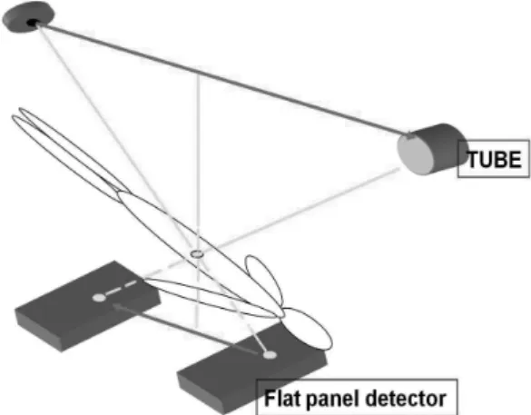
Fig. 1. Mimetic diagram of digital tomosynthesis.
Digital tomosynthesis image acquired by continuous fluoroscopic ex- posure from top to bottom at a range of 40°, and with the target part as the center of the CBD. To focus the CBD, the target point is set at a depth of 120 mm from the skin with 70 mm buffer in all directions and continuous X-ray exposure for 2.5 seconds.
Note.-CBD = common bile duct
CBD stones, while the remaining 28 had no stones. ERCP successfully detected 81 of the 89 stones while digital tomo- synthesis detected 82 stones; 77 stones were detected by both methods, whereas 4 stones were detected only on ERCP and 5 stones only on digital tomosynthesis. There were 6 false- posi- tive cases using ERCP (Fig. 2), but only 2 using digital tomo- synthesis. Moreover, there were 8 false-negative cases on ERCP (Fig. 3), and 7 on digital tomosynthesis (Table 1). The sensi- data were reported herein as the means and standard devia-
tions. A p-value < 0.05 was considered to indicate statistically significance.
RESULTS
On the basis of the final diagnosis obtained from endoscop- ic stone removal, 54 of the 82 patients collectively had 89
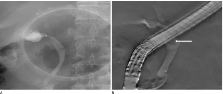
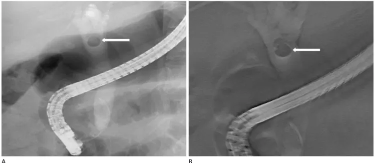
Fig. 2. A 79-year-old man without a CBD stone.
A. ERCP image obtained after injection of contrast material showing a CBD stone (arrow), which is considered to be a score of 5.
B. Although the digital tomosynthesis image is slightly blurred, no definite stones are seen in the CBD.
Note.-CBD = common bile duct, ERCP = endoscopic retrograde cholangiopancreatography
A B
Fig. 3. A 70-year-old man with one stone in the proximal CBD.
A. On ERCP image, no definite CBD stone is visualized.
B. Digital tomosynthesis image showing a proximal CBD stone (arrow) with a score of 3 because the upper margin of the CBD stone is obscured.
Note.-CBD = common bile duct, ERCP = endoscopic retrograde cholangiopancreatography
A B
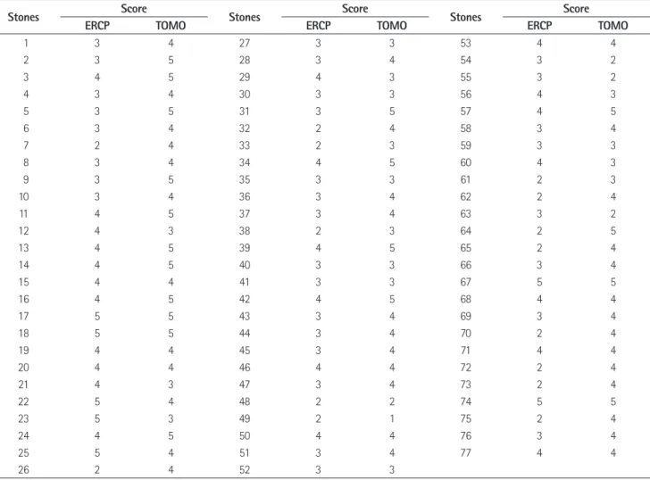
Table 3. Evaluation of Conspicuity of 77 Samely Detected Stones Using the Five-Pointed Scale on ERCP and Digital Tomosynthesis
Stones Score Stones Score Stones Score
ERCP TOMO ERCP TOMO ERCP TOMO
1 3 4 27 3 3 53 4 4
2 3 5 28 3 4 54 3 2
3 4 5 29 4 3 55 3 2
4 3 4 30 3 3 56 4 3
5 3 5 31 3 5 57 4 5
6 3 4 32 2 4 58 3 4
7 2 4 33 2 3 59 3 3
8 3 4 34 4 5 60 4 3
9 3 5 35 3 3 61 2 3
10 3 4 36 3 4 62 2 4
11 4 5 37 3 4 63 3 2
12 4 3 38 2 3 64 2 5
13 4 5 39 4 5 65 2 4
14 4 5 40 3 3 66 3 4
15 4 4 41 3 3 67 5 5
16 4 5 42 4 5 68 4 4
17 5 5 43 3 4 69 3 4
18 5 5 44 3 4 70 2 4
19 4 4 45 3 4 71 4 4
20 4 4 46 4 4 72 2 4
21 4 3 47 3 4 73 2 4
22 5 4 48 2 2 74 5 5
23 5 3 49 2 1 75 2 4
24 4 5 50 4 4 76 3 4
25 5 4 51 3 4 77 4 4
26 2 4 52 3 3
Note.-ERCP = endoscopic retrograde cholangiopancreatography, TOMO = digital tomosynthesis Table 1. Cross-Tabulation of the Number of CBD Stones on ERCP and Digital Tomosynthesis
CBD Stone Positive Negative
ERCP Positive 81 (TP) 6 (FP)
Negative 8 (FN) 25 (TN)
Digital tomosynthesis Positive 82 (TP) 2 (FP)
Negative 7 (FN) 29 (TN)
Note.-CBD = common bile duct, ERCP = endoscopic retrograde cholangiopancreatography, FN = false negative, FP = false positive, TN = true negative, TP
= true positive
Table 2. Sensitivity, Specificity of ERCP and Digital Tomosynthesis for the Detection of CBD Stones
Sensitivity (%) Specificity (%)
ERCP 91.0 80.6
Digital tomosynthesis 92.1 93.5
Note.-CBD = common bile duct, ERCP = endoscopic retrograde cholangiopancreatography
Table 4. The 5-Point Scores of CBD Stones on ERCP and Digital Tomosynthesis
ERCP (n = 77) Digital Tomosynthesis (n = 77) p-value
Average of score (Mean ± SD) 3.29 ± 0.89 3.89 ± 0.88 0.001
Median value of score 3.00 4.00
Note.-CBD = common bile duct, ERCP = endoscopic retrograde cholangiopancreatography, Mean ± SD = mean standard deviation
results are shown in Table 3. The average score was 3.29 for ERCP and 3.89 for digital tomosynthesis. In the evaluation of the stones’ margins, digital tomosynthesis demonstrated a sta- tistically superior conspicuity compared to ERCP (p = 0.001) (Table 4). Forty-four stones were seen more clearly by digital tivity and specificity for CBD stone detection were 91.0% and
80.6% for ERCP, and 92.1% and 93.5% for digital tomosyn- thesis, respectively (Table 2).
The margins of the 77 concurrently detected stones were evaluated for conspicuity according to the 5-point scale. The
Fig. 5. A 74-year-old woman with a CBD stone.
A. ERCP image clearly showing a CBD stone (arrow) with a score of 5.
B. Digital tomosynthesis image showing the presence of a stone (arrow) in the proximal CBD with blurring of the margin, considered to have a score of 4.
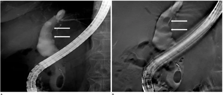
Note.-CBD = common bile duct, ERCP = endoscopic retrograde cholangiopancreatography Fig. 4. A 66-year-old man with two proximal CBD stones.
A. ERCP image obtained after injection of contrast material showing 2 CBD stones (arrows) which are considered to have a score of 2 (upper stone) and 3 (lower stone).
B. Digital tomosynthesis image showing 2 CBD stones (arrows) more clearly compared with the visualization in (A), considered as score to have a 5 for both stones.
Note.-CBD = common bile duct, ERCP = endoscopic retrograde cholangiopancreatography
A A
B B
CT and CTC yield high-resolution images of the biliary tree and adjacent anatomic structures with low invasiveness, but both require relatively high doses of ionizing radiation (11) in addition to intravenous contrast medium (12, 13).
MRCP is not invasive. Moreover, because diagnostic accuracy does not depend on the skill of the operator, more objective results can be obtained. Many studies have confirmed the high sensitivity of MRCP for detecting CBD stones (14-17).
However, MRCP cannot be used in patients who are claustro- phobic or who have implanted devices, such as internal cardio- verters, pacemakers, or metallic clips for cerebral aneurysms.
Furthermore, MRCP has several potential pitfalls including pa- tients with tortuous common bile ducts, compression of the he- patic duct by the right hepatic artery, an origin of the cystic duct mimicking CBD stones, or pneumobilia (17).
For these reasons, ERCP has become the standard diagnos- tic and therapeutic technique in the evaluation of the pancre- aticobiliary tract. However, it is an invasive procedure with a mortality rate of 1-2% (18-23). Despite its invasiveness, ERCP is widely performed, as it not only accurately detects the pres- ence of stones, but also allows for simultaneous treatment, such as extraction.
In our study, during the ERCP procedure, digital tomosyn- thesis was conducted concurrently, without the need for addi- tional preparation of the patient to determine its complemen- tomosynthesis and thus had a higher score than ERCP (4.3 vs
2.9) (Fig. 4). In contrast, twelve stones were detected with a higher score on ERCP than digital tomosynthesis (3.8 vs. 2.8) (Fig. 5), while 21 stones were seen equally well by both ERCP and digital tomosynthesis (Fig. 6).
DISCUSSION
The symptoms and signs of common bile duct stones in- clude right upper quadrant abdominal pain, jaundice, fever, and biochemical abnormality. A more serious complication is cholangitis, which, if progressive, and can lead to septic shock with a high mortality. Thus, the early diagnosis of CBD stones is important, and their subsequent removal is recommended.
Currently, there are several modalities to detect common bile duct stones including abdominal ultrasound, endoscopic ultra- sound, MRI with magnetic resonance cholangiopancreatogra- phy (MRCP), ERCP, and computed tomography (CT), which included computed tomographic cholangiography (CTC).
The traditional first-line modality, abdominal ultrasound, is operator-dependent and has a wide-ranging sensitivity (20- 80%) (6-9). Endoscopic ultrasound is considered as an alter- native method, but it is an invasive procedure that is not only operator-dependent but also accompanied by the potential for endoscopy-related complications (10).
Fig. 6. A 47-year-old woman with a CBD stone.
CBD stone (arrow) clearly seen on both the ERCP image (A) and digital tomosynthesis image (B) with a score of 5.
Note.-CBD = common bile duct, ERCP = endoscopic retrograde cholangiopancreatography
A B
itive cases and a higher conspicuity score. Digital tomosyn- thesis is an effective and complementary diagnostic method to evaluate CBD stones, with clinical usefulness in imaging of the biliary ductal system.
REFERENCES
1. Dobbins JT 3rd, Godfrey DJ. Digital x-ray tomosynthesis:
current state of the art and clinical potential. Phys Med Biol 2003;48:R65-R106
2. Vikgren J, Zachrisson S, Svalkvist A, Johnsson AA, Boijsen M, Flinck A, et al. Comparison of chest tomosynthesis and chest radiography for detection of pulmonary nodules:
human observer study of clinical cases. Radiology 2008;
249:1034-1041
3. Tingberg A. X-ray tomosynthesis: a review of its use for breast and chest imaging. Radiat Prot Dosimetry 2010;139:
100-107
4. Dobbins JT 3rd, McAdams HP. Chest tomosynthesis: tech- nical principles and clinical update. Eur J Radiol 2009;
72:244-251
5. Dobbins JT 3rd, McAdams HP, Godfrey DJ, Li CM. Digital tomosynthesis of the chest. J Thorac Imaging 2008;23:86- 92
6. Pasanen P, Partanen K, Pikkarainen P, Alhava E, Pirinen A, Janatuinen E. Ultrasonography, CT, and ERCP in the diag- nosis of choledochal stones. Acta Radiol 1992;33:53-56 7. Pickuth D, Spielmann RP. Detection of choledocholithiasis:
comparison of unenhanced spiral CT, US, and ERCP. Hepa- togastroenterology 2000;47:1514-1517
8. Rickes S, Treiber G, Mönkemüller K, Peitz U, Csepregi A, Kahl S, et al. Impact of the operator’s experience on value of high-resolution transabdominal ultrasound in the diag- nosis of choledocholithiasis: a prospective comparison us- ing endoscopic retrograde cholangiography as the gold standard. Scand J Gastroenterol 2006;41:838-843
9. Stott MA, Farrands PA, Guyer PB, Dewbury KC, Browning JJ, Sutton R. Ultrasound of the common bile duct in pa- tients undergoing cholecystectomy. J Clin Ultrasound 1991;19:73-76
10. Kondo S, Isayama H, Akahane M, Toda N, Sasahira N, Na- kai Y, et al. Detection of common bile duct stones: com- tary utility in the diagnosis of CBD stones. Tomographic
imaging has several advantages such as improved conspicuity and contrast with local structures which offer more accurate diagnosis of CBD stones during the ERCP procedure (1). A drawback in our patient series was the need for conscious se- dation during ERCP and digital tomosynthesis, which result- ed in the blurring of some images because patients could not perform a spontaneous breath hold during the procedure. In this situation, the higher conspicuity and contrast of local structures achieved with tomographic imaging are likely to improve CBD stone diagnosis.
Our findings demonstrated the sensitivity and specificity of digital tomosynthesis, both of which were slightly higher than those of ERCP with respect to the detection of CBD stones. In terms of conspicuity, the mean score obtained with digital to- mosynthesis was significantly higher than that obtained with ERCP, suggesting that digital tomosynthesis is a superior mo- dality to ERCP in the evaluation of CBD stones. Moreover, our findings showed that digital tomosynthesis can better de- pict the size and extent of CBD stones during the procedure and thus allows a more appropriate treatment choice. Addi- tionally, digital tomosynthesis resulted in fewer false-positive cases than did ERCP, implying that digital tomosynthesis can avoid unnecessary stone-removal procedures. Hence, digital tomosynthesis can be a complementary diagnostic tool for the evaluation of CBD stones during ERCP.
This study has some limitations. First, because some pa- tients were referred for an ERCP examination without a pre- vious non-invasive diagnostic examination, such as MRCP or CT, only 54 of the 82 patients actually had CBD stones. Thus, a relatively small number of patients with CBD stones were evaluated. Further studies with more cases are needed to con- firm the clinical usefulness of digital tomosynthesis for appli- cations involving the biliary ductal system. Second, a method to distinguish air in the CBD from a stone is still lacking. This is potentially problematic, as biliary air might be confused with biliary calculi. However, differentiation is usually possi- ble because biliary air moves into a nondependent position when the patient’s position is changed, whereas calculi tend to localize in a dependent position.
In conclusion, the advantages of digital tomosynthesis over ERCP in the diagnosis of CBD stones include fewer false pos-
parison with ERCP and clinical follow-up. Dig Surg 2003;
20:32-37
18. Kessler RE, Falkenstein DB, Clemett AR, Zimmon DS. Indi- cations, clinical value and complications of endoscopic retrograde cholangiopancreatography. Surg Gynecol Ob- stet 1976;142:865-870
19. Silviera ML, Seamon MJ, Porshinsky B, Prosciak MP, Do- raiswamy VA, Wang CF, et al. Complications related to en- doscopic retrograde cholangiopancreatography: a com- prehensive clinical review. J Gastrointestin Liver Dis 2009;
18:73-82
20. Rieger R, Wayand W. Yield of prospective, noninvasive evaluation of the common bile duct combined with selec- tive ERCP/sphincterotomy in 1390 consecutive laparo- scopic cholecystectomy patients. Gastrointest Endosc 1995;42:6-12
21. Vitale GC, Larson GM, Wieman TJ, Cheadle WG, Miller FB.
The use of ERCP in the management of common bile duct stones in patients undergoing laparoscopic cholecystecto- my. Surg Endosc 1993;7:9-11
22. Cotton PB. Endoscopic retrograde cholangiopancreatogra- phy and laparoscopic cholecystectomy. Am J Surg 1993;
165:474-478
23. Shiozawa S, Kim DH, Usui T, Tsuchiya A, Masuda T, Inose S, et al. Indication of endoscopic retrograde cholangiogra- phy by noninvasive predictive factors of common bile duct stones before laparoscopic cholecystectomy: a pro- spective clinical study. Surg Laparosc Endosc Percutan Tech 2011;21:28-32
parison between endoscopic ultrasonography, magnetic resonance cholangiography, and helical-computed-tomo- graphic cholangiography. Eur J Radiol 2005;54:271-275 11. Breen DJ, Nicholson AA. The clinical utility of spiral CT
cholangiography. Clin Radiol 2000;55:733-739
12. Persson A, Dahlström N, Smedby O, Brismar TB. Three-di- mensional drip infusion CT cholangiography in patients with suspected obstructive biliary disease: a retrospective analysis of feasibility and adverse reaction to contrast ma- terial. BMC Med Imaging 2006;6:1
13. Nilsson U. Adverse reactions to iotroxate at intravenous cholangiography. A prospective clinical investigation and review of the literature. Acta Radiol 1987;28:571-575 14. Griffin N, Wastle ML, Dunn WK, Ryder SD, Beckingham IJ.
Magnetic resonance cholangiopancreatography versus en- doscopic retrograde cholangiopancreatography in the di- agnosis of choledocholithiasis. Eur J Gastroenterol Hepatol 2003;15:809-813
15. Kim TK, Kim BS, Kim JH, Ha HK, Kim PN, Kim AY, et al. Di- agnosis of intrahepatic stones: superiority of MR cholan- giopancreatography over endoscopic retrograde cholangi- opancreatography. AJR Am J Roentgenol 2002;179:429- 434
16. Stiris MG, Tennøe B, Aadland E, Lunde OC. MR cholangio- pancreaticography and endoscopic retrograde cholangio- pancreaticography in patients with suspected common bile duct stones. Acta Radiol 2000;41:269-272
17. Kats J, Kraai M, Dijkstra AJ, Koster K, Ter Borg F, Hazenberg HJ, et al. Magnetic resonance cholangiopancreaticography as a diagnostic tool for common bile duct stones: a com-
총담관결석 평가에서 디지털단층영상합성법과 내시경역행담췌관조영술의 비교 연구1
허지미
1· 백승연
1· 황윤미
1· 이정경
1· 김유경
1· 이선영
2목적: 총담관결석 평가에 디지털단층영상합성법(digital tomosynthesis)과 내시경역행담췌관조영술(endoscopic retrograde cholangiopancreatography; 이하 ERCP)의 상호 보완적인 진단 방법으로서의 가치를 비교하고자 하였다.
대상과 방법: 2008년 12월부터 2010년 5월까지 ERCP와 digital tomosynthesis를 동시에 시행한 환자 중 총담관결석 증 상이 의심되는 96명의 환자를 대상으로 하였다. 이 중 14명이 제외되어 82명의 환자가 연구에 포함되었다. 두 명의 영상 의학과 의사가 ERCP와 digital tomosynthesis 영상을 후향적으로 분석하였다. 먼저 총담관결석의 유무를 평가하였고, 두 검사 방법에서 동시에 결석이 발견된 경우에는 결석의 경계를 1점에서 5점으로 척도를 정하였다.
결과: 82명의 환자 중 54명의 환자가 89개의 총담관결석이 있었으며 28명의 환자는 결석이 없었다. 총담관결석 진단에 서 ERCP의 민감도, 특이도는 91.0%, 80.6%이며 digital tomosynthesis는 92.1%, 93.5%였다. 두 진단 방법에서 동시에 발견된 77개의 총담관결석 경계의 정확도에 대한 평균 점수는 ERCP에서 3.29이며 digital tomosynthesis에서 3.89였다.
총담관결석 경계의 정확도는 digital tomosynthesis에서 통계적으로 유의하게 높았다(p = 0.001).
결론: 총담관결석을 평가하는 데 있어 digital tomosynthesis는 효과적이며 상호 보완적인 진단 방법이 될 수 있다.
이화여자대학교 의학전문대학원 1영상의학과학교실, 2내과학교실