Effects of Scutellarin on MUC5AC Mucin Production Induced by Human Neutrophil Elastase or Interleukin 13 on Airway
Epithelial Cells
Scutellarin is a flavonoid extracted from a traditional Chinese herb, Erigeron breviscapus.
The present study investigated the effect of scutellarin on MUC5AC mucin production and the possible mechanism. Human bronchial epithelial 16 (HBE16) cells were pretreated with scutellarin for 60 min, and then exposed to human neutrophil elastase (HNE) or interleukin (IL)-13 for 12 hr. RT-PCR and ELISA were performed to measure the amount of MUC5AC mucin production. The results showed that scutellarin inhibited MUC5AC expression both in mRNA and protein level induced by HNE in a concentration-dependent manner.
However, scutellarin failed to inhibit MUC5AC mucin production induced by IL-13. To investigate the intracellular mechanisms associated with the effect of scutellarin on MUC5AC mucin production, western blotting was carried out to examine the
phosphorylation of protein kinase C (PKC), signal transducer and activator of transcription 6 (STAT6) and extracellular signal-regulated kinase 1/2 (ERK1/2). The phosphorylation of PKC and ERK1/2 was attenuated after treatment with scutellarin, whereas STAT6 was not significantly affected. Therefore, it is suggested that scutellarin down-regulates MUC5AC mucin production on HBE16 cells via ERK-dependent and PKC-dependent pathways.
Key Words: Scutellarin; MUC5AC; Human Neutrophil Elastase; Interleukin-13; Airway Epithelial Cells
De-Peng Jiang1, Juliy M. Perelman2, Victor P. Kolosov2 and Xiang-Dong Zhou1
1Department of Respiratory Medicine, the Second Affiliated Hospital, Chongqing Medical University, Chongqing, China; 2Far Eastern Scientific Center of Physiology and Pathology of Respiration, Blagoveschensk, Russia
Received: 21 November 2010 Accepted: 15 March 2011 Address for Correspondence:
Xiang-Dong Zhou, MD
Department of Respiratory Medicine, The Second Affiliated Hospital, Chongqing Medical University, No.74, Linjiang Road, Yuzhong District, Chongqing 400010, China
Tel: +86-23-63693207, Fax: +86-23-63711527 E-mail: [email protected]
This work was supported by grant from the National Nature Science Foundation of China (No.81070031), and China-Russia Cooperation Research Foundation (No.81011120108).
DOI: 10.3346/jkms.2011.26.6.778 • J Korean Med Sci 2011; 26: 778-784
INTRODUCTION
The excess mucus production is an important feature of chron- ic inflammatory diseases, including chronic bronchitis, bron- chiectasis and asthma. Mucus overproduction contributes to morbidity and mortality in many patients, particularly in those with more severe disease. During acute exacerbation of chronic obstructive pulmonary disease (COPD), human neutrophil elas- tase (HNE), reactive oxygen species (ROS) and many cytokines may injure the airway and activate mucin gene regulation (1).
Excessive mucus in the airways is linked to an increase in the frequency and duration of infection and a decline in lung func- tion. More than 20 human mucin genes have been deposited in the GenBank. Among all these mucin in mammals, mucin (MUC) 5AC and MUC5B are produced significantly in intrapul- monary airways. Up-regulated MUC5AC expression is the cen- tral event in goblet cell metaplasia (2). HNE has been shown to be a potent secretagogue for goblet cells in vitro and in vivo. HNE increases the transcriptional activity of MUC5AC promoter and MUC5AC mRNA expression through mitogen-activated pro- tein kinase (MAPK)/extracellular signal-regulated kinase (ERK)
signaling pathways. HNE is also known to elevate the MUC5AC mRNA levels by enhancing mRNA stability or inducing ROS (3, 4). Several cytokines including tumor necrosis factor, interleukin (IL)-1β, and IL-13 upregulate MUC5AC expression. IL-13, a cen- tral mediator of airway remodeling in asthma, increases MUC5AC expression by the phosphorylation of signal transducer and ac- tivator of transcription 6 (STAT6) and the suppression of the transcription factor, Forkhead boxA2 (FOXA2) (5).
Mucoregulatory medications decrease mucin secretion or impede the formation of the DNA/F-actin secondary polymer network. The most thoroughly studied mucoregulatory medi- cations are the low-dose macrolide antibiotics. The mechanism of that medication appears to be through the modulation of the MAPK/ERK cascade (6). However, rapid spread of bacterial re- sistance has been observed after overdose medication or the inaccurate treatment. Moreover, the gastrointestinal adverse events of macrolide antibiotics occurred frequently, including gastroesophageal reflux and functional dyspepsia. All these prob- lems have impeded the application of the macrolide antibiotic as an effective treatment for mucus hypersecretion. No current safety and effective mucoregulatory medications is developed
in clinical management of patients with COPD or asthma. In these clinical situations, it is noteworthy to find novel therapeu- tic approaches to decrease mucus hypersecretion.
It was reported that several natural compounds such as genis- tein and curcumin could affect mucus production in the airway.
Ram and his colleagues reported that curcumin attenuated al- lergen induced airway hyperresponsiveness in sensitized guin- ea pigs (7). Also, Heo and his colleagues (8) reported that genis- tein and curcumin suppressed epidermal growth factor induced MUC5AC mucin production and gene expression from human airway epithelial cells. Thus, natural compounds might provide possible approaches to prevent mucus hypersecretion.
Scutellarin (scu) (Fig. 1), a known flavone 7-O-β-D-glucuro- nide with a molecular weight of 462.21, is considered as the pri- mary active ingredient of breviscapine. It has been extensively used to treat ischemic cerebrovascular and cardiovascular dis- eases (9, 10). Scutellarin has been observed to possess compre- hensive pharmacological functions, including dilating vessels, inhibiting platelet aggregation and reducing intracellular free Ca2+ levels (11). Nowadays, various preparations of scutellarin are still widely used clinically to treat cerebrovascular and car- diovascular diseases such as hypertension, angina pectoris, cor- onary heart disease, cerebral ischemia and vascular dementia.
Scutellarin also possesses strong ability to inhibit protein kinase C (PKC) (12, 13), which is involved in the regulation of MUC5AC expression in vivo and in vitro (14, 15). Therefore, we hypothe- size that scutellarin may decrease MUC5AC mucin production in airway epithelial cells.
In this study, the immortalized human bronchial epithelial cell line (HBE-16) were pretreated with scutellarin for 60 min and then exposed to HNE or IL-13 for 12 hr. MUC5AC mucin product was assessed by RT-PCR and ELISA. We investigated intracellular mechanisms and signaling pathways associated with the effects of scutellarin on MUC5AC mucin production.
The mechanism appears to involve the inhibition of PKC and ERK1/2.
MATERIALS AND METHODS Materials
All the antibodies were purchased from Santa Cruz Biotechnol-
ogy (Santa Cruz, CA, USA) unless otherwise specified. All the chemicals and reagents were from Sigma (St. Louis, MO, USA) unless otherwise indicated. Calphostin C (a specific PKC inhibi- tor) and PD98059 (a specific ERK inhibitor) were purchased from Calbiochem (San Diego, CA, USA). A771726 were purchased from Enzo Life Sciences. The HNE inhibitor elastatinal was ob- tained from Calbiochem (La Jolla, CA, USA). Scutellarin (purity
> 95%) was purchased from Wangzilong Pharmaceutical Co.
LTD (Yuxi, Yunnan, China). Scutellarin was dissolved in DMSO and diluted with saline before use. The final concentration of DMSO was less than 0.1%.
Normal human bronchial epithelial cell culture
HBE-16 cells were maintained in RPMI-1640 medium (Gibco- Invitrogen, Carlsbad, CA, USA), supplemented with 10% fetal calf serum (Gibco) and 1% weight per volume penicillin/strep- tomycin (Gibco). Cell cultures were maintained in a humidified atmosphere of 5% CO2 in air at 37°C. The medium was changed till cells were 90% confluent.
Exposure of the cells to HNE or IL-13
The cells were seeded in 6-well plate at 5 × 10⁵ cells/well. After the cells reached 90% confluence, they were serum starved for 24 hr to maintain a low basal level of MUC5AC mucin produc- tion. To determine the effects of HNE (Elastin Products, Owens- ville, MO, USA) or IL-13 (Peprotech, Hill, NJ, USA), cells were exposed to 0.01-1 μM/L HNE or 1-25 ng/mL recombinant hu- man IL-13 for 12 hr (15, 16).
Treatment with scutellarin
To investigate the concentration effect of scutellarin on the cell, the cells were incubated in RPMI-1640 medium containing scu- tellarin with the concentration from 10 μM/L to 100 μM/L for 60 min (17) and then exposed to 0.1 μM/L HNE or 10 ng/mL re- combinant human IL-13 for 12 hr. In the positive control group, Elastatinal (100 μM/L) was added to the medium before expo- sure to HNE and A771726 (10 μM/L) was used as an inhibitor against IL-13. In the negative control group, the cells were incu- bated with medium alone. At the end of stimulation, cells were collected for further evaluations.
RT-PCR
Total RNA was extracted from HBE16 cells after stimulation with HNE or IL-13 for 12 hr using ISOGEN (Gibco-Invitrogen). Oli- gonucleotide primers for PCR were designed according to the published sequence for human MUC5AC (sense: TCA ACG GAG ACT GCG AGT ACA C; antisense: TCT TGA TGG CCT TGG AGC A). The PCR conditions were as follows: a pre-denaturing at 94°C for 10 min, followed by 33 cycles of denaturation at 94°C for 30 sec, annealing at 59°C for 30 sec and extension at 72°C for 30 sec. PCR products were separated by electrophoresis through HOOC
OH OH
O O O
OH OH
OH O
OH
Fig. 1. The structure of scutellarin.
1% agarose gel containing ethidium bromide, and the signal in- tensity was analyzed by BIO-RAD Image. Glyceraldehyde-3- phosphate dehydrogenase (GAPDH) controls were used to stan- dardize the quantification of RNA samples.
ELISA
The MUC5AC protein was measured by enzyme-linked immu- nosorbent assay (ELISA). The cells were lysed in a standard RIPA buffer, containing protease inhibitors. All the samples were in- cubated at 42°C in a 96-well plate for 1 hr and 4°C overnight. The plates were washed three times with PBS and blocked with 2%
BSA for 1 hr at room temperature. After washing the plates for three times with PBS, the wells were incubated in PBS with mouse monoclonal antibody against MUC5AC (NeoMarkers, Fremont, CA, USA) for 1 hr, which was diluted with PBS containing 0.05%
Tween 20. Then, the wells were washed three times with PBS, and 100 μM of horseradish peroxidase-goat anti-mouse IgG conjugate was dispensed into each well for 1 hr. The plates were washed three times with PBS and the color reaction was devel- oped with 3, 3’, 5, 5’-tetramethylbenzidine peroxide solution and stopped with 50 mM/L H2SO4. The absorbance was read at 405 nm wavelengths using a synergy HT multi-detection microplate reader.
Western blot analysis
The phosphorylation PKC, ERK1/2 and STAT6 were examined by western analysis. Cells were collected at the end of exposure.
Protein concentrations were determined by Bio-Rad assays. Cells lysate was fractionated on SDS–PAGE, and proteins were then transferred to a nitrocellulose membrane (Millipore, Billerica, MA, USA). Membranes were blocked with 5% FBS in Tris-buff- ered saline containing 0.1% Tween 20 at room temperature for 1 hr and then incubated overnight at 4°C with the mouse anti- bodies against p-PKC, p-ERK1/2 and p-STAT6. GAPDH protein was used to normalize the total tissue lysate on the same nitro- cellulose membrane. After three washed, membranes were in-
cubated for 1 hr at room temperature with horseradish peroxi- dase conjugate goat anti-mouse IgG antibody. The three washes were repeated, and then protein was visualized with enhanced chemiluminescence kit (Amersham Pharmacia Biotech, Pisca- taway, NJ, USA).
Statistical analysis
The results were expressed as mean ± SEM. Differences were examined for statistical significance using one-way analysis of variance. The post hoc test was Fisher’s protected least signifi- cant difference (PLSD) test. A P value < 0.05 denoted the pres- ence of a statistically significant difference.
RESULTS
Effects of HNE or IL-13 on MUC5AC gene and protein expression
To confirm whether HNE or IL-13 can induce MUC5AC mucin production on HBE-16 cells, we first evaluated the MUC5AC protein expression after addition of HNE or IL-13 to the cells at various concentrations. We found that the stimulation with HNE led to a concentration-dependent increase in MUC5AC produc- tion compared with the control group (Fig. 2). The expression of MUC5AC protein also increased as a result of IL-13 stimula- tion in a concentration dependent manner (Fig. 3).
Scutellarin inhibited MUC5AC mucin production induced by HNE
We next evaluated the effects of scutellarin on MUC5AC expres- sion induced by HNE. RT-PCR and ELISA were used to examine the expression of MUC5AC production regulated by scutellarin.
The cells were pretreated with scutellarin for 60 min before the addition of 0.1 μM/L NHE. In the HNE control group, the cells were stimulated with HNE after treatment with medium only.
As shown in Fig. 4, scutellarin significantly reduced the enhanc- ed expression of MUC5AC induced by HNE at both the mRNA
% of control
250
200
150
100
50
0
*
*
*
Negative control 0.01 µM HNE 0.1 µM HNE 1 µM HNE Fig. 2. Up-regulation of MUC5AC protein induced by human neutrophil elastase (HNE) on HBE16 cells. Cells were collected after exposed to HNE at different concentrations.
The amount of MUC5AC was measured by ELISA. Data represent means ± SEM of four experiments. ﹡P < 0.05 compared with the negative control group.
% of control
250
200
150
100
50
0
*
* *
Negative control 1 ng/mL IL-13 10 ng/mL IL-13 25 ng/mL IL-13 Fig. 3. Up-regulation of MUC5AC induced by IL-13 on HBE16 cells. Cells were collect- ed after exposed to IL-13 at different concentrations. The amount of MUC5AC was measured by ELISA. Data represent means ± SEM of four experiments. ﹡P < 0.05 compared with the negative control group.
and protein levels. In the positive control group, elastatinal great- ly reduced the MUC5AC synthesis induced by HNE (data not shown).
The mRNA and protein expression of MUC5AC decreased as a result of scutellarin inhibition in a concentration dependent manner. Half-maximal inhibition effect was elicited after incu- bation with scutellarin at the concentration of 50 μM/L (Fig. 4B).
Therefore, concentration of 50 μM/L was chosen for the follow- ing experiments.
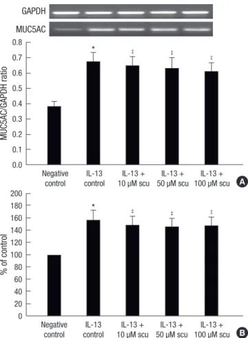
Scutellarin did not reduce MUC5AC mucin production induced by IL-13
To determine whether scutellarin could inhibit mucus produc- tion induced by IL-13, MUC5AC synthesis was assayed using RT-PCR and ELISA. The cells were pretreated with scutellarin for 60 min before the addition of 10 ng/mL IL-13. In the IL-13 control group, the cells were stimulated with IL-13 after treat- ment with medium only. As shown in Fig. 5, scutellarin did not reduce the MUC5AC synthesis induced by IL-13 at both the mRNA and protein levels.
Scutellarin inhibited PKC signaling responding to HNE stimuli
We investigated the possible involvement of PKC in the protec- tive activity of scutellarin against mucus secretion. The cells were preincubated with 0.1 μM/L calphostin C or 50 μM/L scutellarin for 60 min before exposed to 0.1 μM/L HNE for 1 hr. As shown in Fig. 6, PKC activity induced by HNE was attenuated when the cells were pretreated with calphostin C. Compared with the HNE control group, a significant decrease in the phosphorylation of PKC was also observed when the cells were pretreated with scu- tellarin.
The phosphorylation of ERK1/2 and STAT6 after treatment with scutellarin
To study the involvement of ERK1/2 in the protective activity of scutellarin against MUC5AC mucin production induced by HNE, the cells were stimulated with HNE for 1 hr after treatment with 50 μM/L scutellarin or 50 μM/L PD98059 for 60 min. As shown in Fig. 7, Pretreatment with PD98059 resulted in significant de- creases in ERK1/2 phosporylation when exposed to HNE. Com-
Fig. 4. Effects of scutellarin (scu) on MUC5AC mRNA (A) and protein (B) expression after exposed to HNE. The cells were pretreated with scu or medium only for 60 min before the addition of 0.1 µM/L HNE. Total RNAs were reverse transcribed and used for PCR amplication (A). The amount of MUC5AC protein was measured by ELISA (B).
Data represent means ± SEM of four experiments. ﹡P < 0.05 compared with the negative group; †P < 0.05 compared with the HNE control group.
MUC5AC/GAPDH ratio% of control 1 0.9 0.8 0.7 0.6 0.5 0.4 0.3 0.2 0.1 0.0
250
200
150
100
50
0
*
*
†
†
†
†
†
†
Negative HNE HNE + HNE + HNE + control control 10 µM scu 50 µM scu 100 µM scu
Negative HNE HNE + HNE + HNE +
control control 10 µM scu 50 µM scu 100 µM scu A
B GAPDH
MUC5AC
Fig. 5. Effects of scutellarin (scu) on MUC5AC mRNA (A) and protein (B) expression after exposed to IL-13. The cells were pretreated with scu or medium only for 60 min before the addition of 10 ng/mL IL-13. Total RNAs were reverse transcribed and used for PCR amplication (A). The amount of MUC5AC protein was measured by ELISA (B).
Data represent means ± SEM of four experiments. ﹡P < 0.05 compared with the negative control group; ‡P > 0.05 compared with the IL-13 control group.
MUC5AC/GAPDH ratio% of control
0.8 0.7 0.6 0.5 0.4 0.3 0.2 0.1 0.0
200 180 160 140 120 100 80 60 40 20 0
*
*
‡
‡
‡
‡
‡
‡
Negative IL-13 IL-13 + IL-13 + IL-13 + control control 10 µM scu 50 µM scu 100 µM scu
Negative IL-13 IL-13 + IL-13 + IL-13 + control control 10 µM scu 50 µM scu 100 µM scu
A
B GAPDH
MUC5AC
pared with the HNE control group, the phosphorylation of ERK1/
2 also significantly decreased after treatment with scutellarin.
For STAT6 group, the cells were pretreated with 50 μM/L scu- tellarin or 10 μM/L A771726 for 60 min before exposed to 10 ng/
L IL-13 for 1 hr. As shown in Fig. 8, A771726 significantly decreas- es STAT6 phosphorylation responding to IL-13. In contrast, pre- treatment with scutellarin failed to interrupt STAT6 phosphory- lation when exposed to IL-13. These data suggest that MAPK/
ERK signal pathway, not STAT6 pathway, may play an important role in the inhibition of scutellarin on MUC5AC mucin produc- tion induced by HNE.
DISCUSSION
Excessive mucus secretion is problematic in patients with chron- ic airway diseases due to airway obstruction and impairment of gas exchange. In this study, we carried out experiments to examine whether treatment with scutellarin might attenuate mucus production induced by HNE or IL-13. Our study dem- onstrated that: 1) stimulation with HNE or IL-13 increased the expression of MUC5AC; 2) scutellarin significantly attenuated the MUC5AC expression at both the mRNA and protein levels induced by HNE. However, scutellarin failed to inhibit the MU- C5AC expression induced by IL-13; 3) PKC played an important role in mucus secretion and was involved in the inhibition of scutellarin against MUC5AC mucin production induced by HNE; 4) MAPK/ERK, but not STAT6 pathway, was implicated in the effects of scutellarin on mucus production.
Our study demonstrated that HNE and IL-13 induced MU- C5AC expression on HBE-16 cells. HNE is the most widely stud- ied with regard to enhanced mucus secretion. Purified HNE has been shown to provoke secretion of mucin by isolated airway epithelial cells and glands from several species (3, 4, 15). Mucin protein is also upregulated by IL-13, which plays a central role
p-PKC/GAPDH ratio
1.2 1 0.8 0.6 0.4 0.2 0
*
† †
Negative control HNE control HNE + scu HNE + Calphostin Fig. 6. Effects of scutellarin (scu) on the phosphorylation of PKC induced by HNE. The cells were pretreated with scu, Calphostin C or medium only for 60 min before the addition of 0.1 µM/L HNE. The phosphorylation of PKC was measured by western.
Data represent means ± SEM of four experiments. ﹡P < 0.05 compared with the control group; †P < 0.05 compared with the HNE control group.
GAPDH p-PKC
P-ERK/GAPDH ratio
1.2 1 0.8 0.6 0.4 0.2 0
*
† †
Negative control HNE control HNE + scu HNE + PD98059 Fig. 7. Effects of scutellarin (scu) on the phosphorylation of ERK1/2 induced by HNE.
The cells were pretreated with scu, PD98059 or medium only for 60 min before the addition of 0.1 µM/L HNE. The phosphorylation of ERK1/2 was measured by western.
Data represent means ± SEM of four experiments. ﹡P < 0.05 compared with the negative group; †P < 0.05 compared with the HNE control group.
GAPDH P-ERK
P-STAT6/GAPDH ratio
1.0 0.9 0.8 0.7 0.6 0.5 0.4 0.3 0.2 0.1 0.0
* ‡
†
Negative control IL-13 control HNE + scu HNE + A771726 Fig. 8. Effects of scutellarin (scu) on the phosphorylation of STAT6 induced by IL-13.
The cells were pretreated with scu, A771726 or medium only for 60 min before the addition of 10 ng/mL IL-13. The phosphorylation of STAT6 was measured by western.
Data represent means ± SEM of four experiments. ﹡P < 0.05 compared with the negative group; †P < 0.05 compared with the IL-13 control group; ‡P > 0.05 com- pared with the IL-13 control group.
GAPDH p-STAT6
in the pathogenesis of chronic inflammatory diseases. Previous studies have indicated that IL-13 could induce mucus secretion in mouse airways via the expression of both the IL-13 receptor and the IL-13 signaling molecule signal transducer (18). Up- regulated signaling through STAT6 pathway plays an important role in mucus production induced by IL-13 (5, 16).
After the confirmation of the effect of HNE or IL-13 on mucus secretion on HBE16 cells, we investigate the effect of scutellarin on MUC5AC mucin production induced by HNE or IL-13. Com- pared with calphostin C, scutellarin showed an equipotent ef- fect in the inhibition of MUC5AC mucin production induced by HNE. However pretreatment with scutellarin did not reduce IL- 13 induced MUC5AC production. To investigate the reason for this discrepancy with the effect of scutellarin against MUC5AC production induced by HNE or IL-13, we examined the effects of scutellarin on MAPK/ERK signaling transduction and STAT6
signal transduction. Scutellarin attenuated the phosphorylation of ERK1/2, which was significantly phosphorylated after the stimulation of HNE. Pretreatment with scutellarin failed to at- tenuate the phosphorylation of STAT6 induced by IL-13. In con- trast, A771726, a specific STAT6 inhibitor, greatly decreased the phosphorylation of STAT6 and the expression of MUC5AC in- duced by IL-13. Taken together, we identified that scutellarin down-regulated MUC5AC production induced by HNE through MAPK/ERK signal transduction. Scutellarin could not affect mucus production induced by IL-13 and STAT6 pathway was not involved in the mechanism of scutellarin on MUC5AC pro- duction.
Among a variety of signal transduction molecules, MAPK has been shown to play an important role in mucus secretion (19).
Kuwahara demonstrated that a PKC → ERK1/2 → Sp1 pathway was responsible for HNE-activated mucus secretion (20). Our study showed that such pathway might be related to the protec- tive effect of scutellarin against MUC5AC production. Pan et al.
(11) reported that suppression of ERK1/2 activation by scutella- rin represented one of the possible mechanisms accounting for its beneficial effects on impaired cardiac function after myocar- dial infarction. Our results are consistent with the conclusion and clearly reveal that scutellarin blocked ERK pathway activat- ed by HNE.
PKC was known to be involved in secretion of airway mucin responding to various stimuli (14). It has been reported that scu- tellarin has the strong inhibitory effects on PKC activation, which contributes to its protective role in cerebral ischemia and hepat- ic injury during brain-death (12, 13). To confirm whether PKC was involved in the inhibition of scutellarin against mucus pro- duction on HBE16, phosphorylation of PKC was determined by western blotting. Our data clearly revealed that scutellarin blocked PKC activation induced by HNE. Inhibition of PKC at- tenuated HNE-mediated mucus secretion. Therefore, the effect of scutellarin on MUC5AC mucin production can be part as- cribed to its specifically blocking PKC activation.
The mechanism whereby scutellarin inhibit PKC is uncer- tain. Many studies have shown that scutellarin is a Ca2+ chan- nel-blocking agent with the ability to inhibit extracellular calci- um influx (11, 21). Pan et al. (22) reported scutellarin exerted its anti-hypertrophic effects via suppressing the Ca2+-mediated calcineurin. Hong et al. (23) reported that scutellarin protected against hydrogen peroxide-induced cytotoxicity in PC12 cells via reducing intracellular accumulation of Ca2+. Considering calcium as a robust activator of PKC, the inhibition of scutellar- in on PKC phosphorylation might be associated with its ability to block Ca2+ channel. Interestingly, correlation between calci- um channels and mucus expression in airways has suggested a causal relationship. Transfection of hCLCA1 into human mu- coepidermoid cells resulted in up-regulation of the MUC5AC gene (24). Macrolide antibiotics, the most thoroughly studied
mucoregulatory medications, may inhibit mucus secretion via reducing intracellular accumulation of Ca2+ (25). Therefore, the molecular mechanism of scutellarin against mucus secretion might be associated with its ability to block calcium channel.
Further studies are required to investigate the specific mecha- nism.
In conclusion, the present study has demonstrated that scu- tellarin inhibited MUC5AC mucin production via inhibiting PKC activation and diminishing the phosphorylation of ERK1/2.
Our results provide experimental evidence for the use of scutel- larin in the clinic as effective means for treatment of mucus hy- persecretion. Indeed, scutellarin shows promising effect on re- spiratory disease and possesses wide-ranging positive effects on the control of mucus secretion. Importantly, the curative dosage of scutellarin in clinic is safe, without liver or kidney toxicity. It has been used in cardiovascular disease and cerebral infarction with satisfactory tolerance and safety (26). In addition to this, it is supposed that scutellarin is a potential effective and safety candidate in the regulation of mucus secretion.
REFERENCES
1. Caramori G, Di Gregorio C, Carlstedt I, Casolari P, Guzzinati I, Adcock IM, Barnes PJ, Ciaccia A, Cavallesco G, Chung KF, Papi A. Mucin expres- sion in peripheral airways of patients with chronic obstructive pulmo- nary disease. Histopathology 2004; 45: 477-84.
2. Young HW, Williams OW, Chandra D, Bellinghausen LK, Pérez G, Suárez A, Tuvim MJ, Roy MG, Alexander SN, Moghaddam SJ, Adachi R, Blackburn MR, Dickey BF, Evans CM. Central role of Muc5ac expression in mucous metaplasia and its regulation by conserved 5’ elements. Am J Respir Cell Mol Biol 2007; 37: 273-90.
3. Voynow JA, Fischer BM, Malarkey DE, Burch LH, Wong T, Longphre M, Ho SB, Foster WM. Neutrophil elastase induces mucus cell metaplasia in mouse lung. Am J Physiol Lung Cell Mol Physiol 2004; 287: L1293-302.
4. Shao MX, Nadel JA. Neutrophil elastase induces MUC5AC mucin pro- duction in human airway epithelial cells via a cascade involving protein kinase C, reactive oxygen species, and TNF-alpha- converting enzyme. J Immunol 2005; 175: 4009-16.
5. Tanabe T, Fujimoto K, Yasuo M, Tsushima K, Yoshida K, Ise H, Yamaya M. Modulation of mucus production by interleukin-13 receptor alpha 2 in the human airway epithelium. Clin Exp Allergy 2008; 38: 122-34.
6. Shinkai M, Henke MO, Rubin BK. Macrolide antibiotics as immunomod- ulatory medications: proposed mechanisms of action. Pharmacol Ther 2008; 117: 393-405.
7. Ram A, Das M, Ghosh B. Curcumin attenuates allergen-induced airway hyperresponsiveness in sensitized guinea pigs. Biol Pharm Bull 2003; 26:
1021-4.
8. Heo HJ, Lee SY, Lee MN, Lee HJ, Seok JH, Lee CJ. Genistein and curcum- in suppress epidermal growth factor-induced MUC5AC mucin produc- tion and gene expression from human airway epithelial cells. Phytother Res 2009; 23: 1458-61.
9. Gao ZX, Huang DY, Li HX, Zhang LN, Lv YH, Cui HD, Zheng JH. Scutel- larin promotes in vitro angiogenesis in human umbilical vein endotheli-
al cells. Biochem Biophys Res Commun 2010; 400: 151-6.
10. Lin LL, Liu AJ, Liu JG, Yu XH, Qin LP, Su DF. Protective effects of scutella- rin and breviscapine on brain and heart ischemia in rats. J Cardiovasc Pharmacol 2007; 50: 327-32.
11. Pan Z, Zhao W, Zhang X, Wang B, Wang J, Sun X, Liu X, Feng S, Yang B, Lu Y. Scutellarin alleviates interstitial fibrosis and cardiac dysfunction of infarct rats by inhibiting TGFβ1 expression and activation of p38-MAPK and ERK1/2. Br J Pharmacol 2011; 162: 688-700.
12. Xu W, Zha RP, Wang WY, Wang YP. Effects of scutellarin on PKCgamma in PC12 cell injury induced by oxygen and glucose deprivation. Acta Phar- macol Sin 2007; 28: 1573-9.
13. Wang M, Zhang WB, Zhu JH, Fu GS, Zhou BQ. Breviscapine ameliorates hypertrophy of cardiomyocytes induced by high glucose in diabetic rats via the PKC signaling pathway. Acta Pharmacol Sin 2009; 30: 1081-91.
14. Abdullah LH, Bundy JT, Ehre C, Davis CW. Mucin secretion and PKC isoforms in SPOC1 goblet cells: differential activation by purinergic ago- nist and PMA. Am J Physiol Lung Cell Mol Physiol 2003; 285: L149-60.
15. Park JA, He F, Martin LD, Li Y, Chorley BN, Adler KB. Human neutro- phil elastase induces hypersecretion of mucin from well-differentiated human bronchial epithelial cells in vitro via a protein kinase C{delta}- mediated mechanism. Am J Pathol 2005; 167: 651-61.
16. Zhen G, Park SW, Nguyenvu LT, Rodriguez MW, Barbeau R, Paquet AC, Erle DJ. IL-13 and epidermal growth factor receptor have critical but dis- tinct roles in epithelial cell mucin production. Am J Respir Cell Mol Biol 2007; 36: 244-53.
17. Yan L, Huang H, Tang QZ, Zhu LH, Wang L, Liu C, Bian ZY, Li H. Brevis- capine protects against cardiac hypertrophy through blocking PKC-al- pha-dependent signaling. J Cell Biochem 2010; 109: 1158-71.
18. Whittaker L, Niu N, Temann UA, Stoddard A, Flavell RA, Ray A, Homer
RJ, Cohn L. Interleukin-13 mediates a fundamental pathway for airway epithelial mucus induced by CD4 T cells and interleukin-9. Am J Respir Cell Mol Biol 2002; 27: 593-602.
19. Rhee CK, Kang CM, You MB, Yoon HK, Kim YK, Kim KH, Moon HS, Park SH, Song JS. Effect of fudosteine on mucin production. Eur Respir J 2008; 32: 1195-202.
20. Kuwahara I, Lillehoj EP, Lu W, Singh IS, Isohama Y, Miyata T, Kim KC.
Neutrophil elastase induces IL-8 gene transcription and protein release through p38/NF-{kappa}B activation via EGFR transactivation in a lung epithelial cell line. Am J Physiol Lung Cell Mol Physiol 2006; 291: L407-16.
21. Zhu BH, Ma L, Pan XD, Huang YL, Liu J. Scutellarin induced Ca(2+) re- lease and blocked KCl-induced Ca(2+) influx in smooth muscle cells iso- lated from rat thoracic artery. J Asian Nat Prod Res 2008; 10: 583-9.
22. Pan ZW, Zhang Y, Mei DH, Zhang R, Wang JH, Zhang XY, Xu CQ, Lu YJ, Yang BF. Scutellarin exerts its anti-hypertrophic effects via suppressing the Ca2+-mediated calcineurin and CaMKII signaling pathways. Naun- yn Schmiedebergs Arch Pharmacol 2010; 381: 137-45.
23. Hong H, Liu GQ. Protection against hydrogen peroxide-induced cytotox- icity in PC12 cells by scutellarin. Life Sci 2004; 74: 2959-73.
24. Nakanishi A, Morita S, Iwashita H, Sagiya Y, Ashida Y, Shirafuji H, Fuji- sawa Y, Nishimura O, Fujino M. Role of gob-5 in mucus overproduction and airway hyperresponsiveness in asthma. Proc Natl Acad Sci USA 2001; 98: 5175-80.
25. Lu S, Liu H, Farley JM Sr. Macrolide antibiotics inhibit mucus secretion and calcium entry in swine airway submucosal mucous gland cells. J Pharmacol Exp Ther 2011; 336: 178-87.
26. Wang M, Zhang WB, Zhu JH, Fu GS, Zhou BQ. Breviscapine ameliorates hypertrophy of cardiomyocytes induced by high glucose in diabetic rats via the PKC signaling pathway. Acta Pharmacol Sin 2009; 30: 1081-91.
AUTHOR SUMMARY
Effects of Scutellarin on MUC5AC Mucin Production Induced by Human Neutrophil Elastase or Interleukin 13 on Airway Epithelial Cells
De-Peng Jiang, Juliy M. Perelman, Victor P. Kolosov and Xiang-Dong Zhou
Mucus hypersecretion is clinically critical problem of various airway diseases. The present study demonstrates that scutellarin inhibited MUC5AC mucin production via inhibiting PKC activation and diminishing the phosphorylation of ERK1/2. Our results provide experimental evidence for a plausibility of clinical trial of scutellarin treatment for the mucus hypersecretion. Our results suggest that the clinically applicable dosage of scutellarin could be safe, without liver or kidney toxicity. As a whole, we suppose that scutellarin is a potentially effective and safety candidate in the regulation of mucus secretion.