Journal of Korean Arthroscopy Society abbreviation (by Index Medicus): J Korean Arthrosc Soc Volume 16, Number 2, August, 2012
서 론
후방십자인대는 슬관절의 중심축을 이루는 구조물로서, 경 골의 후방전위를 막아주는 일차적인 역할을 하는 중요한 구 조물이다.1)해부학,2)생역학적 연구3)가 많이 보고되면서 후방 십자인대의 중요성이 더욱 부각되고 있으며, 최근 교통사고 의 증가와 스포츠 레저활동이 활발해지면서 후방십자인대의 손상 발생 빈도가 증가되고 있다. 후방십자인대 단일 손상은 보존적 요법을 선호하였는데,4,5)장기 추시에서 불안정증의 발
동
동종 종건 건을 을 이 이용 용한 한 단 단일 일 절 절개 개,, 단 단일 일 다 다발 발 후 후방 방십 십자 자인 인대 대 재 재건 건술 술의 의 비 비교 교
경북대학교 의학전문대학원 정형외과학교실 경희수∙천상호∙박경현∙정재욱
Comparison of Single-Incision, Single-Bundle Posterior Cruciate Ligament Reconstruction Using Allograft Tendon
Hee-Soo Kyung, M.D., Sang-Ho Cheon, M.D., Kyung-Hyun Park, M.D., Jae-Wook Jung, M.D.
Department of Orthopedic Surgery, School of Medicine, Kyungpook National University, Daegu, Korea
Purpose: The purpose of this study was to evaluate the posterior cruciate ligament (PCL) reconstruction with single bundle, sin- gle-incision technique using Achilles tendon and tibialis anterior allograft with ligament remnant preservation.
Materials and Methods: Twenty six patients underwent PCL reconstruction was included. There were 21 males and 5 females.
Mean age was 32 years. Used graft was a fresh frozen Achilles tendon allograft (group I, 14 cases) and tibialis anterior allograft (group II, 12 cases). Arthroscopic PCL reconstruction was performed using transtibial, single-incision and single bundle technique with remnant preserving as possible. For clinical evaluation, range of motion, posterior drawer test, Lysholm score, Tegner activity scale, International Knee Documentation Committee (IKDC) grade and posterior stress radiograph were used. The mean follow-up period was 21.6 months (12-40 months). Associated injuries were 5 medial collateral ligament injuries, which were treated by con- servative method.
Results: Range of motion (ROM) was returned to normal range in 24 cases, but ROM deficit under 10。flexion was 2 cases at final follow-up period. Preoperative posterior drawer test was 17 cases in grade II and 9 cases in grade III. At final follow-up 13 cases returned within normal grade, 7 cases grade I and 6 cases grade II posterior instability. Lysholm mean score was improved from pre- operatively 62 to 90 at final follow-up period. Tegner activity mean scale improved from preoperatively 3.5 to 5.6 at final follow-up period. IDKC grade was grade A was 3 cases, grade B 17 cases, grade C 6 cases. In posterior stress radiograph, posterior displace- ment was improved from mean 12 mm preoperative to 4.5 mm at final follow-up. There were no statistical differences between two groups in clinical evaluations. There were two cases of re-rupture of graft at the bone-tendon junction in group I.
Conclusion: We had successful results of PCL reconstruction with single-incision, single bundle technique using Achilles and tib- ialis anterior allograft without difference between two groups in patients with PCL injury. There were more re-rupture of graft in Achilles tendon group.
KEY WORDS: Posterior cruciate ligament, Allograft tendon, Single bundle
�Address reprint request to Hee-Soo Kyung, M.D.
Department of Orthopaedic Surgery, Kyungpook National University Hospital,
50 Samduk-2ga, Jung-gu, Daegu 700-721, Korea Tel: 82-53-420-5636, Fax: 82-53-422-6605 E-mail: [email protected]
접수일: 2011년 8월 16일 게재심사일: 2011년 11월 27일 게재승인일: 2012년 2월 28일
생과 슬개-대퇴 관절 뿐 아니라 대퇴-경골 관절의 퇴행성 변 화로 보존적 치료 후 장기 추시 결과가 좋지 않았고, 손상 전 의 스포츠 활동을 할 수 있는 운동력을 회복하는데 주된 관심 이 모아져 최근에는 활동이 많은 환자에서 수술적 치료를 선 호하는 경향이 있다.6-8)최근 관절경을 이용한 다양한 후방십 자인대 재건술이 많이 소개되고 있다.
후방십자인대 재건술 시 이식건의 선택에 있어 자가건 또 는 동종건을 사용할 수가 있다. 후방십자인대는 최대 인장 강 도가 3,000-5,000 N 이며, 경도가 381 N/mm 라고 한다.9) 자가건을 사용할 경우 골 합체(incorporation)가 좋고, 질병 전이 및 면역반응 이상이 없는 장점이 있으나, 충분한 강도, 길이, 두께를 얻기가 어렵고, 이미 외상 받은 슬관절에 추가적 인 이환율을 높일 수 있고, 수술 시간이 길어지고 이식건 공여 부의 합병증이 발생될 수 있는 단점이 있다. 이와 반대로 동종 이식건은 질병전이, 면역학적 반응, 살균 시 변성 등의 단점이 있지만, 충분한 길이와 두께를 얻을 수 있고 수술 시간을 줄일 수 있어 널리 사용되고 있다.10)이러한 동종건으로 아킬레스 건(Achilles tendon), 전경골건(tibialis anterior tendon) 등이 널리 사용되고 있다. 아킬레스건은 골편이 있어 골-골 유합으로 대퇴골과의 빠른 유합을 기대할 수 있지만, 대퇴터 널에 간섭 나사로 고정 시 건 손상의 위험이 있다. 전경골건은 골편이 없어 건-골 유합으로 대퇴골과의 유합이 느린 단점이 있지만, 대퇴터널에 EndoButton과 같은 고정으로 간섭 나 사에 의한 건 손상의 위험을 줄일 수 있다. 전경골건은 단일 고리(loop), 이중 가닥(strand)의 생역학적 연구에서 최대인 장 강도가 3,412 N, 경도가 344.3 N/mm 로 알려져 있다.11) 그러나 후방십자인대 재건술 시 이식건에 따른 비교연구가 없었다.
이에 저자들은 후방십자인대 파열 환자 중에서 남아 있는 후방십자인대를 최대한 보존하면서 아킬레스건 및 전경골건 동종건을 이용하여 후방십자인대 재건술을 시행 후 임상적 결과를 비교 분석하였다.
대상 및 방법
2007년 1월부터 2010년 7월까지 후방십자인대 손상으로 진단되어서 최소 3개월 이상 보존적 치료시행 후 grade II 이 상의 불안정성과 만성적으로 활동 시 임상 증상을 호소하여 단일 술자에 의하여 수술적 치료를 시행한 환자 중에서 최소 1년 이상 추시가 가능하였던 26예를 대상으로 후향적으로 분 석하였다. 이식건으로는 아킬레스 동종건 14예(제I군), 전경 골 동종건 12예(제II군)를 사용하였다. 이식건의 선택은 초기 에는 의료 급여 등의 이유로 아킬레스건을 주로 사용하였으 며 그 후 전경골건이 공급되어 이식건 공급자의 제공여부에 따라 적절히 선택하였다. 대부분 여가 활동을 하는 일반인으 로 제I군은 평균나이 33세(18-61세), 남녀 비는 남자 12명, 여자 2명, 수술 부위는 우측이 12예, 좌측이 2예였으며, 평균
20.3개월 추시 관찰하였다. 제II군은 평균나이 32세(17-62 세), 남녀 비는 남자 9명, 여자 3명, 수술 부위는 우측이 9예, 좌측이 3예였으며, 평균 22.9개월 추시 관찰하였다. 양군 간 에 성별, 나이, 활동 정도에는 차이가 없었다. 손상 원인은 전 례에서 자동차 사고에 의한 손상이었다. 동반 손상으로는 내 측측부인대 손상이 제I군 3예, 제II군 2예로 총 5예였으며, 전 례에서 보존적 치료를 시작하였다. 2 등급 이상의 후외측 불 안정성이 동반되거나, 전방십자인대 파열이 있는 경우는 제 외하였다.
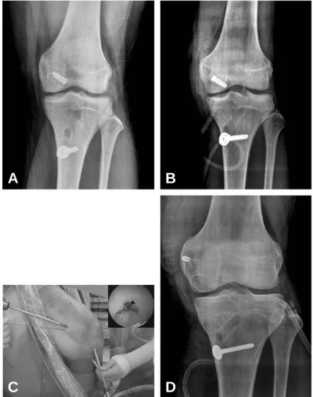
수술 방법은 단일 다발, 경경골터널 방법을 이용하였으며 대퇴터널, 경골터널의 직경은 10 mm로 하였다. 대퇴터널은 11시 또는 1시 방향에서 관절연골 변연부에서부터 약 6 mm 뒤에서 만들었다. 사용된 아킬레스 신선 동결 동종건의 경우 골편이 있는 부분을 직경 10 mm, 길이 25 mm 정도로 만들 어 대퇴골에 금속 간섭 나사를 고정하였다. 이때 초기 10예에 대하여 inside-out 방법으로 하였으며(Fig. 1A), 4예는 modified outside-in 방법으로 전외측 삽입구를 통하여 대 퇴 내과의 후방십자인대 부착부에서 피질골까지 대퇴터널을 만든 후 간섭 나사 지시자(guide pin)를 관절 내에서 밖으로 빼낸 후 약 5 mm의 절개를 가하여 지시자를 따라 관절 밖에 서 안으로 간섭 나사를 고정한다(Fig. 1B-C). 건 부분은 경 골에 와셔/나사 및 흡수성 간섭 나사로 보강하여 고정하였다.
전경골 동종건을 이용한 경우 대퇴터널에 EndoButton CL (Smith & Nephew, Andover, MA, USA)을 이용하여 고 정하였으며(Fig. 1D), 경골터널 고정은 아킬레스 동종건과 같은 방법으로 하였다.
수술 후 재활은 수술 후 슬관절을 완전 신전 시킨 상태로 경 골 근위부에 경골이 후방으로 전위되는 것을 방지하기 위한 경골 후방 지지대를 사용하여 약 4주간 장하지 석고 부목 고 정을 하였다. 수술 직후부터 등척성 근력 강화 운동을 시행하 였다. 그 후 후방십자인대 보호 보조기를 착용하여 부분 체중 부하 보행을 약 3개월 시행하였다. 관절 운동은 수술 후 3개 월에 완전 운동범위를 목표로 수술 후 약 4주에 시작하여 점 차 증가시켰다. 그 후 점진적인 근력 강화 운동을 시행하였으 며, 수술 후 약 1년에 수상 전의 활동을 허용하였다.12,13)
평가는 이학적 평가로 수술 전, 후 슬관절의 운동범위, 후방 전위 검사를 측정하였다. 임상적인 평가로는 Lysholm 점수, Tegner 활동 지수, International Knee Documentation Committee (IKDC) 등급을 수술 전과 후로 비교하였다. 또 한 긴장 방사선 검사로 후방 전위 정도를 비교하였다. 통계 방 법은 paired t-test를 사용하였고, 유의 수준은 95% 이내로 하였다.
결 과
평균 관절 운동범위는 10。이하의 굴곡 제한이 제I군, 제II 군에서 각각 1예, 2예이었으나, 나머지는 모두 슬관절 운동범
위가 정상이었다. 후방전위 검사에서 수술 전 grade II 17예, grade III 9예이었으나 최종 추시 시 grade 0 13예, grade I 7예, grade II 6예로 호전되었으며, 양군 간에 통계적으로 유 의한 차이는 없었다(Table 1).
Lysholm 점수는 수술 전 평균 62±7에서 수술 후 90±4 로 추시 시 통계학적으로 유의하게 향상되었으며, 두 군간의 통계학적 차이는 없었다(P>0.05). Tegner 활동 지수는 점수 는 수술 전 평균 3.5±2.2에서 수술 후 5.6±3.0으로 향상되었 으며, 두 군간의 통계학적 차이는 없었다(P>0.05). IKDC 평 가법 상에서도 수술 전 grade B 3예, grade C 19예, grade D 3예에서 최종 추시 시 grade A 3예, grade B 17예, grade C 6예로 향상되었으며, 두 군간의 유의한 차이는 없었 다(P>0.05, Table 1).
슬관절 90。굴곡 위에서 시행한 후방전위 긴장 방사선 소견 상 수술 전 12±4 mm에서 최종 추시 시 4.5±3 mm로 호전
되었으며, 양군 간에 통계적으로 유의한 차이는 없었다 (Table 1).
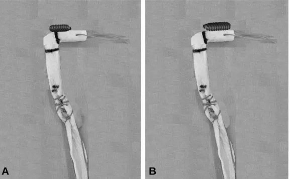
합병증으로 제I군에서 inside-out 방법으로 대퇴터널을 고정한 2예에서 넘어지거나, 추락사고로 골-건 부착(bone- tendon junction) 부위에서 파열이 관찰되어 재 재건술을 실시하였다(Fig. 2).
고 찰
후방십자인대 손상은 슬관절의 후방 불안정성을 초래하고, 나사 회전 운동 기능이 상실되어 슬개-대퇴 압력 및 내측 관 절 압력이 증가되고, 대퇴사두근에 주어지는 힘이 증가되며, 결국에는 연골판과 연골의 손상을 초래하여 퇴행성 변화를 초래하는 것으로 보고 되고 있다.7,14)
일반적으로 단독 손상이고, 슬관절의 후방 불안정성이 심
Fig. 1. (A, B) Femoral fixation methods of Achilles tendon allograft with metal interference screw is showing with (A) inside-out method, and (B) modified outside-in method. (C) This shows modified inside-out femoral screw insertion. Interference screw tip (arrowhead) is visible in inset photo. (D) Femoral fixation method of tibialis anterior allograft tendon with EndoButton CL (Smith & Nephew, Andover, MA, USA) is showing.
C A
D
B
하지 않은 경도 및 중증도의 손상에서는 보존적으로 치료하 여 좋은 결과를 얻을 수 있고, 후방 불안정성이 심한 단독 손 상이거나 다발성 인대 손상과 같이 동반 손상이 같이 있는 경 우는 통상적인 수술적 치료의 적응증으로 생각되어져 왔
다.15,16)본 연구에서도 grade II 이상이고 증상이 있는 환자를
대상으로 후방십자인대 재건술을 실시하였다.
후방십자인대 손상 시 후방 불안정성이 있더라도 수술 전 자기공명영상17)이나 관절경을 보면 후방십자인대가 완전 파 열되어 연속성이 소실되어 보이기보다는 후방십자인대의 일 부나 반월상 연골-대퇴 인대가 잔존해 있는 경우가 대다수이
Table 1. Results of Posterior Cruciate Ligament Reconstruction between Two Groups
Group I Group II Total
Pre-operation Follow-up Pre-operation Follow-up Pre-operation Follow-up Posterior drawer
grade 0 7 6 13
grade I 3 4 07
grade II 09 4 8 2 06 17
grade III 05 4 09
Lysholm score 60±10 89±50 64±90 91±40 62±70 90±40
Tegner activity 3.4±2.1 5.5±3.1 3.6±2.3 5.7±2.7 3.5±2.2 5.6±3.0
scale IKDC grade
A 2 1 03
B 02 9 1 8 03 17
C 10 3 9 3 19 06
D 2 1 03
Posterior laxity (mm) 10±40 4.8±3.1 12±50 5.1±2.1 12±40 4.5±30.
IKDC: International Knee Documentation Committee.
Fig. 2. Thirty-four years old male patient. (A) This shows inside-out femoral fixation of Achilles tendon allograft. This shows rup- tured graft (*) at Achilles tendon-bone junction. (B) Metal interference screw head (arrowhead) is visible in femoral tunnel.
A B
다. 잔여 후방십자인대를 제거하지 않고 후방십자인대 재건 술을 시행 시 좋은 결과가 보고되고 있으며,9)이식 인대와의 혈액 순환 및 잔여 인대와의 접촉 면적이 넓어져서 재혈관화 가 유리할 것으로 생각된다. 또한 잔여 후방십자인대에 의해 서 후방 안정성도 호전될 것으로 사료된다. 본 증례 모두 후방 십자인대 잔여물이 남아 있었으며 최대한 보존하면서 수술을 시행하였다.
후방십자인대 재건하는데 있어서 슬개골-인대-경골결절, 슬괵근, 대퇴사두근건 등의 자가 조직들이 사용되어 좋은 결 과들을 보고하고 있다. 하지만 수술 시간 단축 및 공여부의 이 환율을 줄이고, 튼튼한 고정을 얻을 수 있으며, 최근 많이 시 행되고 있는 해부학적 재건에 사용될 수 있다는 장점 등으로 동종건이 많이 쓰이고 있다. 동종 전경골건과 아킬레스건을 직접 비교한 연구는 보고가 없으며, 각각의 동종건을 이용한 연구는 보고되고 있다.9,18-22) 그 중에서도 아킬레스 동종건을 이용한 재건술이 최근까지도 많이 사용되고 있으며, 자가 조 직들을 이용한 재건술과 비교해 유사한 임상 결과를 나타내 고 있다.9)Pearsall 등11)은 경골건은 인대의 최대 인장 강도 실험에서 비골건, 전방십자인대, 골-슬개건-골, 슬건의 인장 강도보다 높게 나타났고, 전경골건의 경도(stiffness)가 344 N/mm로 후경골건의 경도 302 N/mm보다 높았다고 보고하 고 있다.
본 연구에서 전경골 동종건 재건술이 아킬레스 동종건 재 건술과 유사한 임상 결과를 보였다. 합병증으로 아킬레스 동 종건을 사용한 제I군에서 대퇴골터널에서 골-이식건 주위 파 열이 2예 있었는데, 이는 금속 간섭나사를 inside-out 방법 으로 고정 시 이식건의 손상이 발생할 수 있는데, modified
outside-in 방법으로 간섭나사를 고정한 경우에서는 이식건 파열 등의 합병증을 보이지 않았다(Fig. 3). 전경골근 이식건 의 대퇴골 삽입 고정 시 Endobutton CL에 의해 고정을 한 경우 이식건의 손상 등은 발생하지 않았다. 아킬레스건을 사 용할 경우 대퇴터널에 간섭나사 사용시 건-골 이행부의 파열 이 발생할 수 있으므로 주의를 요한다.
저자들의 경우 신선 동결 아킬레스건 또는 전경골건을 이 용한 후방십자인대 재건술로 양군 간에 차이 없이 좋은 결과 를 관찰하였으나, 더 많은 증례와 장기간의 추시 관찰이 필요 할 것으로 사료된다.
결 론
동종 아킬레스건과 전경골건을 이용하여 단일 절개, 단일 다발 후방십자인대 재건술은 양군 간에 임상적으로 차이 없 이 좋은 결과를 얻었으나 아킬레스건 사용군에서 재파열이 더 많았으며, 더 많은 증례로 장기간의 추시가 필요할 것으로 사료된다.
REFERENCES
01. Hughston JC, Andrews JR, Cross MJ, Moschi A.
Classification of knee ligament instabilities. Part II. The lateral compartment. J Bone Joint Surg Am. 1976;58:173-9.
02. Inderster A, Benedetto KP, Klestil T, Künzel KH, Gaber O. Fiber orientation of posterior cruciate ligament: an experimental morphological and functional study, Part 2.
Clin Anat. 1995;8:315-22.
Fig. 3. (A) Schematic view of inside-out method and (B) modified outside-in method of femoral fixation of the metal interference screw is showing.
A B
03. Wang CJ, Chen HH, Chen HS, Huang TW. Effects of knee position, graft tension, and mode of fixation in poste- rior cruciate ligament reconstruction: a cadaveric knee study. Arthroscopy. 2002;18:496-501.
04. Ahn JH, Lee SH, Choi SH, Wang JH, Jang SW.
Evaluation of clinical and magnetic resonance imaging results after treatment with casting and bracing for the acutely injured posterior cruciate ligament. Arthroscopy.
2011;27:1679-87.
05. Fanelli GC, Beck JD, Edson CJ. Current concepts review:
the posterior cruciate ligament. J Knee Surg. 2010;23:61- 72.
06. Castle TH Jr, Noyes FR, Grood ES. Posterior tibial sub- luxation of the posterior cruciate-deficient knee. Clin Orthop Relat Res. 1992;(284):193-202.
07. Clancy WG Jr, Shelbourne KD, Zoellner GB, Keene JS, Reider B, Rosenberg TD. Treatment of knee joint instabil- ity secondary to rupture of the posterior cruciate ligament.
Report of a new procedure. J Bone Joint Surg Am.
1983;65:310-22.
08. Keller PM, Shelbourne KD, McCarroll JR, Rettig AC.
Nonoperatively treated isolated posterior cruciate ligament injuries. Am J Sports Med. 1993;21:132-6.
09. Höher J, Scheffler S, Weiler A. Graft choice and graft fix- ation in PCL reconstruction. Knee Surg Sports Traumatol Arthrosc. 2003;11:297-306.
10. Bullis DW, Paulos LE. Reconstruction of the posterior cruciate ligament with allograft. Clin Sports Med.
1994;13:581-97.
11. Pearsall AW 4th, Hollis JM, Russell GV Jr, Scheer Z. A biomechanical comparison of three lower extremity ten- dons for ligamentous reconstruction about the knee.
Arthroscopy. 2003;19:1091-6.
12. Edson CJ, Fanelli GC, Beck JD. Postoperative rehabilita-
tion of the posterior cruciate ligament. Sports Med Arthrosc. 2010;18:275-9.
13. Fanelli GC. Posterior cruciate ligament rehabilitation: how slow should we go? Arthroscopy. 2008;24:234-5.
14. Dejour H, Walch G, Peyrot J, Eberhard P. The natural his- tory of rupture of the posterior cruciate ligament. Rev Chir Orthop Reparatrice Appar Mot. 1988;74:35-43.
15. Jung YB, Jung HJ, Park SJ, Kim SJ, Lee YS, Kim KW.
Tensioning of remnant posterior cruciate ligament with a reconstruction of the anterolateral bundle in chronic PCL injuries. J Korean Orthop Assoc. 2006;41:665-74.
16. Maynard MJ, Deng X, Wickiewicz TL, Warren RF. The popliteofibular ligament. Rediscovery of a key element in posterolateral stability. Am J Sports Med. 1996;24:311-6.
17. Shelbourne KD, Jennings RW, Vahey TN. Magnetic reso- nance imaging of posterior cruciate ligament injuries:
assessment of healing. Am J Knee Surg. 1999;12:209-13.
18. Miyamoto RG, Taylor S, Desai P, Bosco J. Histologic pre- sentation of achilles allograft 11 years after its use in pos- terior cruciate ligament reconstruction. Am J Orthop (Belle Mead NJ). 2009;38:E25-7.
19. Min BH, Lee YS, Jin CZ, Son KH. Evaluation of transtib- ial double-bundle posterior cruciate ligament reconstruc- tion using a single-sling method with a tibialis anterior allograft. Am J Sports Med. 2011;39:374-9.
20. Jin CZ, Roh JH, Min BH. Posterior cruciate ligament reconstruction with a single-sling technique using a tibialis anterior tendon allograft. Arthroscopy. 2007;23:323 e1-4.
21. Heinzelmann AD, Barrett GR. Posterior cruciate ligament reconstruction: Achilles tendon allograft, double bundle.
Clin Sports Med. 2009;28:245-57.
22. Borden PS, Nyland JA, Caborn DN. Posterior cruciate lig- ament reconstruction (double bundle) using anterior tib- ialis tendon allograft. Arthroscopy. 2001;17:E14.
목적: 아킬레스건과 전경골건 동종건을 이용하여 인대 잔여물을 최대한 보존하면서 단일 절개, 단일 다발 후방십자인 대 재건술 시행 후 결과를 비교하였다.
대상 및 방법: 후방십자인대 재건술을 받은 26명이 포함되었다. 남자 21명, 여자 5명이었으며 평균 나이는 32세였다.
사용된 이식건은 신선 동결 아킬레스건(제I군, 14예), 전경골건(제II군, 12예)이었으며, 단일 절개, 단일 다발로 인대 잔여 물을 최대한 보존하면서 관절경적 후방십자인대 재건술을 시행하였다. 임상평가는 관절운동범위, 후방전위검사, Lysholm 점수, Tegner 활동지수, International Knee Documentation Committee (IKDC) 등급 및 후방전위 긴장방사선 검사를 이용하여 전위 정도를 평가하였다. 평균 추시기간은 21.6개월(12-40개월)이었다. 동반손상은 내측측부인대 파열 이 5예 있었으나 모두 보존치료로 호전되었다.
결과: 최종 추시 시 관절운동범위는 24예는 정상이었으나 2예에서 10。미만의 굴곡장애가 있었다. 수술 전 후방전위검 사에서 grade II가 17예, grade III가 9예 있었으나, 최종 추시 시 정상 13예, grade I 7예, grade II 6예이었다. Lysholm 점 수는 수술 전 평균 62점에서 최종 추시 시 90점으로 호전되었으며, Tegner 활동지수는 수술 전 평균 3.5에서 최종 추시 시 5.6으로 호전되었다. IKDC 등급은 최종 추시 시 grade A 3예, grade B 17예, grade C 6예이었다. 후방전위 긴장방사선 소견상 수술 전 후방전위는 평균 12 mm에서 수술 후 4.5 mm로 호전되었다. 양군 간에 임상적 결과에는 통계적으로 차 이가 없었다. 합병증으로 아킬레스건을 사용한 군에서 건-골 이행부의 파열이 2예 있었다.
결론: 동종 아킬레스건과 전경골건을 이용하여 단일 절개, 단일 다발 후방십자인대 재건술은 양군 간에 임상적으로 차 이 없이 좋은 결과를 얻었으나 아킬레스건 사용군에서 재파열이 더 많았으며, 더 많은 증례로 장기간의 추시가 필요할 것 으로 사료된다.
색인 단어: 후방십자인대 재건술, 동종건, 단일 다발 초 록