INTRODUCTION
Posterolateral instability of the knee joint refers to the displacement of the lateral plateau of the tibia to the posterolateral side of the lateral condyle of the femur. The displacement results from a combined injury of static and dynamic structures at the posterolateral side. Isolated posterolateral injuries may occur, but many cases are associated with a concurrent cruciate ligament injury. Of the anatomical structures of the posterolateral side, the lateral collateral ligament, popliteofibular ligament, and the popliteus tendon are known to be the most important for knee integrity [1-3]. To date, surgical procedures for
the reconstruction of each structure have been reported.
LaPrade et al. [4], Lee et al. [5], and Yoon et al. [6] have introduced reconstruction procedures for these three key anatomical structures, most of which have been reported to produce good treatment outcomes. Using the allogenous tendon for anatomical reconstruction is advantageous in that a shorter time is required to harvest the tendon, the incidence of complications can be reduced, and sufficient tendon length can be obtained. In our practice, we performed anatomical reconstruction of the popliteofibular ligament, popliteus tendon, and lateral collateral ligament using the Achilles tendon allograft, as described by Lee et al. [5], in patients with posterolateral Background: We evaluated the clinical outcomes of an anatomical reconstruction in patients with posterolateral instability of the knee joint using Achilles tendon as an allograft.
Methods: Twenty-five patients who underwent anatomical reconstruction for grade III posterolateral instability were enrolled. There were 20 men and 5 women with a mean age of 33 years (range, 20 to 42 years). Anatomical reconstruction was achieved by using the Achilles tendon allograft to synchronously reconstruct the lateral collateral ligament, the popliteofibular ligament and the popliteus tendon through a passage of the tibial and fibular tunnels (Lee’s method). The Lysholm scores, the International Knee Documentation Committee (IKDC) grade, the degree of posterolateral instability of the knee joint, and the degree of external rotation of the tibia were measured and analyzed.
Results: The mean follow-up period was 36 months. The mean Lysholom scores were 55 points preoperatively and 88 points postoperatively (P < 0.05). Postoperatively, 80% of patients achieved a grade B (nearly normal) IKDC grade, whereas the remaining 20% exhibited a grade C (abnormal) score. Based on the classifications of Noyes & Barber- Westin, the preoperatively degrees of the posterolateral instability of the knee joint and the external rotation of the tibia were in all cases classified into the ‘Failed’ class. Postoperatively, however, 20 were classified into ‘Functional’, and 5 were classified into ‘Partially functional’ levels (P < 0.05).
Conclusion: In patients with isolated or combined grade III posterolateral ligament injuries of knee, anatomical posterolateral ligament reconstruction using Achilles tendon allograft provided satisfactory clinical outcomes at an average of 36 months follow-up.
Keywords: Posterolateral instability; Anatomical reconstruction; Achilles tendon allograft
Anatomical posterolateral ligament reconstruction of the knee using Achilles tendon allograft
Nam Yong Choi, Eun Jang, Jeong Hun Do, Hyung Seok Kim, Hyun Seok Song
Department of Orthopedic Surgery, St. Paul’s Hospital, The Catholic University of Korea College of Medicine, Seoul, Korea
Copyright © 2014 Korean Arthroscopy Society and Korean Orthopedic Society for Sports Medicine. All rights reserved.
CC This is an open-access article distributed under the terms of the Creative Commons Attribution Non-Commercial License (http://creativecommons.org/licenses/
by-nc/3.0) which permits unrestricted noncommercial use, distribution, and reproduction in any medium, provided the original work is properly cited.
AOSM
Received July 31, 2013; Revised October 4, 2013; Accepted October 10, 2013
Correspondence to: Hyun Seok Song, Department of Orthopedic Surgery, St. Paul’s Hospital, The Catholic University of Korea College of Medicine, 180 Wangsan-ro, Dongdaemun-gu, Seoul 130-709, Korea. Tel: +82-2-958-2288, Fax: +82-2-965-1456, E-mail:
instability of the knee joint (Fig. 1). The purpose of this study was to evaluate the clinical outcomes of anatomical posterolateral ligament reconstruction using Achilles tendon allograft.
METHODS
Subjects
The study protocol was approved by our Institutional Review Board, which waived the requirement for infor- med consent due to the retrospective nature of this study.
The inclusion criteria for reconstruction was a prone tibial external rotation of 15° greater than the contralateral uninjured knee [6], and a grade III posterolateral instability which is associated with marked joint laxity and joint opening of greater than 10 mm compared to the uninjured knee [7]. A grade I & II injury was treated conservatively. Cases with concomitant fractures around the knee were excluded. A total of 25 patients who undertook the reconstruction procedure for the posterolateral complex using the Achilles tendon allograft were enrolled in the current study. Our clinical series of patients were composed of 20 men and 5 women, whose mean age was 33 years (range, 20 to 42 years). A mean of 4.2 months elapsed from the onset of injury until the point of surgery. The mean follow-up period was 36 months (range, 14 to 48 months). There were no loss-to-follow-up cases.
There were 5 isolated cases in which the posterolateral
injury was solely present without concurrent injuries.
There were 4 cases in which there was a concurrent anterior cruciate ligament injury. There were 16 cases of posterior cruciate ligament injury and 20 cases of tears of medial or lateral meniscus.
Surgical techniques
With the knee joint flexed at approximately 90o, a skin incision 5 cm superior to the lateral condyle of the femur was made as a cross-shape between the fibular head and Gerdy’s tubercle. Subsequent dissection of soft tissue exposed the iliotibial band and the biceps femoris muscle.
Then, the peroneal nerve was identified and retracted using the biceps muscle. As the dissection was made between the iliotibial band and the biceps femoris muscle, the region between the lateral head of gastrocnemius muscle, the popliteus tendon and the posterior capsule was also exposed. In the most proximal area to the origin of the popliteus, a femoral tunnel was created using a reamer with a diameter of 10 mm and a length of 20 mm. For popliteus tendon reconstruction, a tunnel with a diameter of 7 mm was made from just beneath the Gerdy’s tubercle up to articular surface of the posterior tibia, leaving 2 cm at the surface periphery and 2 cm at the medial margin of the proximal tibiofibular joint. For reconstruction of the popliteofibular and lateral collateral ligament, a tunnel with a diameter of 6 mm was created in the posteroinferior direction from the anterosuperior region of the fibular head that corresponds to the fibular attachment site of the lateral collateral ligament.
For preparation of the allograft, the bone block of the Achilles tendon allograft was trimmed to a diameter of 10 mm and a length of 20 mm. For the tendon sites of smaller size, as described above, allograft was prepared to a length of 20 mm with a diameter of either 7 mm or 6 mm, respectively. The terminal part of tendon site was sutured using a No.1 PDS and a non-absorbable suture.
The bone block of the Achilles tendon allograft was placed in the pre-formed femoral tunnels and then fixed using an interference screw. Each tendon site was passed below the iliotibial band and the biceps femoris muscle. The anterior bundle of 7 mm in diameter, replacing the popliteus tendon, was passed through a tibial tunnel in a posterior to anterior direction. Then, with a 30o flexion and an internal rotation of the knee joint, it was fixed using a staple below the entry site of the tibial tunnel. The posterior bundle of 6 mm in diameter, used to replace the popliteofibular ligament and the lateral collateral ligament, was passed Fig. 1. A schematic drawing of an anatomical reconstruction of a
posterolateral ligament complex. The split tendon is passed through the tibial and fibular tunnels after bone-block allograft fixation. Modified from Lee et al. [5] with permission from Elsevier.
through a fibular tunnel from the posteroinferior to the anterosuperior direction then below the iliotibial band. It was fixed using a staple with appropriate tension exerted to the anterior part of the femoral origin of the lateral collateral ligament with a 60° flexion [4] (Fig. 2).
Rehabilitation
During a 3-week postoperative period, patients were reco- mmended to apply a brace with full extension of the knee, restrict weight-bearing, and begin to perform quadriceps- setting exercises. Thereafter, patients were allowed greater joint movement as well as partial weight-bearing. Return to sports activity was delayed till 6 months postoperatively.
The same rehabilitation protocol was applied to patient who had isolated posterolateral injuries or combined cruciate ligament injuries.
Evaluation methods
Both preoperative and postoperative Lysholm scores [8]
and the International Knee Documentation Committee (IKDC) grades were measured. The degree of lateral instability of the knee joint and the external rotation of the tibia were measured (Noyes & Barber-Westin’s classification) [9].
Statistical analysis
Statistical analysis was performed using SPSS ver. 10.0 (SPSS Inc., Chicago, IL, USA), for which an independent t-test and chi square test was used. Statistical significance was set at P < 0.05.
RESULTS
Lysholm knee score
Preoperative Lysholm knee scores [8] were of fair grade in 16% of cases (4 of 25) and poor grade in 84% of cases (21 of 25) with a mean value of 55 points. Postoperatively, however, the relative proportions of excellent, good, and fair grade was 16%, 64%, and 20%, respectively with a mean value of 88 points (P < 0.05) (Table 1).
IKDC grade
The proportions of patients who had a preoperative IKDC grades of grade C (abnormal) was 32%, whereas 68%
exhibited grade D (severely abnormal). Postoperatively, the proportion of patients with grade C diminished to 20%, and the rest showed B grade (nearly normal) level. These results indicate that the IKDC grades were significantly improved (P < 0.05) (Table 2).
Noyes & Barber-Westin’s classification
Preoperatively, all cases were classified into the ‘Failed’
category. Postoperatively, however, 80% of total cases
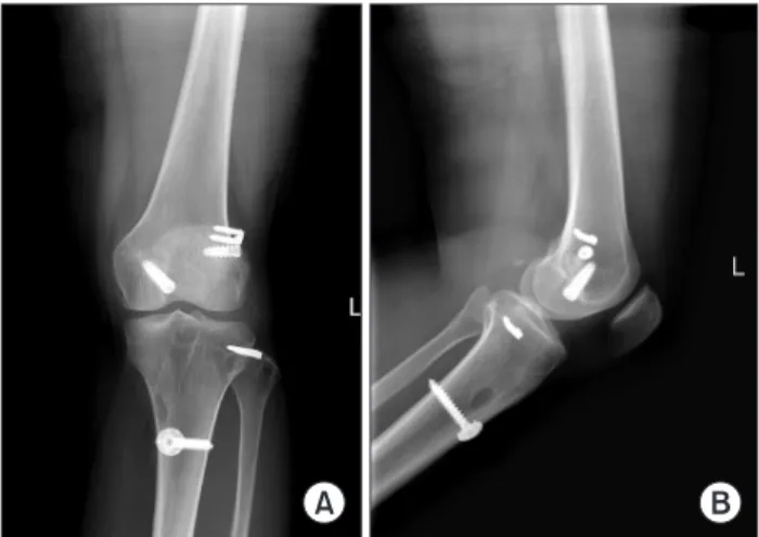
Fig. 2. X-ray at 2 years postoperation showing implants that were used for reconstructing the posterior cruciate ligament and the posterolateral ligament complex in the left knee (anteroposterior (A) and lateral (B) radiography).
Table 1. Lysholm knee score
Score Preoperative Postoperative Excellent (95-100)
Good (84-94) Fair (65-83) Poor (<64)
0 0 4 21
4 16 5 0
Table 2. International Knee Documentation Committee (IKDC) grade
Score Preoperative Postoperative
A (normal) B (nearly normal) C (abnormal) D (severely abnormal)
0 0 8 17
0 20 5 0
Table 3. Noyes & Barber-Westin's classification
Classificationa) Preoperative Postoperative
Functional: lateral opening < 3 mm & external rotation < 5o
Partially functional: lateral opening 3-5 mm & external rotation 5o-10o Failed: lateral opening > 5 mm & external rotation > 10o
0 0 25
20 5 0
a)Difference between the contralateral knee.
were classified as ‘Functional’, and 20% were classified as
‘Partially functional’ (P < 0.05) (Table 3).
Complications
There was no postoperative complication and neuro va- scular injury. None of the patients showed any significant limitation of range of motion of the knee joint.
DISCUSSION
Recent studies based on a biomechanical selective cutting test have shown that the popliteus muscle-tendon unit and the lateral collateral ligament are the two most essential structures for the posterolateral instability of the knee joint [10]. Of these, increasing interest has been given to the importance of the popliteofibular ligament.
Loss of function of the lateral collateral ligament was regenerated with the use of biceps tenodesis by Clancy.
There are articles reporting good clinical outcomes using biceps tenodesis [11-13]. However, this method does not anatomically recreate the popliteus tendon or the popliteofibular ligament and does not pay attention to isometricity [11]. Kanamori et al. [14] conducted a study to compare between the treatment outcomes of biceps tenodesis and the arthroscopic reconstruction of the popliteofibular ligament. The authors reported that the latter approach produces a more effective reconstruction of the posterolateral biomechanics of the knee joint and is more resistant to posterior and external rotational forces of the knee joint.
Methods for synchronous reconstruction of the pop- liteofibular ligament and the lateral collateral ligament exist. Examples of allografts used in such methods include;
a bone-patellar tendon-bone allograft of Latimer et al.’s method [15], and the hamstring tendon (semitendinous tendon and gracilis tendon) of Larson’s [16]. The Achilles tendon allograft was used by Veltri and Warren [17] to synchronously reconstruct the popliteofibular ligament, the lateral collateral ligament and the popliteus tendon, but the anatomical attachments on the femoral and fibular side of the posterolateral structures were not performed. However, Veltri and Warren [17] and LaPrade et al. [4] noted that the anatomical restoration is ideal and enchanced treatment outcomes are expected, as exemplified by the reconstruction of the anterior and posterior cruciate ligament of the posterolateral complex.
In recent years, LaPrade et al. [4], Lee et al. [5], and Yoon
et al. [6] have introduced methods for reconstructing the popliteofibular ligament, the lateral collateral ligament, and the popliteus tendon both synchronously and anato- mically. Lee et al. [5] and Yoon et al. [6] use an approach to fix the bone block of the Achilles tendon allograft to the femoral attachment of the popliteus tendon, whereas LaPrade et al. [4] individually fixed the two allograft tendons to a single tibial tunnel. However, when the resolution of the instability between the femur and the tibia-fibula is the primary concern, as in this clinical condition, LaPrade et al.’s approach [4] to reconstruct the popliteofibular ligament through the passage of the posterolateral tibial tunnel from the posteromedial aspect of the fibular head, may not be the most suitable means for posterolateral reconstruction. By contrast, the methods used by Lee et al. [5] and Yoon et al. [6] ana- tomically fixed the attachments of the femur and tibia, thus, anatomical reconstructions were attempted to an extent. Further, the surgical methods used by these two groups are advantageous in that the key posterolateral structures can be synchronously reconstructed requiring no additional skin incisions, thus are beneficial from an aesthetic perspective and the reconstructed popliteus tendon can be made to its original location.
In our clinical series of patients, all 25 cases were treated with this single type of surgery. All cases were monitored until the final follow-up was complete without any lost- to-follow-up. Based on the classifications of Noyes &
Barber-Westin, there were good clinical outcomes at the final follow-up. But limitations to the current study exist.
There was no control group, no objective or quantitative evaluations that allowed us to compare the degree of the postoperative improvement of the posterolateral instability as well as that of the posterolateral instability of the knee joint. Because the cases of combined injury were included, the potential effects of other pathologies must be taken in consideration. However, we could not find any significant differences of clinical outcomes between the isolated and combined posterolateral instability. Finally, the mean follow-up period was 36 months, which is a relatively short-term follow-up study.
Thus, in the future, it is necessary to devise methods to objectively measure and evaluate the posterolateral instability of the knee postsurgery, and to perform long- term follow-up studies.
In patients with isolated or combined grade III post- erolateral ligament injuries of knee, anatomical post-
erolateral ligament reconstruction using Achilles tendon allograft provided the satisfactory clinical outcomes at the average of 36 months follow-up.
CONFLICT OF INTEREST
No potential conflict of interest relevant to this article was reported.
1. Maynard MJ, Deng X, Wickiewicz TL, Warren RF. The popli- teofibular ligament: rediscovery of a key element in posterolateral stability. Am J Sports Med 1996;24:311-6.
2. Shahane SA, Ibbotson C, Strachan R, Bickerstaff DR. The popli- teofibular ligament: an anatomical study of the posterolateral corner of the knee. J Bone Joint Surg Br 1999;81:636-42.
3. Veltri DM, Deng XH, Torzilli PA, Maynard MJ, Warren RF. The role of the popliteofibular ligament in stability of the human knee: a biomechanical study. Am J Sports Med 1996;24:19-27.
4. LaPrade RF, Johansen S, Wentorf FA, Engebretsen L, Esterberg JL, Tso A. An analysis of an anatomical posterolateral knee reconstruction: an in vitro biomechanical study and development of a surgical technique. Am J Sports Med 2004;32:1405-14.
5. Lee MC, Park YK, Lee SH, Jo H, Seong SC. Posterolateral reconstruction using split Achilles tendon allograft. Arthroscopy 2003;19:1043-9.
6. Yoon KH, Bae DK, Ha JH, Park SW. Anatomic reconstructive surgery for posterolateral instability of the knee. Arthroscopy 2006;22:159-65.
7. Covey DC. Injuries of the posterolateral corner of the knee. J Bone Joint Surg Am 2001;83:106-18.
8. Tegner Y, Lysholm J. Rating systems in the evaluation of knee ligament injuries. Clin Orthop Relat Res 1985;198:43-9.
9. Noyes FR, Barber-Westin SD. Surgical restoration to treat chronic deficiency of the posterolateral complex and cruciate ligaments of the knee joint. Am J Sports Med 1996;24:415-26.
10. Nielsen S, Helmig P. The static stabilizing function of the popliteal
tendon in the knee: an experimental study. Arch Orthop Trauma Surg 1986;104:357-62.
11. Kim SJ, Shin SJ, Choi CH, Kim HC. Reconstruction by biceps tendon rerouting for posterolateral rotatory instability of the knee:
Modification of the Clancy technique. Arthroscopy 2001;17:664- 7.
12. Kim JG, Ha JG, Lee YS, Yang SJ, Jung JE, Oh SJ. Posterolateral corner anatomy and its anatomical reconstruction with single fibula and double femoral sling method: anatomical study and surgical technique. Arch Orthop Trauma Surg 2009;129:381-5.
13. Albright JP, Brown AW. Management of chronic posterolateral rotatory instability of the knee: surgical technique for the posterolateral corner sling procedure. Instr Course Lect 1998;47:369-78.
14. Kanamori A, Lee JM, Haemmerle MJ, Vogrin TM, Harner CD. A biomechanical analysis of two reconstructive approaches to the posterolateral corner of the knee. Knee Surg Sports Traumatol Arthrosc 2003;11:312-7.
15. Latimer HA, Tibone JE, ElAttrache NS, McMahon PJ. Recon- struction of the lateral collateral ligament of the knee with patellar tendon allograft: report of a new technique in combined ligament injuries. Am J Sports Med 1998;26:656-62.
16. Larson RV. Isometry of the lateral collateral and popliteofibular ligaments and techniques for reconstruction using a free semi- tendinosus tendon graft. Oper Tech Sports Med 2012;20:65-71.
17. Veltri DM, Warren RF. Operative treatment of posterolateral instability of the knee. Clin Sports Med 1994;13:615-27.