Volume 14, Number 1, June, 2011
※ 통신저자: 원 예 연
경기도 수원시 영통구 원천동 산5 아주대학교 의과대학 정형외과학교실
TEL: 031) 219-5220 FAX: 021) 219-5229 E-mail: [email protected] 접수일: 2011년 4월 28일, 게재확정일: 2011년 5월 13일
* 본 논문은 (주)유유제약의 임상연구비 지원을 받아 이루어졌음.
Alendronate와 Calcitriol 고정용량복합제의 장기간의 복용이 난소가 제거된 백서의 해면골의 미세구조에 미치는 효과
아주대학교 의과대학 정형외과학교실
지형민・Surej G. Nair・원예연・김진호・손명아
= Abstract =
Effect of a Long-term, Oral Fixed Dose with a Combination of Aledronate and Cacitriol on the Cancellous Bone
Microarchitecture in Ovariectomized Rats
Hyung-Min Ji, M.D., Surej G. Nair MS DNB, Ye-Yeon Won, M.D. Ph.D., Jinho Kim, M.D., Myeong-A Son, M.D.
Department of Orthopedics, Ajou University School of Medicine, Suwon, Korea
Purpose:To study the effect of a long-term, oral, fixed dose with a combination of alendronate and calcitriol on the cancellous bone microarchitecture in an ovariectomized rat model.
Materials and Methods:Twenty eight female Sprague-Dawley rats were divided into 2 equal groups: a non-medication group (OVX), and a medication group (ALD). The ALD group was treated with an oral daily fixed dose with a combination of alendronate and calcitriol for six months, starting from 4 weeks after ovariec- tomy, while the OVX group was given only a placebo. After six months, all animals were sacrificed, and an in vitro micro-CT analysis of the the distal femur was performed. The bone volume fraction (BV/TV), trabecular thickness (Tb.Th), trabecular separation (Tb.Sp), trabecular number (Tb.N), structure model index (SMI), con- nectivity density (Conn.D), and bone mineral density (BMD) were assessed.
Results:The ALD group had significantly higher BV/TV, Tb N, BMD and Conn.D and it also had signifi- cantly lower Tb Sp and SMI than the OVX group.
Conclusion:A long term, daily, oral fixed dose with a combination of alendronate and calcitriol could sig- nificantly reduce the osteoporotic changes in this ovariectomized rat model.
Key Words:Microarchitecture, Cancellous bone, Ovariectomized rat, Alendronate, Calcitriol, Long-term, Oral, Combination
Introduction
Post-menopausal osteoporosis is a major health problem in the elderly women, with substantial morbidity and disability.
It is characterized by low bone mass, and structural deterioration of bone tissue, leading to increased risk of fragility frac- tures1,7,29). However, pharmacological inter- vention can curb these osteoporotic changes and reduce the incidence of these fractures. Several agents are available and oral bisphosphonates, with concomi- tant calcium and vitamin D is currently the most common1,5,7,14,18,28)
. Alendronate, a nitrogen containing bisphosphonate, is a powerful and selective inhibitor of osteo- clast mediated bone resorption. It reverses the progression of osteoporosis, increases bone mineral density (BMD), and decreas- es the incidence of osteoporotic fractures.
Many studies have reported that vitamin D analogues like calcitriol and alfacalcidol can be considered for combination treat- ments with antiresorptives like bisphos- phonates1,7,12,18,24,25)
. The rationale for this combination is that, most elderly osteo- porotic patients who are prescribed alen- dronate, tend to have Vit D deficiency, which in turn adversely affects the antiresorptive efficacy of bisphospho- nates7,14).
The objective of our study was to assess the efficacy of a long term, oral, fixed dose with a combination of alendronate and calcitriol in reverting the osteoporotic changes in an ovariectomized rat model, since it represents the most important clinical features of estrogen deficiency- induced or post-menopausal bone loss in the adult human.
Materials and Methods
Twenty eight female Sprague-Dawley rats were selected for the study. At 12 weeks of age, they were anesthetized and an ovariectomy (OVX) was performed.
Four weeks after OVX, they were ran- domly divided into two groups of 14 each;
a non-medication group (OVX, n=14), and a medication group (ALD, n=14). The mean body weight of the ALD group was 375g (range, 334~428 g), and of the OVX group was 368g (range, 324~ 416 g). The rats were housed in individual cages, for the convenience of administering the oral medication. The room temperature was maintained at 24°C and humidity main- tained at 30%. Food pellets (20% protein, 3.5% lipid, 8% fiber, 8% ash, 5% calci- um, and 1.5% phosphorous) and tap water were being given ad libitum, except during the overnight fasting period, before medication. The medication for the ALD group was started 4 weeks after OVX. In our study, we have used a novel method to orally administer medicine, in the form of a sugar coated pill containing a fixed dose with a combination of alen- dronate and calcitriol. Each pill was refor- mulated to a rat dose to be administered in 400 microgram of alendronate and 0.04 microgram of calcitriol. It was calculated from the daily recommended dose for a 60 kg adult and adopting it to 373 g, which was the average weight of the rats. After an overnight fasting period, a single pill was kept in a glass dish in each cage, and half an hour after the pill was consumed, the food pellets were put back in the cages. Though initially, the animals were hesitant to consume the pills, within a few days, they readily started taking the
지형민 등・Alendronate와 Calcitriol 경구용 고정용량복합제의 효과
pills as soon as it was being given to them. The pills were being given daily for six months. The OVX group was given only a placebo, which was also adminis- tered in a similar manner. All animals were treated according to the practice code of the Ajou University Medical School- Institutional Animal Care and Use Com- mittee (AUSM-IACUC), with the approval number AMC63, dated 14/9/2009.
Scanning was done using the micro-CT scanning machine (Skyscan model 1173, Skyscan, Belgium)(Fig. 1). Image acquisi- tion was done with a source voltage of 130 KV and a source current of 30 μA. The
aluminum filter used for beam hardening artifact reduction was 1 mm thick. The image pixel size was 12.16 μm, the num- ber of rows and columns was 1120 each, vertical object position was 35 mm, and averaging frames 3. The exposure time was 250 ms, and the rotation step was 0.3°. The total scanning time was 20 min.
The scanning region was centered on the distal femur including the femoral condyles and metaphyseo-diaphyseal region. After scanning, the images were reconstructed using the reconstruction software (NRecon,V1.4.1 Skyscan, Kon- tich, Belgium)(Fig. 2). After image recon-
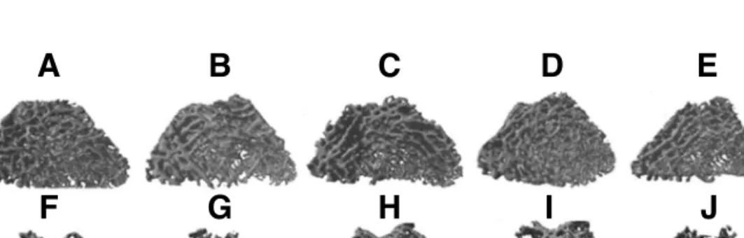
Fig. 1.2D cross sectional images of the cancellous area of distal femur of five rats each, in the ALD group (A, B, C, D, E), and the OVX group (F, G, H, I, J). The OVX group is showing a much lesser number of trabeculae, with increased trabecular separation than the ALD group.
A
F G H J
B C E
Fig. 2. 3D micro-CT reconstruction models of the volume of interest (VOI) of cancellous bone of distal femur of five rats each, in the ALD group (A, B, C, D, E) shown in blue, and the OVX group (F, G, H, I, J) shown in green. It is obvious from the 3D models that the ALD group is having a higher trabecular bone volume, with increased number of trabeculae, and lesser trabecular separation than the OVX group.
A
F G H I J
B C D E
I
D
struction, using the image analysis soft- ware (CTAn V1.10.1.3, Skyscan, Kontich, Belgium), analysis of the reconstructed images were done after selecting a volume of interest (VOI) containing exclusively cancellous bone. The VOI included 200 cross sections extending from 1 mm above the proximal end of the growth plate, proximally for a height of 2 mm.
During analysis, the BMD, and the fol- lowing structural parameters were calcu- lated over each VOI of cancellous bone;
Bone volume fraction (BV/TV), trabecular thickness (Tb.Th), trabecular separation (Tb.Sp), trabecular number (Tb.N), and structure model index (SMI), and connec- tivity density (Conn.D). The BV/TV was assessed using the marching cubes method, calculating the bone volume (BV) over the volume of the VOI i.e, the tissue volume (TV)16). The Tb.Th and Tb.Sp, are measures of the average thickness of the cancellous bone, and the average diameter of the marrow cavities, respectively. They were calculated using the local sphere-fit- ting method8). The Tb.N, which gives the number of trabecular plates per unit length was calculated using the formula Tb.N=(BV/TV)/Tb.Th20,21). SMI is a para- meter calculated using a differential analysis of the triangulated surface of a structure, which gives an estimate of the ratio of the number of plates to the num- ber of rods constituting the 3D struc-
ture9,22). Its values range from 0 to 3, with
0 indicating an ideal plate-like structure and 3 indicating an ideal rod-like struc- ture, and the intermediate values repre- senting a structure composed of both plates and rods. Conn.D is a topological parameter that estimates the number of trabecular connections per cubic
millimeter19).
Statistical analysis was done using SPSS version 12 (Chicago, IL, USA). All the parameters with the exception of Tb.N and Conn.D, had a normal distribution (Shapiro-Wilk test, p>0.05 for all the examined parameters). So, the variables BV/TV, SMI, Tb.Th, Tb.Sp, and BMD were tested by independent T-Test, while the variables Tb.N and Conn.D variables were tested by Nonparametric Test. For all the comparisons, differences were deemed to be statistically significant at p<0.05.
Results
The mean and standard deviations of the various microstructural parameters and the BMD, of the 2 groups are given in table1. All the 28 rats had completed the study. The mean body weight at the end of the treatment period, when the animals were sacrificed was 428 g (range, 388~455 g) in the OVX group, and 368 g (range, 382~562 g) in the ALD group.
Thus the OVX group had higher mean weight gain (90 g), in comparison to the ALD group (53 g) (p value-0.04). Both the OVX and the ALD groups showed sig- nificant difference with respect to the BV/TV, Tb N, Tb Sp, BMD, SMI, as well as the Conn.D. The ALD group had sig- nificantly higher BV/TV, Tb N, BMD and Conn.D and a significantly lower Tb Sp and SMI, in comparison with the OVX group. This indicated that the ALD group had a higher bone mass, increased thick- ness of cancellous bone, increased number of trabecular plates per unit length, as well as higher number of trabecular con- nections per unit length, when compared
지형민 등・Alendronate와 Calcitriol 경구용 고정용량복합제의 효과
to the OVX group. Though both the groups had intermediate values for the SMI (between 0 and 3), the lower SMI value for the ALD group meant that it had a more plate-like structure when compared to the OVX group. However, there was no significant difference in the Tb Th between the two groups.
Discussion
Many recent reports have appeared in literature on the early and late effects of Zoledronic acid injections in ovariectomized rats, assessed by in vivo micro-CT3,23). There have also been previous reports on the combined, as well as comparative effects of alendronate and alfacalcidol in ovariectomized rats11,12), where mechanical testing and bone histomorphometry were used to assess the mechanical properties of bone, alendronate had been administered subcutaneously, and medication was administered for not more than three months. This is the first study to deter- mine the effect of a long-term (6 months
medication period), oral, fixed dose with a combination of alendronate and calcitriol, administered daily, in improving the dete- rioration of the cancellous bone microstruc- ture, assessed by micro-CT, in an ovariec- tomized rat model.
The rat OVX model represents the most important clinical features of estrogen deficiency-induced or post-menopausal bone loss in the adult human13,27). Two weeks post OVX, there is increased bone turnover and bone resorption with signifi- cant decrease in trabecular bone volume in OVX rats. Though the rat OVX model is suitable for evaluating agents for osteo- porosis, it has limitations that restrict the evaluation to cancellous bone sites13,27). The evaluation of the cortical bone response may not be appropriate since the rat OVX model does not mimic postmenopausal women in this respect27). We did the study assessing the effect of alendronate and calcitriol on the cancellous bone of rat dis- tal femur.
There are studies showing that there is a short time window during which the Table 1.Summary of the comparison of the various microstuctural parameters, and the BMD, between the two
groups with the corresponding p values.
*Parameter ALD group (14) OVX group (14) P value
BV/TV 15.029 (±2.978) 07.939(±2.320) 0.0000
SMI 01.379(±0.292) 01.869(±0.337) 0.0004
Tb.Th 08.264(±0.449) 08.372(±0.397) 0.6638
Tb.N 00.018(±0.004) 00.009(±0.003) 0.0000
Tb.Sp 55.027±(7.816) 70.260(±9.959) 0.0001
Conn.D 0.00021(±6.57041E-05) 0.00009(±6.08728E-05) 0.0003
BMD 0.042(±0.037) 0.005(±0.009) 0.0009
Depicted are the mean values, with the standard deviation (SD) in parentheses (±SD).
*TV-total volume (pixel2), BV-bone volume (pixel2), BV/TV-Bone volume fraction (%), SMI-structure model index, Tb.Th-trabecular thickness (pixel), Tb.N-trabecular number (1/pixel), Tb.Sp-trabecular separation (pixel), and Conn.D-connectivity density (1/pixel2) and BMD-bone mineral density (g/cm2).
One pixel size=12.16 micrometer.
microstructural parameters undergo the largest magnitude of change, post-ovariec- tomy2,4). Moreover, since this happens within the first three months post- ovariectomy, they suggest, it is preferable to start antiresorptive treatment early.
Perilli et al23), in a longitudinal study using in vivo micro-CT on ovariectomized rats, have shown that full restoration of the microstructural parameters to the baseline values is possible even after starting treatment 2 weeks post-ovariecto- my. In our study, treatment was started 4 weeks after ovariectomy, when most of the microstructural changes following ovariectomy would be quite well estab- lished according to the previous studies.
We decided to give the medication for a period of only six months because, with longer evaluation periods, cancellous bone turnover indices are found to return to the value of sham controls, with a new steady state in bone remodeling27).
The OVX group had significantly higher mean gain in body weight (90 g) than the ALD group (53 g) (p < 0.05), which might probably be due to a significantly higher body fat content27). The ALD group had a significantly higher BV/TV, Tb.N, and a significantly lower Tb.Sp and SMI in com- parison to the OVX group. The BV/TV plays an important role in the mechanical properties of subchondral trabecular bone29). The higher BV/TV seen in the treatment group in our study, may be attributed to an increase in the Tb.N along with a reduction in the Tb.Sp6,29). Though BV/TV has been found to posi- tively correlate with Tb.Th also29), in our study, there was no significant difference between the two groups in Tb.Th. Post- ovariectomy, a reduction in trabecular
thickness is normally expected, associated with a significant reduction in BV/TV.
However variable findings on trabecular thickness in OVX rats have been reported in literature. While some studies have found a reduction in Tb.Th over time fol- lowing ovariectomy2,4), some others have reported either no change, or an increase3,6,10). But since all of these studies have not used the same strain of animal, which could also possibily influence the change in Tb.Th, more studies may be needed to clarify this issue. It is possible that, in the OVX rats in our study, the bone loss seen was probably due to a pref- erential removal of the thinner trabecu- lae2,4), resulting in a reduction in the tra- becular number and increased trabecular separation, without any significant change in Tb.Th.
The SMI gives an idea about the propor- tion of the number of plates to that of rods in the three dimensional structure of trabecular bone, and is a good indicator of the mechanical strength of trabecular bone. Siu et al26), in a study on ovariec- tomized goats, found that osteoporotic bone tends to have higher SMI values, and that SMI also had a negative correla- tion with BMD. Though the mean SMI value in our study was significantly high- er in the OVX rats, both groups had intermediate values (between 0 and 3);
with a mean value of 1.38 for the med- ication group, and 1.87 for the OVX group (p=0.0004). This meant that the ALD group had a larger fraction of tra- becular plates, which in turn has been found to make a much larger contribution to the bone’s elastic behavior than trabec- ular rods15). Specimens with predominant trabecular plates have been shown experi-
지형민 등・Alendronate와 Calcitriol 경구용 고정용량복합제의 효과
mentally to have lesser deformation than those with predominant rods17).
The Conn.D, which represents the num- ber of trabecular connections per cubic mm, has a positive association with the Tb.N and correlates with mechanical strength and mass of trabecular bone6,29). It is found to be decreased in the osteo- porotic ovariectomized rat model and has a role in predicting bone quality of human being. The medication group in our study had a significantly higher Conn.D (p=0.0003) than the OVX group, repre- senting a higher bone mass and strength.
Our study has limitations. We did not have a sham control group, to provide us baseline values to determine the actual degree of bone recovery following medica- tion. Besides, we also did not have compar- ison groups, either for assessing different dosage schedules, or for the simultaneous assessment of the individual effects of alendronate and calcitriol. Further longitu- dinal studies using in-vivo micro-CT, and with more comparison groups, would defi- nitely help derive more distinct conclusions.
Conclusion
To conclude, in this study, we have attempted to assess with micro-CT, the improvement in cancellous bone microar- chitecture, in ovariectomized rats, follow- ing long-term administration of an oral fixed dose with a combination of alen- dronate and calcitriol. Starting medication 4 weeks following ovariectomy, we could find a significant reduction in the deterio- ration of cancellous bone microarchitec- ture, as well as a significant improvement in the BMD after 6 months of treatment.
Disclosure
The authors have received outside fund- ing in support of this research from Yuyu Pharma Inc, Suwon, South Korea.
Authors have full control of all primary data and agree to allow the journal to review data if requested.
Acknowledgements
The authors thank Jung, Sun Jang of Genoss Co. Ltd, Korea, for the technical help in scanning and analysis of the speci- mens, and Ms Bak, Jean Kyung for her help with the statistical analysis.
REFERENCES
1) Binkley N, Ringe JD, Reed JI, et al: Alen- dronate/vitamin D3 70 mg/2800 IU with and without additional 2800 IU vitamin D3 for osteo- porosis: Results from the 24-week extension of a 15-week randomized, controlled trial. Bone, 44:
639-647, 2009.
2) Boyd SK, Davison P, Muller R and Gasser JA:
Monitoring individual morphological changes over time in ovariectomized rats by in vivo micro-computed tomography. Bone, 39: 854-862, 2006.
3) Brouwers JE, Lambers FM, Gasser JA, van Rietbergen B and Huiskes R: Bone degenera- tion and recovery after early and late bisphospho- nate treatment of ovariectomized wistar rats assessed by in vivo micro-computed tomography.
Calcif Tissue Int, 82: 202-211, 2008.
4) Campbell GM, Buie HR and Boyd SK: Signs of irreversible architectural changes occur early in the development of experimental osteoporosis as assessed by in vivo micro-CT. Osteoporos Int, 19: 1409-1419, 2008.
5) Dell RM, Greene D, Anderson D and Williams
K: Osteoporosis Disease Management: What Every Orthopaedic Surgeon Should Know. J Bone Joint Surg Am, 91: 79-86, 2009.
6) Dempster DW, Birchman R, Xu R, Lindsay R and Shen V: Temporal changes in cancellous bone structure of rats immediately after ovariec- tomy. Bone, 16: 157-161, 1995.
7) Frediani B, Allegri A, Bisogno S and Marco- longo R: Effects of Combined Treatment with Calcitriol plus Alendronate on Bone Mass and Bone Turnover in Postmenopausal Osteoporosis:
Two Years of Continuous Treatment. Clinical Drug Investigation, 15: 235-244, 1998.
8) Hildebrand T and Ru¨egsegger P: A new method for the model-independent assessment of thickness in three-dimensional images. Journal of Microscopy, 185: 67-75, 1997.
9) Hildebrand T and Ruegsegger P: Quantifica- tion of Bone Microarchitecture with the Structure Model Index. Comput Methods Biomech Biomed Engin, 1: 15-23, 1997.
10) Hornby SB, Evans GP, Hornby SL, Pataki A, Glatt M and Green JR: Long-term zoledronic acid treatment increases bone structure and mechanical strength of long bones of ovariec- tomized adult rats. Calcif Tissue Int, 72: 519- 527, 2003.
11) Iwamoto J, Seki A, Takeda T, Sato Y, Yama- da H and Yeh JK:Comparative effects of alen- dronate and alfacalcidol on cancellous and corti- cal bone mass and bone mechanical properties in ovariectomized rats. Exp Anim, 55: 357-367, 2006.
12) Iwamoto J, Takeda T, Matsumoto H, Sato Y and J KY: Beneficial effects of combined administration of alendronate and alfacalcidol on cancellous bone mass of the tibia in orchidec- tomized rats: a bone histomorphometry study. J Nutr Sci Vitaminol (Tokyo), 54: 11-17, 2008.
13) Kalu DN: The ovariectomized rat model of post- menopausal bone loss. Bone Miner, 15: 175-191, 1991.
14) Kennel KA and Drake MT: Adverse effects of bisphosphonates: implications for osteoporosis management. Mayo Clin Proc, 84: 632-637; quiz 638, 2009.
15) Liu XS, Sajda P, Saha PK, Wehrli FW and Guo XE:Quantification of the roles of trabecu- lar microarchitecture and trabecular type in determining the elastic modulus of human tra- becular bone. J Bone Miner Res, 21: 1608-1617, 2006.
16) Lorensen WE and Cline HE: Marching cubes:
A high resolution 3D surface construction algo- rithm. In Proceedings of the 14th annual confer- ence on Computer graphics and interactive tech- niques, pp. 163-169. Edited, 163-169, ACM, 1987.
17) Muller R, Gerber SC and Hayes WC: Micro- compression: a novel technique for the nonde- structive assessment of local bone failure. Tech- nol Health Care, 6: 433-444, 1998.
18) Nakamura Y, Hayashi K, Abu-Ali S, Naito M and Fotovati A: Effect of preoperative com- bined treatment with alendronate and calcitriol on fixation of hydroxyapatite-coated implants in ovariectomized rats. J Bone Joint Surg Am, 90:
824-832, 2008.
19) Odgaard A: Three-dimensional methods for quantification of cancellous bone architecture.
Bone, 20: 315-328, 1997.
20) Parfitt AM, Drezner MK, Glorieux FH, et al:
Bone histomorphometry: standardization of nomenclature, symbols, and units. Report of the ASBMR Histomorphometry Nomenclature Com- mittee. J Bone Miner Res, 2: 595-610, 1987.
21) Parfitt AM, Mathews CH, Villanueva AR, Kleerekoper M, Frame B and Rao DS:Rela- tionships between surface, volume, and thickness of iliac trabecular bone in aging and in osteo- porosis. Implications for the microanatomic and cellular mechanisms of bone loss. J Clin Invest, 72: 1396-1409, 1983.
22) Perilli E, Baruffaldi F, Bisi MC, Cristofolini L
지형민 등・Alendronate와 Calcitriol 경구용 고정용량복합제의 효과
and Cappello A: A physical phantom for the calibration of three-dimensional X-ray microto- mography examination. Journal of Microscopy, 222: 124-134, 2006.
23) Perilli E, Le V, Ma B, Salmon P, Reynolds K and Fazzalari N: Detecting early bone changes using in vivo micro-CT in ovariectomized, zole- dronic acid-treated, and sham-operated rats.
Osteoporosis International, 21: 1371-1382, 2010.
24) Ringe JD, Farahmand P, Schacht E and Rozehnal A: Superiority of a combined treat- ment of Alendronate and Alfacalcidol compared to the combination of Alendronate and plain vita- min D or Alfacalcidol alone in established post- menopausal or male osteoporosis (AAC-Trial).
Rheumatol Int, 27: 425-434, 2007.
25) Ringe JD and Schacht E: Improving the out- come of established therapies for osteoporosis by adding the active D-hormone analog alfacalcidol.
Rheumatol Int, 28: 103-111, 2007.
26) Siu WS, Qin L, Cheung WH and Leung KS: A study of trabecular bones in ovariectomized goats with micro-computed tomography and peripheral quantitative computed tomography.
Bone, 35: 21-26, 2004.
27) Thompson DD, Simmons HA, Pirie CM and Ke HZ:FDA Guidelines and animal models for osteoporosis. Bone, 17: 125S-133S, 1995.
28) Weaver MJ, Miller MA and Vrahas MS: The orthopaedic implications of diphosphonate thera- py. J Am Acad Orthop Surg, 18: 367-374, 2010.
29) Zhang ZM, Li ZC, Jiang LS, Jiang SD and Dai LY:Micro-CT and mechanical evaluation of subchondral trabecular bone structure between postmenopausal women with osteoarthritis and osteoporosis. Osteoporos Int, 21: 1383-1390, 2010.