on by Human Periodontal Ligament Stem Cells both in vitro and in vivo Analysis
2
1)Department of Periodontology, Research Institute for Periodontal Regeneration, College of Dentistry, Yonsei University, Seoul, Republic of Korea
2)Department of Periodontology, College of Dentistry, Dankook University, Cheonan, Republic of Korea
3)Department of Periodontology, Research Institute for Periodontal Regeneration, College of Dentistry, BK Plus 21, Yonsei University, Seoul, Republic of Korea
Jae-Kook Cha1), Sang-Yeob Oh1), Jung-Chul Park2), Dong-Jun Kim1), So-Yon Park1), Kyoo-Sung Cho1), Chang-Sung Kim1, 3)*
The Effect of Enamel Matrix Derivatives on the Collagen Formation by Human Periodontal Ligament
Stem Cells both in vitro and in vivo Analysis
사
사람람 치치주주인인대대줄줄기기세세포포의의 교교원원질질 형형성성에에 대대한한 법법랑랑기기질질 유유도도체체의의 영영향향
1)연세대학교 치과대학 치주과학교실, 치주조직재생연구소, 2)단국대학교 치과대학 치주과학교실
3)연세대학교 BK plus 21 치과대학 치주과학교실, 치주조직재생연구소 Jae-Kook Cha1), Sang-Yeob Oh1), Jung-Chul Park2), Dong-Jun Kim1),
So-Yon Park1), Kyoo-Sung Cho1), Chang-Sung Kim1, 3)*
목적: 법랑기질 유도체(EMD)가 사람 치주인대 줄기세포(hPDLSC)의 조직 형성능에 미치는 영향을 in vitro와 in vivo 분석 모델을 이용해 평가 한다.
재료 및 방법: hPDLSC를 배양하여 운반체와 함께 면역 억제된 쥐 등에 이식하였다; (1)대조군: EMD 처치하지 않은 운반체에 심어진 hPDLSC 군 (EMD-/hPDLSC+), (2)실험군: EMD 처치한 운반체에 심어진 hPDLSC군 (EMD+/hPDLSC+). 각 군당 5마리씩 시행하고 8주 후 희생하였다. 조 직학적, 조직계측학적 분석을 통해 형성된 백악질의 면적과 백악세포의 수 그리고 샤피 섬유의 수를 계측하였으며 면역조직화학적 분석을 통해 백 악질과 교원질 형성을 평가하였다. 또한 in vitro에서 hPDLSC의 수용성 교원질과 glycosaminoglycan 형성에 대한 EMD의 효과를 분석하였다.
결과: 조직학적 분석에서 교원질성 치주 인대 조직이 실험군에서 현저하게 많이 생성된 것을 관찰할 수 있었다. 형성된 백악질의 면적과 백악세 포의 수는 군 간 차이가 없었으나, 새롭게 형성된 샤피 섬유의 수는 실험군에서 대조군보다 유의하게 많았다(p<0.05). 교원질 형성에 대한 면역조직 화학적 분석 결과, 실험군에서 I, III형 교원질과 hydroxyproline의 발현이 높았다. 또한 in vitro에서 hPDLSC에 의한 수용성 교원질과 glycosaminoglycan 형성이 EMD의 농도에 비례하여 증가하였다 (p<0.05).
결론: EMD는 hPDLSC에 의한 샤피 섬유 및 교원질 생성을 증가시키고, 이는 새로운 백악질의 기능적 부착과 치주조직 재생에 중요한 역할을 한다.
Key words : enamel matrix proteins, mesenchymal stromal cells, periodontal ligament, periodontium, regeneration, tissue engineering
ABSTRACT
Corresponding Author Chang-Sung Kim
Department of Periodontology, Research Institute for Periodontal Regeneration, College of Dentistry Yonsei University, 50 Yonsei-ro, Seodaemun-gu, Seoul and 120752, Republic of Korea
Email : dentall@yuhs.ac, Tel : +82-2-22283186, Fax : +82-2-3920398
Ⅰ. Introduction
Enamel matrix derivatives(EMD) are extracts of porcine enamel matrix proteins secreted during the development of tooth-supporting tissues. Emdogain(Institute Straumann, Basel, Switzerland), a commercialized EMD product introduced in 1997 by Hammarstrom, has been used as the one of the current clinical treatment for periodontal tissue regeneration1, 2). The desired outcome from the clinical application of Emdogain in adult periodontal defect is tissue regeneration that mimics the natural process of tissue development3).
A number of researches have been reported that the application of EMD yielded positive results to conventional periodontal treatment in various type of defect in animal model including intrabony, furcation or dehiscence defects2, 4~6). They demonstrated that clinical attachment level and radiographic bone fill were enhanced, especially the histological analysis has clearly shown that EMD promotes the regeneration of cementum, alveolar bone, and periodontal ligament following application of EMD. Along with the preclinical studies, the positive effect of EMD also has been represented in a number of clinical studies showing comparable results with guided tissue regeneration(GTR)7). Moreover, the use of EMD would be favored by clinicians due to low risk of clinical complication shown in
the GTR technique.
Despite these promising clinical achievements, the precise cellular mechanism underlying the effect of EMD has not been clarified yet. The possible rationale of EMD for periodontal regeneration is that active ingredient such as amelogenin which plays important role during development would stimulate the progenitor to differentiate into cementoblast, osteoblast and collagen producing fibroblast.
Diverse in vitro studies have demonstrated that EMD and amelogenins stimulate the growth of multiple mesenchymal cell types, including fibroblasts, cementoblasts, osteoblasts, and periodontal ligament stem cells(PDLSCs)8~10). Also, it was reported that the epithelial and connective tissue down growth was inhibited by the EMD which determine the attachment gain in histologic analysis11). The EMD may help the periodontal ligament(PDL) cell to be attached with diseased root surface3, 12). When the PDL cells were treated with EMD, the increased collagen formation could be observed as well as the enhanced osteogenic differentiation potential, suggesting that the EMD also have an effect on the new bone and new cementum formation2). While a number of basic researches on the effect of EMD, the precise cellular mechanism underlying the effect of EMD on PDLSCs for periodontal regeneration still remains unclear.
Acknowledgement
This work was supported by the National Research Foundation of Korea (NRF) grant funded by the Korea government(MSIP) (No.2012R1A2A4A01007124) and the Bio&Medical Technology Development Program of the National Research Foundation (NRF) funded by the Ministry of Science, CT & Future Planning (No. 2012M3A9B2052521).
on by Human Periodontal Ligament Stem Cells both in vitro and in vivo Analysis Regeneration of the Sharpey’s fiber in PDL
should be considered as one of the critical point for the periodontal regeneration. It was reported that the human PDLSCs(hPDLSCs) could produce not only a new cementum on the matrix, but also a collagenous ligament fiber resembling Sharpey’s fiber which was responsible for attachment between the cementum and bone13). Although the collagenous ligament fiber formation by PDLSCs has been known accomplishing critical role of attachment, there is lack of information on the effects of EMD on collagenous ligament fiber formation by PDLSCs.
In our previous studies, we have utilized an ectopic transplantation model which has been a straightforward and effective tool to investigate the in vivo behaviors of stem cells of interest. By this model, the sequence of regeneration by hPDLSc could be successfully evaluated in the previous study13). Thus, we assumed that the application of EMD to the current study model will help toward gaining a deeper understanding of the effect of EMD on hPDLSCs.
The purpose of the present study was thus to determine the effect of EMD on the tissue forming activity of hPDLSCs, especially the collagenous periodontal ligament formation which is critical composition of periodontal attachment, using in vitro and in vivo analysis models.
Ⅱ. Materials and Methods
1.hPDLSCsculture
hPDLSCs were isolated and cultured according to a modification of previously reported protocols14). The experimental protocol was approved by the Institutional Review Board of Yonsei university(2-2010-0016). The subjects were provided informed consent to participate.
Four healthy teeth were extracted for orthodontic purposes from one healthy patient(female, 28 years old). hPDLSCs were isolated from the root surface of the 4 extracted premolars using a scalpel, and then cut into pieces under 1 mm diameter.
The small pieces of tissue were digested four or five times at 20-min intervals in -minimum essential medium( -MEM; GIBCO, Grand Island, NY, USA) containing 3 mg/ml collagenase type I(WAKO, Tokyo, Japan) and 4 mg/ml dispase(GIBCO) at 37 . Single-cell suspensions were obtained by passing the mixture through a strainer with a pore size of 70 (Falcon, BD Labware, Franklin Lakes, NJ, USA), and the cells(5x10⁵) were seeded on to T75 cell culture dishes containing -MEM supplemented with 15% fetal bovine serum(GIBCO), 100 L-ascorbic acid 2-phos phate(Sigma-Aldrich, St. Louis, MO, USA), 2 mML-glutamine(GIBCO), 100 U/ml penicillin, and 100 /ml streptomycin(GIBCO), and incubated at 37 in an atmosphere containing 5% CO2. The third or fifth passage of cells was used for the study. Various experiments including in vitro and in vivo assay were conducted to confirm whether these group of cells possessed
the characteristics of mesenchymal stem cells.
2.hPDLSC transplantation into an ectopic subcutaneous transplant ation model using a carrier treated withorwithoutEMD
Hydroxyapatite/ -tricalcium phosphate(HA/ - TCP) powder 80 mg[macroporous biphasic calcium phosphate(MBCP); Biomatlante, Vig neux, France) was used as a carrier for the hPDLSCs. The animals were treated under the following two experimental conditions: (1) hPDLSCs seeded onto an untreated MBCP carrier(EMD-/hPDLSC+ group), and (2) hPDL SCs seeded onto an EMD-pretreated MBCP carrier(EMD+/hPDLSC+group). The results from the negative control group with MBCP are not shown in this study since the results are previously reported in our previous studies13, 15, 16). Carrier was either pretreated or not with EMD, by soaking MBCP carriers overnight with Emdogain gel at a concentration of 30 mg/ml.
The mice were treated using a protocol that was approved by the Animal Care and Use Committee, Yonsei Medical Center, Seoul, Korea. hPDLSCs (6 106 per carrier) were pre cultured for 1.5 h with the carrier at 37 in a 5%
CO2 atmosphere before transplantation. The carriers were loaded subcutaneously on each side(left and right, respectively) into the dorsal region of 5-week-old male CB17 severe combined immunodeficiency(SCID) mice(n=5 per group, with two ectopic transplantations in each animal). The animals were allowed to heal
for 8 weeks, and then they were sacrificed.
3. Histologic and histometric analysis
Samples of the transplant tissue were fixed in 4% formalin for 3 days and then decalcified with 5% EDTA(pH 8.0), dehydrated in ethanol, and embedded in paraffin. The central sections were reduced to a thickness of 5 and then stained with hematoxylin and eosin(H-E). Light and polarized-light microscopy(Olympus BX50, Olympus Optical, Tokyo, Japan) were used for the histological analyses. The number of cells was counted using an automated image-analysis system(Image-Pro Plus, Media Cybernetics, Silver Spring, MD, USA). The formation and organization of Sharpey’s fibers resembling tissue in the collagen were observed following Picrosirius staining.
4.Immunohistochemicalanalysis
For immunohistochemical analysis, sections were immersed in 0.3% hydrogen peroxide to block endogenous peroxidase activity, and then incubated with primary antibodies diluted in PBS (1:200-1:500). A human-specific mitochondrial antibody(mitochondrial ribosomal protein L11, hMito; Abcam, Cambridge, UK) diluted to 1:100 was used to confirm the origin of the human cells. Analysis of markers related to collagen formation was achieved using the following primary antibodies: collagen type I(Col I, Collagen I antibody; Abcam), collagen type III(Col III, Collagen III antibody; Abcam), and
on by Human Periodontal Ligament Stem Cells both in vitro and in vivo Analysis hydroxyproline(hydroxyproline antibody;
Bioss). A commercially available kit(Zymed SuperPicTure polymer detection kit, Zymed, Invitrogen, Carlsbad, CA, USA) was used to detect the antibodies according to the manufacturer’s protocol. Slides were then counterstained with hematoxylin. Intensity of immunohistochemical staining at interested point was evaluated according to our previous study12).
5.Effect of EMD treatment on soluble collagenandsulfatedglycosamino- glycans(GAG) formation by hPDLSCs in vitro; Sircol collagen assay and BlyscansulfatedGAGassay
When the culture of hPDLSCs came to the subconfluent stage, hPDLSCs were cultured in medium with EMD at various test concentra tions: 0, 100, and 200 /ml. After 5 days, the cell supernatants were collected by centrifugation at 260 g for 5 min for total soluble collagen forma tion. Total soluble collagen in the cell superna tant was measured using the Sircol collagen assay kit(Biocolor, Newtownabbey, UK). Briefly, a 200- aliquot of the supernatant was added to 1?ml of the dye reagent provided with the kit, and the mixture was incubated for 30 min at room temperature. After centrifugation at 9,300 g for 10 min, the separated suspensions were discarded and the remnant pellets were dissolved in 1 ml of the alkali reagent provided with the kit. The relative absorbance was then measured at 555 nm.
For the detection of the sulfated GAG by hPDLSCs the cell supernatants were collected by
centrifugation at 260 g for 5 min, and then1 ml of the dye reagent was added to a 100- aliquot of the supernatant. The tubes were centrifuged at 13,400xg for 10 min and a dissociation reagent was then added(0.5 ml) to each tube. After ensuring thorough dissolution, the mixture was loaded onto 96-well plates, which were placed into a microplate reader and the absorbance was measured at 656 nm.
6. Statistical analysis
All experiments repeated 3 times and the data were presented as mean and standard deviation.
Student’s t test was used to analyze the statistical significance of differences between the EMD- /hPDLSC+and EMD+/hPDLSC+groups. One-way ANOVA was used to compare the soluble collagen and GAG among the three EMD concentration conditions(i.e., 0, 100, and 200 /ml). The statistical analysis was carried out using SPSS for Windows version 18.0(SPSS, Chicago, IL, USA). The cutoff for statistical significance was set at P<0.05.
Ⅲ. Results
1.Effect of EMD on the formation of cementum-like tissue, cementocytes, andSharpey
’sfibersresemblingtissue by hPDLSCs: a histological and histometricanalysisofaninvivomodel
The clinically using dose of EMD(30 mg/ml) was applied in vivo model, because the PDLSCs
from human were used for transplantation.
Histologic evaluation revealed that there was no any significant adverse effect or inflammatory reaction in both experiment groups.
Newly formed mineralized/cementum-like tissue was observed in association with the transplanted hPDLSCs along the HA/ -TCP particles. Newly formed cementum-like tissue was present along the periphery of the HA/ -TCP
carriers, and a high degree of mineralization was observed in both groups(Fig.1A-D).The new cementum-like tissue of both group was characterized by a highly mineralized collagen matrix and numerous cementocytes were embedded within the mineralized tissue. The histologic phonotype of newly formed cemen tum-like tissue was different from the bone like tissue showing rare lamella structure in line with
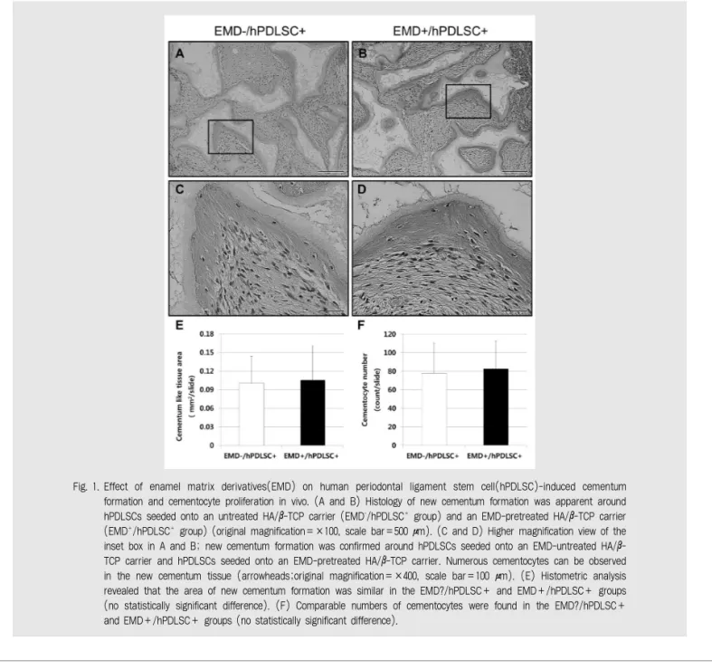
Fig. 1. Effect of enamel matrix derivatives(EMD) on human periodontal ligament stem cell(hPDLSC)-induced cementum formation and cementocyte proliferation in vivo. (A and B) Histology of new cementum formation was apparent around hPDLSCs seeded onto an untreated HA/β-TCP carrier (EMD-/hPDLSC+group) and an EMD-pretreated HA/β-TCP carrier (EMD+/hPDLSC+group) (original magnification=×100, scale bar=500 μm). (C and D) Higher magnification view of the inset box in A and B; new cementum formation was confirmed around hPDLSCs seeded onto an EMD-untreated HA/β- TCP carrier and hPDLSCs seeded onto an EMD-pretreated HA/β-TCP carrier. Numerous cementocytes can be observed in the new cementum tissue (arrowheads;original magnification=×400, scale bar=100 μm). (E) Histometric analysis revealed that the area of new cementum formation was similar in the EMD?/hPDLSC+ and EMD+/hPDLSC+ groups (no statistically significant difference). (F) Comparable numbers of cementocytes were found in the EMD?/hPDLSC+
and EMD+/hPDLSC+ groups (no statistically significant difference).
on by Human Periodontal Ligament Stem Cells both in vitro and in vivo Analysis the previous studies17). Interestingly, in the bone-
like tissue, multinucleated osteoclast-like cells were regularly observed in relation to the osteoblast lining, however, in the formation cementum-like tissue, such appearances were seldom found. Also, the cell rich zone composed of well-organized collagenous ligament fiber like tissue and characteristic finding of histology produced by PDLSC was clearly observed in both groups13). However, there was no significant histological difference according to the treatment of EMD in histologic analysis.
Likewise, the H-E staining revealed that the total area of newly formed cementum-like tissue was similar in the EMD-/hPDLSC+ and EMD+/hPDLSC+ groups and there was no statistically significant difference between groups(Fig. 1E)18). A number of cementocytes were observed in the lacunae of the newly formed cementum-like tissue(Fig. 1C and D), and the total number of cementocytes was counted. The number of cementocytes was also comparable in the EMD-/hPDLSC+ and EMD+/hPDLSC+ groups without any statistically significant difference(Fig. 1F).
When microscopic observation under the Picrosirius stain the formation of Sharpey’s fiber resembling tissue could be observed clearly(Fig.
2A and B). The Sharpey’s fiber resembling tissue was well associated with new cementum-like tissue running into the PDL spaces mimicking histologic pattern shown in the natural periodontal tissue. The insertion of Sharpey’s fiber resembling tissue into the cementum-like tissue originated from the collagenous tissues is
the typical finding in the regenerated mineralized tissue by hPDLSCs as reported in the previous study13). It seems that longer healing period may have resulted a difference in cementum-like tissue formation. Inserted Sharpey’s fibers resembling tissue were also frequently observed under the polarized light microscope(Fig. 2C and D). The total number of inserted Sharpey’s fiber resembling tissue was measured using the polarized light images, and the numbers per unit area was significantly greater in the EMD+/hPDLSC+ group than in the EMD- /hPDLSC+ group(P<0.05; Fig. 2E). In addition, the Shapey’s fiber in the EMD+/hPDLSC+ group appeared longer and progressed into the PDL pace. This suggests that EMD has no significant effect on the formation of mineralized tissue but may provide a beneficial effect on the collagenous PDL fiber which has a pivotal role in tooth attachment.
2.Immunohistochemicalanalysis
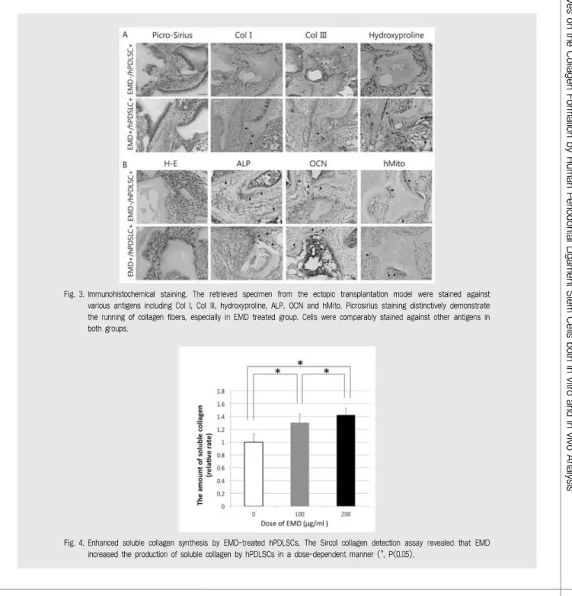
Based on the histological analysis of new cementum-like tissue and fiber formation, immunohistochemical staining was performed to further understand the mechanism of cementogenesis and collagenous ligament fiber formation(Fig. 3). In previous study, cemento genic differentiation was preceded by collage nous ligament fiber formation13). Therefore, the representative cementogenic makers were used:
ALP for earlier differentation and OCN for later stage. As a result, ALP showed slightly increased intensity in the EMD group but there was no
difference in the intensity of OCN between the two groups. Positively stained cells against hMito in both experimental groups revealed that the newly regenerated cementum-like tissue is originated from the transplanted human cells. Col I and III which are distinctive collagen subtypes comprising the PDL were strongly expressed in the EMD treated group along with the histologic analysis which revealed increase in collagenous ligament fiber formation. This could be also
confirmed through hydroxyproline staining.
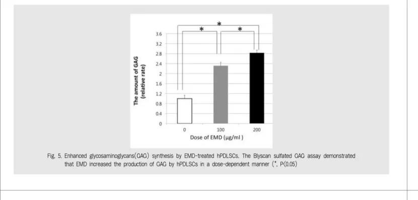
3.In vitro analysis of collagen synthesis : Sircol collagen assay, Blyscans GAG assay
Collagens and GAG are major components of connective tissue, and GAG interacts with collagen in life structure19). Therefore, the effect of EMD on the regenerative potential of
Fig. 2. Effect of EMD on hPDLSC-induced Sharpey’s fiber resembling tissue in vivo. Sharpey’s fiber resembling tissue was observed lining the surface of the carrier and running into the new cementum. (A and B) Newly formed Sharpey’s fiber resembling tissue were confirmed around EMD-untreated HA/β-TCP carrier cultured with hPDLSCs (EMD-/hPDLSC+ group) and an EMD-pretreated HA/β-TCP carrier (EMD+/hPDLSC+ group) (arrowheads; original magnification=×400, scale bar=100 μm). (C and D) The polarized light microspoic images showed numerous insertion of Sharpey’s fiber resembling tissue running parallel into the cementum tissue. (E) The number of new Sharpey’s fiber resembling tissue was significantly greater in the EMD+/hPDLSC+ group than in the EMD-/hPDLSC+group (*, P<0.05).
on by Human Periodontal Ligament Stem Cells both in vitro and in vivo Analysis hPDLSCs regarding collagen and GAG
production was assessed in vitro. Sircol collgen assay is an established evaluation tool for the measurement of collagen formation15). The results
showed that collagen and GAG formation by the hPDLSCs was significantly enhanced by EMD in a dose-dependent manner(Figs. 4 and 5). Statistic ally significant differences in the degree of
Fig. 3. Immunohistochemical staining. The retrieved specimen from the ectopic transplantation model were stained against various antigens including Col I, Col III, hydroxyproline, ALP, OCN and hMito. Picrosirius staining distinctively demonstrate the running of collagen fibers, especially in EMD treated group. Cells were comparably stained against other antigens in both groups.
Fig. 4. Enhanced soluble collagen synthesis by EMD-treated hPDLSCs. The Sircol collagen detection assay revealed that EMD increased the production of soluble collagen by hPDLSCs in a dose-dependent manner (*, P<0.05).
collagen and GAG formation were observed between each of the three EMD concentration groups(i.e., 0, 100, and 200 /ml).
Ⅳ. Discussion
The main function of PDLSC is the formation of cementum and collagenous ligament fibers responsible for the critical role of attachment.
The periodontal regenerative effects of EMD have been well documented, but the cellular mechanisms are yet unknown. Based on the PDLSC technology, the results of the current study composed of in vitro and in vivo assays showed that EMD application had no effect on PDLSCs’ cementogenic potential, but through enhancement of collagen and GAG formation, potential for formation of collagenous ligament fiber may be increased, which has a pivotal role in periodontal attachment. This may bring forth a
cellular mechanism of EMD’s beneficial effect on the enhancement of periodontal regeneration.
The results of this study indicated that the area of new cementum-like tissue formation in vivo was comparable in the EMD-/hPDLSC+ and EMD+/hPDLSC+ groups. It has been previously reported that EMD enhanced mineralization in association with an up-regulation of bone markers in human mesenchymal stem cells (hMSCs) in bone marrow during osteogenic differentiation,20) and it appears to significantly induce proliferation of hMSCs and early-stage osteoblast differentiation in a concentration- dependent manner.21) The number of cemento cytes was also similar regardless of the treatment of EMD on hPDLSCs. In the present study, the results showed an interesting revelation that EMD did not induce any difference in the formation of cementum-like tissue in vivo, which may be due to the relatively brief observation period of 8 weeks. As our previous study
Fig. 5. Enhanced glycosaminoglycans(GAG) synthesis by EMD-treated hPDLSCs. The Blyscan sulfated GAG assay demonstrated that EMD increased the production of GAG by hPDLSCs in a dose-dependent manner (*, P<0.05)
on by Human Periodontal Ligament Stem Cells both in vitro and in vivo Analysis showed13), the healing of periodontal tissue
follows a certain dynamic time process, and cementum formation may be preceded by the formation of collagens. We have demonstrated that the collagen fibers are regenerated first, then followed by the cementum formation induced by cementogenic signals. It was assumed that the formation of collagen fibers appear to be very important for the regeneration of cementum-like tissues in vivo. Therefore, it would be inappropriate to conclude that the treatment of EMD has failed to regenerate the cementum-like tissue, but it should be interpreted that the EMD has significantly induced the formation of collagen tissue such as Sharpey’s fiber resembling tissue, which may be deeply involved in the formation of cementum in later stage and the functional attachment of Sharpey’s fibers.
Although we have followed only 8 weeks of healing period, the results may be different in the longer term of healing period, and the numerous clinical studies demonstrate that the healing period makes a very important difference in the maturation of the periodontal tissues22). Further studies are warranted to fully elucidate the effect of healing period on the regeneration of periodontal tissues treated by EMD.
The hPDLSCs were seeded onto a HA/ -TCP carrier in an ectopic transplantation model using immunocompromised mice, which has been utilized in our previous studies to evaluate the biologic potential of transplated cells23, 24). It was reported that the surface of HA/ -TCP possess number of specially designed micropores and unique ionic microenvironment which is
favorable for cellural attahcement, proliferation and differentiation25, 26). Although the MBCP is known to exhibit osteoinductive capacity, previous studies reported that such osteoind uctive effects were not observed in this model by using MBCP alone27), thus the negative control group was not included in this study design.
Additionally, we have confirmed that the formation of Sharpey’s fiber resembling tissue was significantly up-regulated in the presence of EMD. To elucidate this phenomenon, we investigated the effect of EMD on soluble collagen formation using the Sircol collagen assay in vitro, and the results showed that soluble collagen formation by hPDLSCs in vitro was significantly enhanced by EMD in a dose- dependent manner. There are several reports of EMD enhancing the proliferation of PDL cells and increasing collagen production in vitro; thus, EMD appear to play an important role in periodontal tissue repair by the regeneration of collagen fibers12, 28, 29). Gestrelius et al. reported that EMD enhanced the proliferation of PDL cells and promoted mineralized nodule formation by PDL cells, but did not affect the migration or attachment in vitro12). Cattaneo et al. showed that EMD enhanced the proliferation of PDL fibroblasts in a time-dependent manner28). In the present study, EMD significantly enhanced the formation of collagen fibers both in vivo and in vitro, and these results are in line with previous studies. The authors assume that the increased formation of collagen fibers and collagenous tissues in vitro and in vivo can play a critical role in the periodontal regeneration, and seem to be
deeply involved in the formation of cement tissue as shown in our previous study13) and further studies are warranted to clearly elucidate the underlying mechanism.
The results of this study also revealed that GAG formation by hPDLSCs was significantly enhanced by EMD in a dose-dependent manner.
GAG, which vary in molecular size and characteristics, have many functions in the extracellular matrix, such as connective tissue matrix formation, regulation of cell growth, cell adhesion, and binding of growth factors30). Kirkham et al. reported that the GAG content of PDL is related to the maintenance of the unmineralized state of the PDL tissue31), and it appears that GAG are very important for the stability of PDL tissue. Furthermore, EMD have also been shown to affect collagen fiber regeneration and the production of GAG19, 32, 33), and it was reported that EMD have the potential to modulate GAG in a manner consistent with the early regenerative period19). Since collagen fibers and GAG play an important role in periodontal tissue regeneration, we could assume that the increased formation of collagen fibers and GAG by hPDLSCs play a critical role in the periodontal regeneration.
Up to date, research into the effects of EMD on hPDLSCs is lacking and controversial if any.
Previous studies have shown that EMD could induce the regeneration and formation of acellular extrinsic fibercementum(AEFC)34~36),
and it is well established that AEFC plays an important role in attachment during periodontal regeneration. However, some studies have demonstrated the EMD-induced formation of cellular intrinsic fiber cementum37, 38). In the present study, we could observe a number of cementocytes within the newly formed minera lized tissue, and it appears that the new cementum-like tissue formed under the treatment with EMD was mainly cellular, rather than acellular cementum. The present findings are thus more in line with those of the latter studies.
Collectively, the results of this study demonstrated that the EMD significantly enhanced the collagen formation by hPDLSCs, which may be deeply involved in the functional attachment of PDL. Although there was no difference of the cementum-like tissue formation according to the aid of EMD in 8 weeks, the results might be different in the longer term of healing period. These findings from the current study provide insights into the mechanisms underlying periodontal regeneration by hPDLSCs and a potential treatment strategy for periodontal defects.
CONFLICT OF INTEREST
No potential conflict of interest relevant to this article was reported
on by Human Periodontal Ligament Stem Cells both in vitro and in vivo Analysis 1. Hammarstrom L. Enamel matrix, cementum
development and regeneration. J Clin Periodontol 1997;24(9 Pt 2):658-668
2. Heijl L, Heden G, Svardstrom G, Ostgren A. Enamel matrix derivative (EMDOGAIN) in the treatment of intrabony periodontal defects. J Clin Periodontol 1997;24(9 Pt 2):705-714
3. Gestrelius S, Lyngstadaas SP, Hammarstrom L.
Emdogain--periodontal regeneration based on biomimicry. Clin Oral Investig 2000;4(2):120-125 4. Donos N, Sculean A, Glavind L, et al. Wound healing
of degree III furcation involvements following guided tissue regeneration and/or Emdogain. A histologic study. J Clin Periodontol 2003;30(12):1061-1068 5. Casati MZ, Sallum EA, Nociti FH, Jr., et al. Enamel
matrix derivative and bone healing after guided bone regeneration in dehiscence-type defects around implants. A histomorphometric study in dogs.
J Periodontol 2002;73(7):789-796
6. Hammarstrom L, Heijl L, Gestrelius S. Periodontal regeneration in a buccal dehiscence model in monkeys after application of enamel matrix proteins.
J Clin Periodontol 1997;24(9 Pt 2):669-677
7. Esposito M, Grusovin MG, Papanikolaou N, et al.
Enamel matrix derivative (Emdogain) for periodontal tissue regeneration in intrabony defects. A Cochrane systematic review. Eur J Oral Implantol 2009;2(4):247-266
8. Lyngstadaas SP, Lundberg E, Ekdahl H, et al.
Autocrine growth factors in human periodontal ligament cells cultured on enamel matrix derivative.
Journal of clinical periodontology 2001;28(2):181-188 9. Warotayanont R, Zhu D, Snead ML, Zhou Y.
Leucine-rich amelogenin peptide induces osteogenesis in mouse embryonic stem cells.
Biochemical and biophysical research communications 2008;367(1):1-6
10. He J, Jiang J, Safavi KE, et al. Emdogain promotes osteoblast proliferation and differentiation and stimulates osteoprotegerin expression. Oral Surgery, Oral Medicine, Oral Pathology, Oral Radiology, and Endodontology 2004;97(2):239-245
11. Kawase T, Okuda K, Yoshie H, Burns DM.
Cytostatic action of enamel matrix derivative (EMDOGAIN) on human oral squamous cell carcinoma-derived SCC25 epithelial cells. J Periodontal Res 2000;35(5):291-300
12. Gestrelius S, Andersson C, Lidstrom D, et al. In vitro studies on periodontal ligament cells and enamel matrix derivative. J Clin Periodontol 1997;24(9 Pt 2):685-692
13. Kim YT, Park JC, Choi SH, et al. The dynamic healing profile of human periodontal ligament stem cells: histological and immunohistochemical analysis using an ectopic transplantation model. J Periodontal Res 2012;47(4):514-524
14. Seo B-M, Miura M, Gronthos S, et al. Investigation of multipotent postnatal stem cells from human periodontal ligament. The Lancet 2004;364 (9429):149-155
15. Jung IH, Park JC, Kim JC, et al. Novel application of human periodontal ligament stem cells and water-soluble chitin for collagen tissue regeneration: in vitro and in vivo investigations.
Tissue Eng Part A 2012;18(5-6):643-653
16. Park JC, Kim JM, Jung IH, et al. Isolation and characterization of human periodontal ligament (PDL) stem cells (PDLSCs) from the inflamed PDL tissue: in vitro and in vivo evaluations. J Clin Periodontol 2011;38(8):721-731
17. Bosshardt DD, Selvig KA. Dental cementum: the dynamic tissue covering of the root. Periodontol 2000 1997;13:41-75
18. Houshmand B, Behnia H, Khoshzaban A, et al.
Osteoblastic differentiation of human stem cells derived from bone marrow and periodontal ligament under the effect of enamel matrix derivative and transforming growth factor-beta. Int J Oral Maxillofac Implants 2013;28(6):e440-450 19. Haase HR, Bartold PM. Enamel matrix derivative
induces matrix synthesis by cultured human periodontal fibroblast cells. J Periodontol 2001;72(3):341-348
20. Tanimoto K, Huang YC, Tanne Y, et al. Amelogenin
참 고 문 헌
enhances the osteogenic differentiation of mesenchymal stem cells derived from bone marrow. Cells Tissues Organs 2012;196(5):411-419 21. Jue SS, Lee WY, Kwon YD, et al. The effects of
enamel matrix derivative on the proliferation and differentiation of human mesenchymal stem cells.
Clin Oral Implants Res 2010;21(7):741-746
22. Park JC, Wikesjo UM, Koo KT, et al. Maturation of alveolar bone following implantation of an rhGDF- 5/PLGA composite into 1-wall intra-bony defects in dogs: 24-week histometric observations. J Clin Periodontol 2012;39(6):565-573
23. Park JC, Kim JC, Kim YT, et al. Acquisition of human alveolar bone-derived stromal cells using minimally irrigated implant osteotomy: in vitro and in vivo evaluations. J Clin Periodontol 2012;39(5):495-505
24. Park JC, So SS, Jung IH, et al. Induction of bone formation by Escherichia coli-expressed recombinant human bone morphogenetic protein-2 using block-type macroporous biphasic calcium phosphate in orthotopic and ectopic rat models. J Periodontal Res 2011;46(6):682-690
25. Park JC, Lee JB, Daculsi G, et al. Novel analysis model for implant osseointegration using ectopic bone formation via the recombinant human bone morphogenetic protein-2/macroporous biphasic calcium phosphate block system in rats: a proof- of-concept study. J Periodontal Implant Sci 2012;42(4):136-143
26. Rohanizadeh R, Trecant-Viana M, Daculsi G.
Ultrastructural study of apatite precipitation in implanted calcium phosphate ceramic: influence of the implantation site. Calcif Tissue Int 1999;64(5):430-436
27. Miramond T, Corre P, Borget P, et al.
Osteoinduction of biphasic calcium phosphate scaffolds in a nude mouse model. J Biomater Appl 2014;29(4):595-604
28. Cattaneo V, Rota C, Silvestri M, et al. Effect of enamel matrix derivative on human periodontal fibroblasts: proliferation, morphology and root surface colonization. An in vitro study. J Periodontal
Res 2003;38(6):568-574
29. Palioto DB, Coletta RD, Graner E, et al. The influence of enamel matrix derivative associated with insulin-like growth factor-I on periodontal ligament fibroblasts. J Periodontol 2004;75(4):498-504 30. Larjava H, Hakkinen L, Rahemtulla F. A
biochemical analysis of human periodontal tissue proteoglycans. Biochem. J 1992;284:267-274 31. Kirkham J, Brookes S, Shore R, et al. The effect
of glycosylaminoglycans on the mineralization of sheep periodontal ligament in vitro. Connective tissue research 1995;33(1-3):23-29
32. Bosshardt DD. Biological mediators and periodontal regeneration: a review of enamel matrix proteins at the cellular and molecular levels. J Clin Periodontol 2008;35(8 Suppl):87-105
33. Nokhbehsaim M, Winter J, Rath B, et al. Effects of enamel matrix derivative on periodontal wound healing in an inflammatory environment in vitro. J Clin Periodontol 2011;38(5):479-490
34. Hammarström L, Heijl L, Gestrelius S. Periodontal regeneration in a buccal dehiscence model in monkeys after application of enamel matrix proteins. Journal of Clinical Periodontology 1997;24(9 Pt 2):669
35. Hammarstrom L. The role of enamel matrix proteins in the development of cementum and periodontal tissues. Ciba. Found. Symp, 1997:246-255.
36. Bosshardt DD, Sculean A, Donos N, Lang NP.
Pattern of mineralization after regenerative periodontal therapy with enamel matrix proteins.
European journal of oral sciences 2006;114(s1):225-231
37. Sculean A, Donos N, Windisch P, et al. Healing of human intrabony defects following treatment with enamel matrix proteins or guided tissue regeneration. Journal of Periodontal Research 1999;34(6):310-322
38. Bosshardt DD, Sculean A, Windisch P, et al.
Effects of enamel matrix proteins on tissue formation along the roots of human teeth. Journal of periodontal research 2005;40(2):158-167
참 고 문 헌