유근피 추출물의 피부개선효과
김영옥*†·서용창**·이현용**·오명숙***·이상원*·김형돈*
*국립원예특작과학원 인삼특작부, **강원대학교 의료·바이오신소재융복합연구센터, ***경희대학교 약학대학 한약학과
Anti-Wrinkle Effect of Ulmus davidiana Extracts
Young Ock Kim*†, Yong Chang Seo**, Hyeon Yong Lee**, Sook Myung Oh***, Sang Won Lee* and Hyung Don Kim*
*Department of Herbal Crop Research, NIHHS, RDA, Eumseong, 369-873, Korea.
**Medical & Bio-Material Research Center, Kangwon National University, Chuncheon 200-701, Korea.
***Department of Oriental Pharmaceutical Science, College of Pharmacy, Kyung Hee University, Seoul 130-701, Korea.
ABSTRACT : The bark of the root and stem of Ulmus davidiana var. japonica has been used as a traditional Korean medi- cine to treat inflammatory disorders. This plant reportedly shows antioxidant, anticancer, and anti-inflammatory effects. In this study, we investigated the protective effects of Ulmus davidiana var. japonica ethanolic extract (UDE) on UVB irradia- tion-induced wrinkle in hairless mice. We evaluated for their free radical-scavenging activities against 1,1-diphenyl-2-picryl- hydrazyl (DPPH) and the anti-elastase activities, and for their anti-matrix metalloproteinase-1 (MMP-1) activity in human skin fibroblast cells. In the wrinkle measurement and image analysis of skin replicas, the results showed that UDE signifi- cantly inhibited wrinkle formation caused by chronic UVB irradiation. These results suggest that UDE has anti-wrinkle activity.
Key Words : Anti-Wrinkle, Ulmus davidiana, UVB, DPPH, MMP-1
INTRODUCTION
Many skin changes are mere cutaneous senescence and cumulative environmental insults (Zimbler et al., 2001). The relationship observed between sun exposure and decreased skin elasticity (Takema et al., 1998). Available evidence suggests that there are at least two types of elastases in the skin, neutrophil elastase and skin fibroblast elastase (Godeau et al., 1988). The overproduction of elastases induced by ultraviolet (UV) irradiation affects the elastic-fiber network. The fact that the exposure of animal skin to UV light at less than a suberythemal dose also causes wrinkles, despite the lack of inflammatory cell infiltration including neutrophils (Learn et al., 1991).
When the skin is exposed to UV or visible light, it leads to wrinkle formation are recognized as major environmental factors deleterious skin (Ha et al., 2010). It’s changes in the dermal elastic fibers attributed to a loss of linearity and curling
(Imokawa et al., 1995). UV irradiation-induced injury to the skin can be photodamage (Farkas et al., 2002).
The effects of UV on the skin is the use of antioxidants scavenging and quenching reactive oxygen species (ROS) (Bae et al., 2009). Oxidative stresses can be generated in the connective tissues and the skin cells by photodamage and inflammatory processes (Jacknon et al., 1999)
Excessive MMP activity, which causes the collapse of the meshwork in the extracellular matrix, produces UV irradiation- like skin damage, including wrinkling, loss of elasticity, and dilation of surface microcapillary vessels (Bolognia, 1993)
Collagen and elastin provide suppleness and elasticity to the skin and reinforce the fibers of the two fundamental elements which constitute the supporting capacity of the cutaneous layer.
Collagen and elastin contribute to form a network under the epidermis and significantly reduce the lines and wrinkles (Berthod et al., 2001). Elastase is an enzyme associated with the
†Corresponding author: (Phone) +82-43-871-5585 (E-mail) [email protected]
Received 2011 December 15 / 1st Revised 2011 December 22 / 2nd Revised 2011 December 15 / Accepted 2011 December 23
deterioration of collagenous fibers, keratins and proteoglycans, important components of the skin matrix (Wiedow et al., 1990).
Its activity may be negatively regulated by a family of naturally occurring tissue inhibitors (Murphy et al., 1992). Therefore, inhibition of elastase activity could be an excellent method for protection against skin aging (Jeong et al., 2009).
UDE is a deciduous tree which is widely distributed in Korea and has been used for treatment of edema, mastitis, gastric cancer and inflammation in oriental medicine (Jin et al., 2006). The UDE also has analgesic and antileukocyte migration (Hong et al., 1990) and antioxidative activity (Han et al., 2006)
Then, we hypothesized that UDE is a skin care cosmetics agent because of having antioxidative activity. Therefore, the main purpose of this study was to determine the biological activities such as antioxidant, anti-elastase, and anti-wrinkle activity of the UDE in vivo for further industrial applications.
MATERIALS AND METHODS
1. Preparation of UDE
UD (Ulmus davidiana) was collected from Kunsan in Korea (August 2004). A voucher specimen (HPR-207) was deposited at the herbarium of Herbal Crop Research Institute (Eumsung, Republic of Korea). Ulmus davidiana extract (UDE) was prepared as dried powder from the consecutive extractions with diluted ethanol, and provided by Kangwon National university.
Specifically, UD (100 g) was extracted with 600㎖ of 55%
ethanol at 50-53℃ for 24 h with stirring. The extraction procedure was repeated three times. The pooled filtrates were subjected to vacuum evaporation at 50-60℃ to yield 50 g of solid content, which was further purified with 500㎖ of 88% ethanol at 5-10℃ overnight and centrifuged at 5000 × g for 10 min. The supernatants were evaporated and spray dried to yield 17 g of UDE.
2. Cell Culture
Human dermal fibroblasts (HDFs, derived from newborn skin) were purchased from American Type Culture Collection (ATCC). Fibroblast cells were grown in Dulbecco’s modified Eagle’s medium (DMEM; Hyclone) supplemented with 10%
fetal bovine serum (FBS; Gibco) and antibiotics (100 U/ml of penicillin and 100㎍/㎖ of streptomycin). Cells were plated in 75㎠ culture flasks and placed in an incubator at 37oC with a humidified atmosphere containing 5% carbon dioxide. When the cells reached 80-90% confluence, they were subcultivated to 60
㎜ culture dishes.
3. Determination of Anti-oxidant Activity
The DPPH assay was performed as described previously (Lee et al., 2006). The antioxidant reaction was carried out in 99.8%
ethanol containing 0.1 mM/L DPPH and UDE. The scavenging effect against DPPH radical was assessed at room temperature for 10 min. The change in the absorbance at 520㎚ as measured in a 96-well reader.
4. Determination of MMP-1 Secretions by ELISA
HDFs were seeded in 100㎜ culture dishes at density of 2 × 10⁶ cells per dish, and then irradiated with UVB (25 mJ/㎠).
Following 24 h of incubation, the culture supernatants were collected and centrifuged at 10,000 × g for 5 min to remove the particulate matter, and stored at −80℃ in fresh tubes. In supernatants, the protein concentration was determined using the Bradford method. The active MMP-1 in culture supernatants were quantified by fluorescent assay, using the Fluorokine E Human Active MMP-1 Fluorescent Assay Kit (R&D Systems, Minneapolis, USA) in the cell culture supernatants was then determined using Quantikine ELISA kits (R&D Systems, Minneapolis, USA), according to the manufacturers protocol.
5. Elastase Inhibition Activity
Elastase inhibition activity was determined by the method of Tschesche and colleagues (Tschesche et al., 1992). In detail, 0.1
㎖ of a 0.2 M Tris-HCl buffer (containing 1% albumin), 0.025 ㎖ of a substrate solution [10 mM MAAPVN(N-(methoxysuccinyl)- ala-ala-pro-val 4-nitroanilide)], and 0.05 mL of a sample were mixed, and then 0.025㎖ of elastase (3 units/㎖, Green Elastase Assay Kit) was added. The reaction mixtures were incubated in a 25℃ water bath for 20 minutes, and the inhibition rate was measured by an enzyme-linked immunosorbent assay (ELISA) reader (Molecular Devices Toronto, Canada). Inhibition rate (%)
= [1 – (C – D) / (A – B)]× 100, where A indicates the absorbance at 410㎚ without a test sample after incubation, B indicates the absorbance at 410㎚ without a test sample before incubation, C indicates the absorbance at 410㎚ with a test sample after incubation, and D indicates the absorbance at 410㎚ with a test sample before incubation.
6. Mice
Male SKH-1 hairless mice (20 g each, SLC, Japan,) were acclimated for 1 week under standard laboratory conditions at room temperature of 22~26℃ and humidity of 45~55% under a
12-h light/12-h dark cycle. The mice had free access to tap water and to a commercial standard mice chow throughout the experimental period. This study was conducted according to the
“Guiding Principles for the Care and Use of Laboratory Animals”, and all procedures were approved by the Animal Care and Use Committee of Kyung Hee University Medical Center.
7. UV Irradiation
UVB was supplied by an array of five G5T5 Sankyo Denki sunlamps containing 30% UVA (Kanagawa, Japan). UVB radiation was applied to the backs of the mice three times a week for 8 weeks. The amount of irradiation was progressively increased, from 100 mJ/㎠ per exposure at week 1 (1 minimal erythematous dose = 100 mJ/㎠) to 400 mJ/㎠ at week 8.
8. UDE Treated
The mice were divided into three groups (n = 10) using a randomized block design in accordance with body weight. Each mouse in the group was topically applied for 300㎎/㎖ of UDE daily for 8 weeks. The control group was treated with 300µL distilled water.
9. Wrinkle Measurement
After 8 weeks, impressions were made of the back skin of nine unrestrained mice, using Aphrodite-III (Enhanced Image Technologies, LLC, Dallas, USA). We set the impression of wrinkles on the sample stand so that the measurement surface was wrinkled. Dorsal skin wrinkling caused by chronological suberythemal dose UVB exposure was graded each week, as described (Bissett et al., 1990). As follows: grade 0, no coarse wrinkles; grade 1, a few shallow coarse wrinkles; grade 2, some coarse wrinkles; grade 3, deep coarse wrinkles. Image analysis of wrinkles was performed on a 10 X10 mm area. The percentage of area of wrinkles in the image analysis area was then calculated as previously described (Takema et al., 1997). That percentage has been established to be closely related to wrinkle scores (Imokawa et al., 1993).
10. Statistical Analysis
The wrinkle grading score and wrinkle area (%) were expressed as mean± SD which were made in triplicate experiments. The paired t-test was used for comparisons between control group and UDE treated group. All analyses were performed using an SPSS 16 system (SPSS institute, Chicago, IL, USA).
RESULTS AND DISCUSSION
In this study, topical application of UDE, which inhibits skin fibroblast-derived elastase, decreased UVB-induced wrinkle formation (evaluated by visible score and replica image) in mouse dorsal skin.
1. Free radical scavenging activity
It has been reported that free radical scavenging capacity againg (Guanglong et al., 2004). Assays of the free radical scavenging capacity were carried out by the DPPH method. The results being shown in Table 1. The free radical scavenging capacity is expressed as SC50, the concentration needed to reduce 50% of DPPH radical. UDE had the radical scavenging activity (SC50= 8.6㎍/㎖). This extract showed very high free radical scavenging activity compared to BHT (di-t-hydroxytoluene:
SC50= 28.6㎍/㎖) which was used as a positive control.
2. Inhibition of the elastase activity
Table 2 showing the results of elastase activities. UDE was found to have the elastase inhibition activity (IC50= 54.7 ug/㎖) compared to oleanolic acid (IC50= 85.4 ug/㎖) which was used as a positive control. This result suggested that UDE would have potential as an anti-wrinkle agent for use in cosmetic products.
3. UDE inhibites the UVB- induced MMP-1 expression and secretions in HDFs
UVB activates the secretion of MMPs, which is the hallmark
Table 1. Free radical scavenging activity of UDE.
DPPH radical scavenging activity (%)
SC50 (㎍/㎖)*
100㎍/㎖ 50㎍/㎖ 10㎍/㎖ 1㎍/㎖
UDE − 92.53±0.79 675.4±14.31 9.16±0.6*** 8.55±1.19***,**
BHTb 86.56±6.66 73.07±7.87* 35.75±4.82*** 669.4±1.97*** 67.1±4.0***
*SC50 indicates the concentration (㎍/㎖) at which the percentage inhibition of DPPH radical scavenging activity was 50%. BHTb (di-t-butyl hydroxyl toluene). **Those with different superscript letters are significantly at *p < 0.05, *** p < 0.001.
of skin aging (Pillai et al., 2005). We examined effects of UDE on UVB-induced MMP expression. UDE reduced MMP-1 protein expression in irradiation of HDFs with UVB (25 mJ/㎠).
We also determined the effect of UDE on UVB-induced MMP secretion by ELISA. UVB irradiation of HDFs resulted in an increase in the secretion of MMP-1, and UDE significantly diminished the UVB-induced MMP-1 secretions (Fig. 1). UDE itself had no effects on expression and secretion of MMP-1 in HDFs. This result indicates that UDE inhibits UVB-induced MMP-1 expressions and secretions in HDFs.
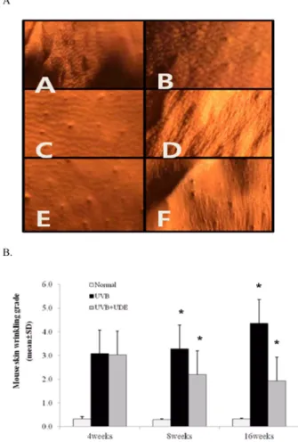
4. Inhibitory activity of UDE on wrinkle formation As early as 4 weeks after UV irradiation there were visible signs of wrinkling on the dorsal skin of hairless mice, and the wrinkles became distinct at week 8 of irradiation in contrast to the absence of wrinkle formation in the agematched unirradiated controls (Fig. 2A, B). When UDE was topically applied daily for 8 weeks at a concentration of 300㎎/㎖ to dorsal skins of hairless mice immediately after each suberythemal UV irradiation, the formation of wrinkles was obviously diminished compared with the water-treated controls (Fig. 2A). Comparison
of wrinkle scores revealed that UDE significantly decreased wrinkle formation by 13-16 weeks of irradiation compared with the water-treated controls (Fig. 2B). UVB radiation is one of the most environmental hazardous effects that can cause acute and chronic response in human skin (Bissett et al., 1990) and Table 2. Elastase inhibition activity of UDE.
Inhibiton (%)
IC50(㎍/㎖)*
100㎍/㎖ 50㎍/㎖ 10㎍/㎖
UDE 78.57±6.09 61.25±16.72 10.33±0.47*,** 654.6±23.27
Oleanolic acid 664.6±9.87 648.55±16.91* 66.73±3.79*** 85.42±8.44
*IC50 indicates the concentration (㎍/㎖ ) at which the pecentage inhibition of elastase activity was 50%. **Those with different superscript letters are significantly at *p < 0.05. ***p < 0.001.
Fig. 1. Effect of UDE on UVB-induced MMP-1 expressions in HDFs.Cells were stimulated with UVB (25 mJ/㎠) and the indicated concentrations of UDE for 24 h. The presence of MMP-1 in the cell-free culture supernatants was measured using a commercially available ELISA kit as described in Materials and Methods. Each value represents the mean±SD of three independent experiments. *p <
0.05 (vs. control control + UDE)
Fig. 2. Effects of UDE treated mice with UV B on skin wrinkles.
(A) Photographs after UVB irradiation (three times a week for 8 weeks.) of hairless mouse skin at last day after UVB irradiation. A, C, E : UVB (8 weeks) +UDE (topically application for 8 weeks) , B, D, F : UVB irradiation (16 weeks, No application of UDE). (B) Visual scoring (scale ¼ 0-3) of wrinkles during UVB exposure of mice. The scoring was performed by the method of Bissett et al,. after UVB irradiation. Data represent mean±SD. *p < 0.05 (vs. control control + UDE)
stimulate synthesis of elastin, which is highly related to the elasticity and renewal of skin, and induces wrinkles and a lack of elasticity (Schwartz et al., 1995). The degeneration of elastic fibers is estimated to be due to an increase in fibroblast elastase, an enzyme produced and secreted by dermal fibroblasts that degrades elastin (Tsukahara et al., 2001). The connective tissue elements collagen and elastin as known to be degraded by collagenase and elastase produced by imflammatory cells (Hastly et al., 1982) and fibroblast (Welgus et al, 1982). Moloney et al.
(1992) reported that visible signs of wrinkling were occur after approximately six weeks of UVB irradiation and were very obviously after ten weeks of irradiation. The progress of wrinkle formation noticed in our experiment was similar to that in the above report. The timing of the inhibition of wrinkles seemed to be similar. In the present study, comparison of skin properties between UDE treated group after UVB irradiation and sham- operated hairless mice without UVB irradiation demonstrated that UDE treated group at week 4 appeared no difference in skin elasticity compared with sham-operated group. The reduced skin elasticity in the UVB irradiation group is accompanied by a significant increase in the activity of elastase in the skin. In addition, during the experiments with UVB irradiation, redness and exfoliation were observed in the skin of many mice only in the UVB irradiation.
Our study demonstrated that elastase activity in UVB- irradiated mouse skin, was not changed following 4 weeks of UVB irradiation, but was markedly increased following 8 weeks of UV irradiation. Our study observations demonstrated that fine elastic fibers were markedly decreased after 16 weeks of UVB irradiation (Fig. 2A.).
Photoaging (UV irradiation) causes human skin aging through activation of MMPs, which are responsible for the degradation of collagen. It is conceivable that the degeneration of elastic fibers observed in the UVB exposed skin is mainly due to the increased activity of skin fibroblast elastase. In this report, the wrinkle formation by the elastase inhibitor UDE was accompanied by a marked decrease in degradation of the dermal fine elastic-fiber network.
In Korea, UDE has been used to treat various skin problems, including atopy, skin aging, and pimples. Eom et al. (2006) studied that UDE recovered from photo-induced damage after UVA irradiation (3 J/㎠) with 2 times higher than that of positive control and UDE showed approximately 48% of the increased cell viability of the control. UDE has been popular in cosmetic compositions and found to have beneficial functions similar to
those investigated in this study. However, no side effects were reported until now. Therefore, the UDE, a natural agent for human skin, would have more benefits than chemical treatments.
ACKNOWLEDGEMENTS
This work was performed with the support of the Cooperative Research Program for Agriculture Science & Technology Development (PJ007479022011), Rural Development Admini- stration, Republic of Korea
LITERATURE CITED
Bae JY, Lim SS, Kim SJ, Choi JS, Park J, Ju SM, Han SJ, Kang IJ and Kang YH. (2009). Bog blueberry anthocyanins alleviate photoaging in ultraviolet-B irradiation-induced human dermal fibroblasts. Molecular Nutrition & Food Research.
53:726-738.
Berthod F, Germain L, Li H, Xu W, Damour O and Auger F.
(2001). Collagen fibril network and elastic system remodeling in a reconstructed skin transplanted on nude mice. Matrix Biology. 20:463-473.
Bissett DL, Chatterjee R and Hannon DP. (1990).
Photoprotective effects of superoxide-scavenging antioxidants against ultraviolet radiation-induced chronic skin damage in the hairless mouse. Photodermatology, Photoimmunology & Photo- medicine. 7:560-562.
Bolognia JL.(1993). Dermatologic and cosmetic concerns of the older woman. Clinics in Geriatric Medicine. 9:209-229.
Eom SY, Kim YS, Lee CJ, Lee CH, Kim YD and Kim H.
(2006). Cosmeceutical properties of polysaccharides from the root bark of Ulmus davidiana var. japonica. Journal of Cosmetic Science. 57:355-367
Farkas B, Magyarlaki M, Csete B, Nemeth J, Rabloczky G, Bernath S, Literáti Nagy P and Sümegi B. (2002). Reduction of acute photodamage in skin by topical application of a novel PARP inhibitor. Biochemical Pharmacology. 63:921-932.
Godeau G and Hornebeck W. (1988). Morphometric analysis of the degradation of human skin elastic fibers by human leukocyte elastase (EC 3-4-21-37) and human skin fibroblast elastase (EC 3-4-24). Pathologie Biologie. 36:1133-1138.
Ha JH, Kwon MC, Kim SS, Jeong MH, Hwang B and Lee HY. (2010). Enhancement of skin-whitening and UV-protective effects of Centella asiatica L. urban by ultrasonification process. Korean Journal of Medicinal Crop Science. 18:79-85.
Han SH, Woo NRY, Lee SD and Kang MH. (2006).
Antioxidative and antibacterial activities of endemic plants extracts in Korea. Korean Journal of Medicinal Crop Science.
14:49-55.
Hastly KA, Jeffrey JJ, Hibbs MS and Welgus HG. (1987). The collagen substrate specificity of human neutrophil collagenase.
Journal of Biological Chemistry. 262:10048-10052.
He G, Kutala VK, Kuppusamy P and Zweier JL. (2004). In vivo measurement and mapping of skin redox stress induced by
ultraviolet light exposure. Free Radical Biology and Medicine.
36:665-672.
Hong ND, Kim NJ and No YS. (1990). A study on efficacy of Ulmi cortex. Korean Journal of Pharmacognosy. 21:217-222.
Imokawa G, Takema Y, Yorimoto Y, Tsukahara K, Kawai M and Imayama S. (1995). Degree of ultraviolet-induced tortuosity of elastic fibers in rat skin is age dependent. Journal of Investigative Dermatology. 150:254-258.
Jeong SJ, Ha JH, Kim Y, Oh SH, Kim SS, Jeong MH and Lee HY. (2009). Effects of Rubus coreanus extracts on ultraviolet-A irradiated cultured human skin fibroblasts. Korean Journal of Medicinal Crop Science. 17:321-327.
Jin UH, Lee DY, Kim DS, Lee IS and Kim CH. (2006).
Induction of mitochondria-mediated apoptosis by methanol fraction of Ulmus davidiana Planch (Ulmaceae) in U87 glioblastoma cells. Environmental Toxicology and Pharmacology.
22: 136-141.
Learn DB and Moloney SJ. (1991). Numbers of murine dermal mast cells remain unchanged during chronic ultraviolet B irradiation. Photodermatology Photoimmunology & Photome- dicine. 8:195-199.
Lee IS , Wei C, Thuong PT, Song KS, Seong YH and Bae KH.
(2006). Antioxidant constituents from the leaves of Cedrela sinensis A. Juss. Korean Journal of Medicinal Crop Science.
14:267-272.
Moloney SJ, Edmonds SH, Giddens LD and Learn DB. (1992).
The hairless mouse model of photoaging: evaluation of the relationship between dermal elastin, collagen, skin thickness and wrinkles. Photochemistry and Photolobiology. 56:505-511.
Murphy G and Docherty AJ. (1992). The matrix metallo- proteinases and their inhibitors. American Journal of Respiratory Cell and Molecular Biology. 7:120-125
Pillai S, Oresajo C and Hayward J. (2005). Ultraviolet radiation and skin aging: roles of reactive oxygen species, inflammation
and protease activation, and strategies for prevention of inflammation-induced matrix degradation a review. International Journal of Cosmetic Science. 27:17-34.
Schwartz E, Feinberg E, Lebwohl M, Mariani TJ and Boyd CD. (1995). Ultraviolet radiation increases tropoelastin accumulation by a post-transcriptional mechanism in dermal fibroblasts. Journal of Investigative Dermatology. 105:65-69.
Ta Takema Y, Nishijima A, Ohsu H, Fujimura T and Hattori M. (1997). Age-related discontinuous changes in the in vivo fluorescence of human facial skin. Journal of Dermatological Science. 15:55-58
Takema, Y. and Imokawa G. (1998). The effects of UVA and UVB irradiation on the viscoelastic properties of hairless mouse skin in vivo. Dermatology. 196:397-400.
Tschesche H, Bakowski B, Schettler A, Knäuper V and Reinke H. (1991) Leukodiapedesis, release of PMN leukocyte proteinases and activation of PMNL procollagenase. Acta Biomedica Biochimica. 50:755-761
Tsukahara K, Takema Y, Moriwaki S, Tsuji N, Suzuki Y, Fujimura T and Imokawa G. (2011). Selective inhibition of skin fibroblast elastase elicits a concentration-dependent prevention of ultraviolet B-induced wrinkle formation. Journal of Investigative Dermatology. 117:671-677.
Welgus HG, Jeffrey JJ and Eisen AZ. (1981). The collagen substrate specificity of human skin fibroblast collagenase. The Journal of Biological Chemistry. 256:9511-9515.
Wiedow O, Schröder JM, Gregory H, Young JA and Christophers E. (1990). Elafin: an elastase-specific inhibitor of human skin. Purification, characterization, and complete amino acid sequence. The Journal of Biological Chemistry. 265:
14791-14795.
Zimbler MS, Kokoska MS and Thomas JR. (2001). Anatomy and pathophysiology of facial aging. Facial Plastic Surgery Clinics of North America. 9:179-187.