Development of a Simple and Reproducible Method for Removal of
Contaminants from Ginseng Protein Samples Prior to Proteomics Analysis
Ravi Gupta1, So Wun Kim1, Chul Woo Min1, Gi-Ho Sung2, Ganesh Kumar Agrawal3,4, Randeep Rakwal3,4,5, Ick Hyun Jo6, Kyong Hwan Bang6, Young-Chang Kim6, Kee-Hong Kim6 and Sun Tae Kim1*
1Department of Plant Bioscience, College of Natural Resources and Life Sciences, Pusan National University, Miryang 627-707, Korea
2Institute for Bio-Medical Convergence, International St. Mary's Hospital, College of Medicine, Catholic Kwandong University, Incheon 404-834, Korea
3Research Laboratory for Biotechnology and Biochemistry, Kathmandu, Nepal
4Global Research Arch for Developing Education (GRADE) Academy Pvt. Ltd, Birgunj, Nepal
5Organization for Educational Initiatives, University of Tsukuba, Tsukuba, Japan
6Ginseng Research Division, Department of Herbal Crop Research, NIHHS, RDA, Eumseong 369-873, Korea Received June 23, 2015 /Revised July 7, 2015 /Accepted July 7, 2015
This study describes the effects of activated charcoal on the removal of salts, detergents, and pigments from protein extracts of ginseng leaves and roots. Incubation of protein extracts with 5% (w/v) acti- vated charcoal (100-400 mesh) for 30 min at 4°C almost removed the salts and detergents including NP-40 as can be observed on SDS-PAGE. In addition, analysis of chlorophyll content showed sig- nificant depletion of chlorophyll (~33%) after activated charcoal treatment, suggesting potential effect of activated charcoal on removal of pigments too along with the salts and detergents. 2-DE analysis of activated charcoal treated protein samples showed better resolution of proteins, further indicating the efficacy of activated charcoal in clearing of protein samples. In case of root proteins, although not major differences were observed on SDS-PAGE, 2-DE gels showed better resolution of spots after char- coal treatment. In addition, both Hierarchical clustering (HCL) and Principle component analysis (PCA) clearly separated acetone sample from rest of the samples. Phenol and AC-phenol samples al- most overlapped each other suggesting no major differences between these samples. Overall, these re- sults showed that activated charcoal can be used in a simple manner to remove the salts, detergents and pigments from the protein extracts of various plant tissues.
Key words : Activated charcoal, ginseng, plant pigment, SDS-PAGE, 2-Dimensional Electrophoresis
*Corresponding author
*Tel : +82-55-350-5505, Fax : +82-55-350-5509
*E-mail : [email protected]
This is an Open-Access article distributed under the terms of the Creative Commons Attribution Non-Commercial License (http://creativecommons.org/licenses/by-nc/3.0) which permits unrestricted non-commercial use, distribution, and reproduction in any medium, provided the original work is properly cited.
Journal of Life Science 2015 Vol. 25. No. 7. 826~832 DOI : http://dx.doi.org/10.5352/JLS.2015.25.7.826
Introduction
In the last decade, proteomics has emerged as a method of choice for analyzing the whole set of proteins present in a given tissue at a particular time. Protein extraction and sample preparation are the most crucial steps for the pro- teome analysis. Protein extraction involves crushing or breaking of cells in order to release the proteins from it. In particular, extraction of plant proteins is relatively difficult due to the presence of a rigid cell wall, which causes hin- drance in the release of proteins from the plant cells.
Moreover, presence of secondary metabolites and different
pigments also interfere with the protein resolution on SDS-PAGE and two dimensional gel electrophoresis (2-DE).
Furthermore, detergents like Triton X-100 and NP-40, which are used in the protein extraction buffer for solubilization of membrane proteins, also interfere with the protein reso- lution on PAGE. Therefore, clearing of plant protein samples is essential prior to utilizing the samples for proteome analysis. Conventional techniques which are being used for removal of salts and detergents from the protein samples include dialysis [12], and ion-exchange chromatography [2, 7], however, both of these techniques are laborious and time consuming and therefore, can lead to the protein degrada- tion.
Activated charcoal or activated carbon is a form of carbon having low volume pores which increases the absorption surface area for chemical reactions. Previously, it was in- dicated that addition of activated charcoal led to the removal of detergents including SDS [10] and Triton X-100 [11] from protein samples. In addition, the potential effect of activated - Note -
charcoal in combination with ion exchange processes was also shown in the removal of phenolic compounds from soy protein isolates [3]. However, there is no report of use of activated charcoal for proteome analysis. Additionally, it is also unknown whether activated charcoal is compatible with the downstream proteomics techniques like SDS-PAGE and high resolution 2-DE, or not, which would be critical for its use in proteome analysis.
In this direction, here we report an activated charcoal based method for removal of salts, detergents and pigments before proteome analysis for the better resolution of proteins on SDS-PAGE and 2-DE.
Materials and Methods
Plant material
Ginseng (Panax ginseng) used for this experiment was cul- tivated at the field of National Institute of Horticultural and Herbal Science (NIHHS) of Rural Development Administra- tion (RDA) in Eumsung (127 45' 13.14" E, 36 56' 36.63" N), Republic of Korea.
Protein isolation
Roots and leaves of 4-year-old ginseng plants were har- vested and grinded well using mortar and pestle. Total pro- teins from ginseng leaves and primary roots were isolated as described previously [4]. For the isolation of leaf proteins, 1 g of leaves were homogenized in 10 ml of Tris-Mg-NP-40 [0.5M Tris-HCl (pH 8.3), 2% (v/v) NP-40, 20 mM MgCl2] buffer followed by centrifugation at 12,000 g for 15 min.
Supernatant thus obtained was used as total protein extract.
For the isolation of ginseng root proteins, 5 g of roots were homogenized in 5 ml of Tris-Mg-NP-40 buffer containing 2%
β-mercaptoethanol and then centrifuged to obtain the total proteins as supernatant.
Activated charcoal treatment
Total proteins in Tris-Mg-NP-40 buffer were incubated with 5% activated charcoal (100-400 mesh size, Agilent Technologies, Wilmington, DE) for 30 min at 4°C with con- stant stirring. After incubation, the mixture was centrifuged at 12,000 g for 10 min to precipitate the activated charcoal and supernatant was used for further analysis. Proteins in the supernatant were then precipitated using either of the following reagents by incubating it at -20 C for 1 hr: four volumes of 12.5% w/v TCA/acetone [5], or 80% acetone,
or one volume water-saturated phenol [6] and then washed with 80% acetone three times.
Chlorophyll estimation
For the estimation of chlorophyll content, total leaf pro- teins, isolated in Tris-Mg-NP-40 buffer, before and after char- coal treatment was used to measure the chlorophyll content.
Total chlorophyll, chlorophyll-a and chlorophyll-b contents were measured spectrophotometrically as described pre- viously [1].
Two dimensional gel electrophoresis
For 2-DE analysis, with (ACT) and without activated char- coal treated (UT) protein pellets were dissolved in the rehy- dration buffer containing 7 M Urea, 2 M Thiourea, 4% (v/v) CHAPS, 2 M DTT, and 0.5% (v/v) IPG buffer pH 4-7 (GE Healthcare, Waukesha, WI, USA). 2D-Quant kit (GE Healthcare) was used to quantify the proteins in each frac- tion and a total of 600 µg of protein of each sample was loaded on the 24 cm IPG strips, pH 4-7 by passive rehydra- tion loading overnight at 20 °C. Iso-electric focusing was car- ried out using following protocol: 50 V for 4 hr, 100 V for 1 hr, 500 V for 1 hr, 1,000 V for 1 hr, 2,000 V for 1 hr, 4,000 V for 2 hr, 8,000 V for 5 hr, 8,000 V for 9 hr, and 50 V for 6 hr on IPGphore II platform (GE Healthcare). Prior to the second dimensional separation, the strips were reduced in an equilibration buffer [6 M urea, 30% (v/v) glycerol, 2%
(w/v) SDS, 50 mM Tris-HCl (pH 6.8), and 0.1 mg/ml bromo- phenol blue] containing 1% DTT as the first step and then alkylated by 2.5% iodoacetamide as the second step. The sec- ond dimension separation of proteins was carried out on 13% SDS-polyacrylamide gels using EttanDalt twelve (GE Healthcare), after which the gels were stained with colloidal Coomassie Brilliant Blue (CBB). A total of three biological replicates were performed for each data set [6].
Image acquisition and data analysis
Images of the colloidal CBB stained 2DE gels were ac- quired using a transmissive scanner (PowerLook 1120, UMAX) with a 32 bit pixel depth, 300 dpi resolution, and brightness and contrast set to default. For the analysis of the 2DE gels, raw tiff image files were imported in the ImageMaster 2D Platinum software (ver. 6.0, GE Healthcare) and spots were detected from three biological replicates and averaged. For the quantitative analysis, the volume of each spot was normalized as an average of the volume of spots on the gel and then spot volumes were calculated to de-
A B
C
Fig. 1. (A) Overview of activated charcoal method. (B) SDS-PAGE gel of leaf and root proteins showing removal of contaminants after activated charcoal treatment. (C) Estimation of chlorophyll-a, chlorophyll-b and total chlorophyll after activated charcoal treatment. Abbreviations: UT, untreated; ACT, activated charcoal treated.
termine the relative abundance of proteins in the ex- perimental samples.
Principal component analysis and hierarchical clustering
For principal component analysis (PCA) and hierarchical clustering (HCL), percentage volume of variable spots were calculated using ImageMaster2DPlatinum software and sub- sequently log transformed to base 2 to equalize their expression. PCA was then carried out using NIA array soft- ware while for HCL, MeV software was used.
Results and Discussion
Activated charcoal absorbs detergents and salts from protein samples
In this study, we describe a simple and rapid method for the removal of salt, detergents and pigments using activated charcoal. In order to test the efficacy of activated charcoal in removal of detergents like NP-40 and pigments, total pro- teins were isolated from ginseng leaves and roots in Tris- Mg-NP-40 buffer and incubated with 5% activated charcoal
for 30 min at 4°C. The flow chart for the methodology used is shown in Fig. 1A. Separation of leaf proteins on SDS- PAGE, before and after activated charcoal treatment, clearly showed a better resolution of proteins after charcoal treat- ment (Fig. 1B). Before charcoal treatment, lane widening and very less distinct bands were observed on SDS-PAGE, how- ever, after charcoal treatment, the bands were very clear with no lane widening (Fig. 1B, lanes 2 and 3). These results can be explained in terms of absorption of salts, detergents and pigments from the protein samples by activated char- coal, which otherwise interfered with the protein resolution on SDS-PAGE. In case of ginseng root proteins, no major differences were observed on SDS-PAGE except for some lane widening which was observed in without charcoal treated sample (Fig. 1B, lanes 4 and 5).
Activated charcoal treated samples showed decreased chlorophyll
In order to check the efficacy of activated charcoal in ab- sorption of plant pigments, total, chlorophyll-a and chlor- ophyll-b contents were measured after activated charcoal treatment. Interestingly, both chlorophyll-a and chlorophyll-
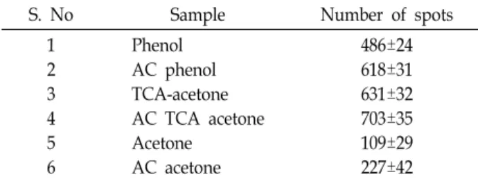
Table 1. Average number of spots detected in the 2-DE gels of different samples using ImageMaster2DPlatinum software (ver. 6.0). Abbreviations: AC; activated char- coal, TCA; Trichloroacetic acid
S. No Sample Number of spots
1 2 3 4 5 6
Phenol AC phenol TCA-acetone AC TCA acetone Acetone
AC acetone
486±24 618±31 631±32 703±35 109±29 227±42 b content were reduced after charcoal treatment and approx-
imately 33% reduction in the total chlorophyll content was observed in charcoal treated samples suggesting the poten- tial effects of activated charcoal in absorption of plant pig- ments (Fig. 1C).
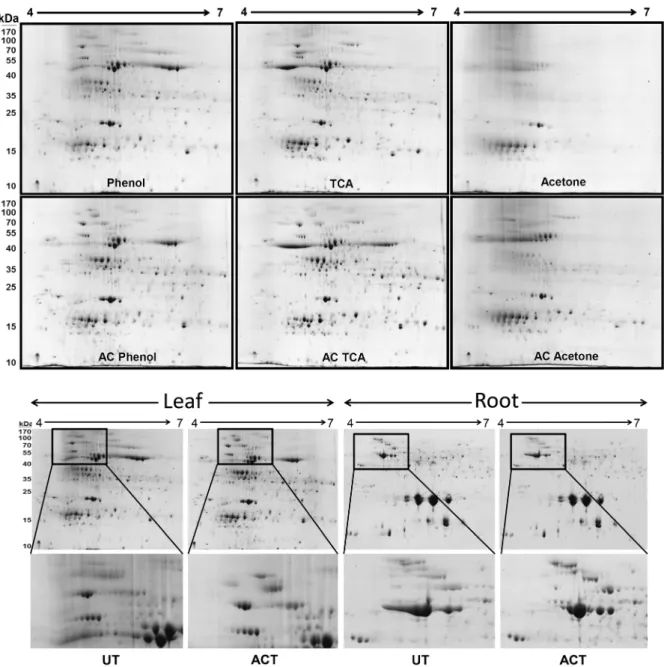
2-DE analysis further confirmed better resolution of spots after charcoal treatment
To further cross check the results obtained on SDS-PAGE, and to investigate the compatibility of activated charcoal with the proteome analysis, high resolution 2-DE gels were carried out. For precipitation of proteins before 2-DE, phe- nol, TCA-acetone and acetone methods were tried and tested for their efficacy in protein precipitation after activated char- coal treatment. Out of the 3 methods tried, phenol method gave best results for protein precipitation followed by TCA-acetone, in terms of better protein resolution and lesser background staining (Fig. 2). ImageMaster2DPlatinum soft- ware was used to detect the total number of spots in each sample (Table 1). Maximum number of spots were observed in TCA-acetone precipitated sample followed by activated charcoal treatment (AC TCA-acetone) suggesting a better resolution of proteins in this sample. However, in this sam- ple, a long trail (horizontal streaking) of RuBisCO protein was observed suggesting that RuBisCO is not completely fo- cused during the iso-electric focusing. Presence of con- taminants in the sample is one of the major causes of hori- zontal streaking in the 2-DE gels. Therefore, it can be con- cluded that although the number of spots were higher in the 2-DE gels of AC TCA-acetone precipitation samples, it still show horizontal streaking because of presence of con- taminants in the protein samples. In contrast, AC phenol method precipitated samples did not show any streaking and better resolution of spots in the 2-DE gels, suggesting that the AC-phenol method is the best condition for the iso- lation, precipitation and separation of proteins, rich in contaminants. In case of acetone precipitation method, very few proteins spots (109±29) and more background staining were observed as compared to other precipitation methods.
Ginseng leaves contain lots of phenolics which interfere with the protein resolution and our results show that acetone method is less effective than phenol and TCA-acetone meth- ods in removal of these phenolics from the protein samples.
Even though the 2-DE profiles were less clear in acetone precipitated samples, 2-DE gel was clearer after activated charcoal treatment, suggesting the potential of charcoal in
removal of phenolics too, however to lesser extent. In case of leaves, similar to the results of SDS-PAGE, 2-DE gels also showed improved protein resolution after charcoal treat- ment (Fig. 2). After charcoal treatment, the background was less and spots were more resolved and clearer (Fig. 2). In case of root proteins, although not major differences were observed on SDS-PAGE, 2-DE gels showed better resolution of spots after charcoal treatment (Fig. 2). In the 2-DE gels of root samples, the major differences were observed on high molecular weight region while in the 2-DE gels of leaf sam- ples, prominent differences were observed at both high as well as low molecular weight levels (Fig. 2).
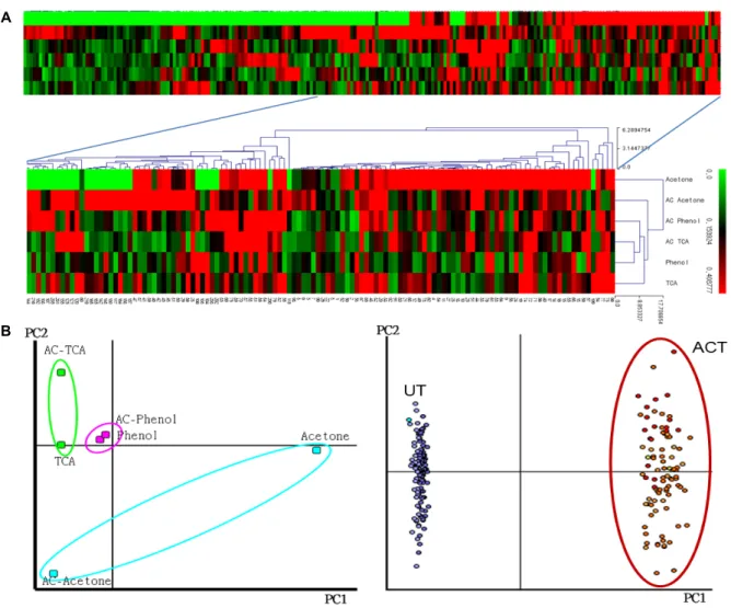
In order to further pronounce the results obtained from the 2-DE, HCL and PCA analyses were carried out. A total of the 189 spots showing visible differences in the 2-DE gels of with and without charcoal treated samples were used for the analysis (Fig. 3). Percentage volumes were calculated for these spots using ImageMaster2DPlatinum software and log 2 transformed to equalize the expression. HCL analysis clearly showed a long trail of missing spots in the acetone fraction (indicated by green color, Fig. 3A). In addition, both HCL and PCA analyses clearly separated acetone sample from rest of the samples (Fig. 3B). Phenol and AC-phenol samples almost overlapped each other suggesting no major differences between these samples. In case of TCA and AC-TCA sample, a significant difference was observed.
These results showed that activated charcoal treatment can significantly clear the protein profiles prior to the acetone and TCA-acetone precipitation of the plant proteins. In addi- tion to the salts, pigments and secondary metabolites, it is possible that a small percentage of the proteins can also bind to the activated charcoal resulting in loss of some proteins.
However, the major portion of the protein remains unbound and can be recovered after centrifugation. Previous reports have also shown the potential effects of activated charcoal
Fig. 2. The effect of different methods for leaf protein precipitation after activated charcoal treatment including phenol, TCA-acetone and acetone methods (upper panel). 2-DE analysis of activated charcoal treated samples of leaf and root proteins (lower panel). Boxed regions indicate the 2D gel areas which are more resolved and clearer after activated charcoal treatment.
Abbreviations: UT, untreated; ACT, activated charcoal treated.
in removal of detergents like Triton X-100 and SDS, prior to immunological assays and staining of gels [2, 10]. In this study, we used activated charcoal of size 400 mesh, which was very effective in removal of detergents and salts from the protein samples. Marcus and Prusky reported the effect of particle size on absorption of Triton X-100 by activated charcoal [11]. It was shown that charcoal of smaller particle size (250-350 mesh) is more effective in absorption of Triton X-100 at a ratio of 0.23 g Triton per g of activated charcoal.
The beauty of this activated charcoal based removal of de-
tergents or salts is that, it neither removes protein sig- nificantly nor affects the enzymatic activities [11].
Taken together, our results show that the activated char- coal based method for removal of salts, detergents and pig- ments from plant protein samples is easy, rapid and inexpensive. This method effectively removes the con- taminants from the proteins samples which could interfere with the protein resolution on SDS-PAGE and 2-DE, without affecting the proteins concentration and activity.
A
B
Fig. 3. Hierarchical clustering (HCL) and principal component analysis (PCA) of ginseng leaf proteins. A total of 189 spots were selected from the 2D gels showing significant changes after activated charcoal treatment and used for both HCL (upper panel) and PCA (middle and lower panel). Middle panel shows the PC plots while the lower panel shows the expression profiles of the proteins selected for analysis. Abbreviations: UT, untreated; ACT, activated charcoal treated.
Acknowledgment
This work was supported by grants from National Agenda Programs for Agricultural R&D (FTA, grant#: PJ01010401), Rural Development Administration (RDA), Republic of Korea.
References
1. Arnon, D. I. 1949 Copper enzymes in isolated chloroplasts, polyphenoxidase in Beta vulgaris. Plant Physiol. 24, 1-15.
2. Duhamel, R. C., Meezan, E. and Brendel, K. 1981 A charcoal cartridge for the removal of anionic detergent and electro- phoresis stains. J. Biochem. Biophys. Methods 4, 73-80.
3. How, J. S. L. and Morr, C. V. 1982 Removal of phenolic compounds from soy protein extracts using activated carbon. J. Food Sci. 47, 933-940.
4. Kim, S. W., Min, C. W., Gupta, R., Jo, I. H., Bang, K. H., Kim, Y. C., Kim, K. H. and Kim, S. T. 2014. Proteomics anal- ysis of early salt-responsive proteins in Ginseng (Panax gin- seng C. A. Meyer) leaves. Kor. J. Med. Crop Sci. 22, 398- 404.
5. Kim, Y. J., Lee, H. M., Wang, Y., Wu, J., Kim, S. G., Kang, K. Y., Park, K. H., Kim, Y. C., Choi, I. S., Agrawal, G. K., Rakwal, R. and Kim, S. T. 2013 Depletion of abundant plant RuBisCO protein using the protamine sulfate precipitation method. Proteomics 13, 2176-2179.
6. Kim, Y. J., Wang, Y., Gupta, R., Kim, S. W., Min, C. W., Kim, Y. C., Park, K. H., Agrawal, G. K., Rakwal, R., Choung, M. G., Kang, K. Y. and Kim, S. T. 2015 Protamine sulfate precipitation method depletes abundant plant seed-storage proteins: Legume plants as a case study. Proteomics 15, 1760- 1764.
7. Kramer, R. and Heberger, C. 1986 Functional reconstitution of carrier proteins by removal of detergents with a hydro- phobic ion-exchange column. Biochim. Biophys. Acta. 863,
초록:활성탄을 이용한 불순물제거에 의한 효과적인 인삼 조직 단백질체 분석 방법 개선 연구
굽타 라비1․김소운1․민철우1․성기호2․아그라왈 가네시 쿠마르3,4․락왈 랜딥3,4,5․조익현6․방경환6․ 김영창6․김기홍6․김선태1*
(1부산대학교 식물생명과학과, 2가톨릭관동대학교 의과대학, 3네팔 생명공학 실험실, 4네팔 글로벌 교육 연구소
5일본 쯔꾸마 대학, 6농촌진흥청 국립원예특작과학원 인삼특작과학부)
본 연구는 인삼의 잎과 뿌리 단백질 추출물에서 활성탄을 이용하여 염, 계면활성제, 색소를 제거하여 단백질체 분석 연구에 미치는 잠재적인 효과에 대한 평가를 기술하고 있다. 5%(w/v) 활성탄(100-400 mesh)과 함께 단백질 추출물을 30분간 4°C에서 반응시켜 염과 계면활성제를 제거한 후 SDS-PAGE를 분석하여 단백질의 양상을 관찰하 였다. 엽록소 함량의 분석은 활성탄 처리 후 엽록소의 상당한 양(~33%)이 제거되는 것을 보여주었고, 이 분석은 염, 계면활성제 제거만이 아닌 색소의 제거에서도 활성탄의 잠재적 효과가 있음을 보여 주고 있다. 활성탄을 처리 한 단백질 시료를 이용하여 이차원 전기영동과 PCA 통계분석을 시행한 결과 단백질은 gel에서 더 나은 해상도를 보여주었으며 단백질 시료의 정제에서도 활성탄의 효과가 있음을 확인하였다. 종합적으로, 이 결과들은 활성탄을 이용한 간단한 방법으로 다양한 식물 조직의 단백질 추출물에서 염, 계면활성제, 색소를 제거함으로써 고해상도의 단백질체 분석에 적용될 수 있을 것으로 기대된다.
289-296.
8. Leamml, U. K. 1970 Cleavage of structural proteins during the assembly of the head of bacteriophgae. Nature 227, 680- 685.
9. Lee, H. M., Gupta, R., Kim, S. H., Wang, Y., Rakwa,l R., Agrawal, G. K. and Kim, S. T. 2015 Abundant storage pro- tein depletion from tuber proteins using ethanol precip- itation method: Suitability to proteomics study. Proteomics 15, 1765-1769.
10. Malhas, A. N., Abuknesha, R. A. and Price, R. G. 2002 Removal of detergents from protein extracts using activated
charcoal prior to immunological analysis. J. Immunol.
Methods 264, 37-43.
11. Marcus, L. and Prusky, D. 1987 Effect of particle size of activated charcoal on separation of Triton X-100 from pro- tein, liver cytosol, and lipoxygenase extracts. Anal. Biochem.
163, 112-116.
12. Schwendener, R. A., Asanger, M. and Weder, H. G. 1981.
n-Alkyl-glucosides as detergents for the preparation of high- ly homogenous bilayer liposomes of variable sizes applying defined rates of detergent removal by dialysis. Biochem.
Biophys. Res. Commun. 100, 1055-1062.