A Study on Antioxidant and Anti-inflammatory Effects Based on Analysis of Functional Components of Cornus officinalis Siebold & Zucc.
Hyun Hwangbo
1,2, Ji-Suk Jeung
3, Min Young Kim
1,2, Seon Yeong Ji
1,2, Seonhye Yoon
4, Tae Hee Kim
5, Sung Ok Kim
6and Yung Hyun Choi
1,7*
1Anti-Aging Research Center, Dong-eui University, Busan 47340, Korea
2Department of Molecular Biology, College of Natural Sciences, Pusan National University, Busan 46241, Korea
3Gurye-gun Agricultural Center and Gurye Wild Flower Institute, Gurye 57660, Korea
4R&D Center, Naturetech Co. Ltd., Jincheon 27858, Korea
5R&D Center, Hamsoapharm Central Research, Jinan 55442, Korea
6Department of Food Science and Nutrition, College of Life and Health Sciences, Kyungsung University, Busan 48434, Korea
7Department of Biochemistry, Dong-eui University College of Korean Medicine, Busan 47227, Korea Received September 7, 2020 /Revised September 21, 2020 /Accepted September 22, 2020
Cornus officinalis Siebold & Zucc. is traditionally used as an edible and medicinal plant in many coun-
tries in East Asia. Previous studies have shown the pharmacological potential of extracts and compo- nents of C. officinalis, but comparative analysis of the composition of the leaf, stem, and fruit extracts has been insufficient to date. In the present study, the content of active antioxidant and anti-in- flammatory ingredients was verified in different C. officinalis parts (under-ripe sansuyu, ripe sansuyu, seed, leaf, stem, and dried sansuyu). One active component, morroniside, was high in fruit (under-ripe and ripe sansuyu), while loganin was high in fruit (under-ripe sansuyu) and cornin was high in seeds.
Total polyphenol contents were highest in fruit (ripe sansuyu) and flavonoids were highest in leaves.
DPPH radical scavenging activity was highest in leaves, followed by seeds and then ripe sansuyu extract. The anti-inflammatory efficacy of leaf extracts of C. officinalis (LCO) was further investigated by measuring their effects on levels of nitric oxide (NO) and the pro-inflammatory cytokines inter- leukin (IL)-1β and IL-6 in RAW 264.7 macrophages. Non-cytotoxic concentrations of LCO effectively decreased the lipopolysaccharide (LPS)-induced expression of inducible NO synthase, resulting in de- creased NO production. LCO also significantly suppressed LPS-induced production and expression of IL-1β and IL-6. Taken together, the present findings suggest that C. officinalis leaves have potential as natural materials for the development of antioxidant and anti-inflammatory agents.
Key words : Active components, anti-inflammation, antioxidant, Cornus officinalis
*Corresponding author
*Tel : +82-51-890-3319, Fax : +82-51-893-3333
*E-mail : [email protected]
This is an Open-Access article distributed under the terms of the Creative Commons Attribution Non-Commercial License (http://creativecommons.org/licenses/by-nc/3.0) which permits unrestricted non-commercial use, distribution, and reproduction in any medium, provided the original work is properly cited.
서 론
생체 내 에너지 생성을 위한 정상적인 대사과정에서도 활성 산소종(reactive oxygen species, ROS)이 발생하며, 이는 불안 정하고 반응성이 높아 지나치게 많이 생성되거나 항산화 시스 템에 의해 제거되지 못하면 산화적 스트레스(oxidative stress) 를 유발하게 된다[2, 28, 41]. 지속적인 산화적 스트레스의 자극 은 세포막, 핵산, 지질 및 단백질을 포함한 세포 구조물을 손상 시킴으로써 조직에 비가역적인 변화를 유도하여 노화를 촉진 시킬 뿐만 아니라 암, 치매, 관절염, 자가면역질환 등의 질병을
유발하거나 촉진시킨다고 알려져 있다[13, 32]. 그러나 항산화 효소(catalase, glutathione-peroxidase, superoxide dismu- tase 등)의 활성 증대뿐만 아니라 항산화 활성을 가지는 식의 약 소재의 적용을 통하여 ROS를 제거함으로써 산화적 스트레 스로부터 보호할 수 있다[11, 14].
염증(inflammation)은 물리, 화학적 손상 또는 세균 감염
등의 다양한 외부 자극에 대해 신체를 보호하기 위한 면역방
어기전으로, 면역세포가 이를 인지하고 염증반응이 일어나는
동안 염증성 사이토카인(pro-inflammatory cytokine)과 염증
성 매개 인자(pro-inflammatory mediator) 등이 생성된다[16,
26]. 그러나 방어기전에 의한 염증반응이 아닌 병리적인 원인
에 의해 발생한 과도하고 지속적인 염증반응은 다양한 질환의
발병 또는 진행을 촉진하는 요인으로 작용한다[9, 12]. 예를
들어 미분화된 대식세포(macrophage)가 그람음성 세균(Gram
negative bacteria)의 외막에 존재하는 지질다당류(lipopoly-
saccharide, LPS)에 과도하게 노출되면 대식세포로 활성화되
면서 일련의 염증반응이 촉발된다[27, 33]. 특히 LPS에 노출된
Table 1. HPLC/DAD operating condition for morroniside, loganin, cornin and extracts from different parts of C. officinalis
Parameter Condition
Instrument Detector Column Wavelength Oven temperature
Flow rate Run time Injection volume
Agilent Technologies 1,200 series Diode array detector
Myghtysil RP-18GP (250×4.6 mm, 5 μm) 240 nm
25℃
0.6 ml/min 45 min
5 μl
Mobile phase Time (min) Acetonitrile (%) 0.1% Phosphoric acid in DDW (%)
Gradient conditions
0 23 30 35 35.1
45
0 21 70 70 0 0
100 79 30 30 100 100
세포는 전사인자인 nuclear factor-κB (NF-κB)가 핵 내로 이동
하여 활성화되어 interleukin-1β (IL-1β), IL-6, tumor necrotic factor-α (TNF-α) 등과 같은 염증성 사이토카인의 발현과 생성 을 촉진하며, 염증성 매개 인자인 nitric oxide (NO)와 prosta- glandin E
2(PGE
2)를 생성하는 inducible NO synthase (iNOS) 와 cyclooxygenase-2 (COX-2)의 발현을 유도한다[23, 27].
층층나무과에 속하는 산수유 나무(Cornus officinalis Siebold
& Zucc.)의 열매인 산수유(山茱萸, Corni Fructus)는 우리나라 를 비롯하여 중국과 일본 등에서 오랫동안 다양한 질환의 예 방과 치료의 목적으로 사용되어왔다[7, 15, 17, 29]. 최근 연구 들에 의하면 산수유 나무의 추출물은 면역증강, 혈당감소, 항 부정맥 및 항균 활성을 포함한 다양한 약리적 효능이 있음이 보고된 바 있으며, 그 열매인 산수유 또한 항고혈당증, 노화방 지, 항산화, 신장 및 신경보호 효과 등을 가지고 있다고 알려져 있다[17, 25, 38, 46]. 그럼에도 불구하고 산수유 나무의 잎, 줄 기, 열매에 대한 체계적인 성분 분석은 여전히 미비한 실정이 다. 따라서 본 연구에서는 산수유 나무의 부위별 유효성분 함 량의 분석과 항산화 및 항염증 활성을 재검증하여 향후 산수 유의 기능성 평가를 위한 기초 자료를 제시하고자 한다.
재료 및 방법
시료 제조
본 연구에서 사용된 산수유의 잎, 줄기, 산수유 열매는 전라 남도 구례군 산동면에서 채취한 것으로 구례군 야생화연구소 로부터 제공받았다. 잎과 줄기는 2017년 9월 첫 주에 채취 후 세척하여 동결건조(EYELA FDU-2100, Tokyo Rikakikai Co.
Ltd., Tokyo, Japan)하여 사용하였으며, 산수유 열매는 9월 첫 주에 덜 익은 산수유 열매(이하 청산수유로 약함)와 12월 첫 주에 익은 산수유 열매(이하 적산수유로 약함)를 직접 채취하
여 씨를 제거한 과육만 동결건조 하였고, 건조 과육(이하 건피 로 칭함)은 농가에서 제조하는 방식으로 예비건조, 데치기, 씨 앗분리기를 활용한 씨 분리 및 열풍건조의 과정으로 제조하였 다. 씨앗은 적산수유의 과육을 제거한 이후 수거하여 열풍건 조 분쇄(Laboratory Mill 120, Perten Instruments, Sweden) 하였다. 부위별 분말 10 g을 70% ethanol 200 ml와 혼합한 후 1시간씩 3회 반복하여 환류 냉각 추출하였다. 추출된 용액 은 여과지(No. 2, Advantec Co. Ltd, Tokyo, Japan)로 여과한 후 감압농축기(EYELA SB-1000, Tokyo Rikakikai Co. Ltd.)로 농축하여 동결건조 후 각각의 수율을 조사하고, 초저온냉동고 (MDF-U53V, Sanyo, Japan)에 보관하면서 실험에 사용하였다.
Morroniside, loganin 및 cornin 함량 분석
산수유의 대표 기능물질인 morroniside, loganin 및 cornin 의 함량은 선행 방법[19, 35]을 참고하여 high-performance liquid chromatography(HPLC, Agilent 1200 series, Agilent Technologies, Santa Clara, CA, USA)를 이용해 동시 분석하 였다. 시험용액은 각 시료 100 mg을 100 ml 정용플라스크에 정밀히 칭량하여 3차 증류수를 가하고 약 30분간 초음파 추출 하였다. 방냉 후 10배 희석하여 0.50 μm polytetrafluoroethyl- ene hydrophilic syringe filter (Advantec Co., Tokyo, Japan) 로 여과한 후 HPLC를 이용하여 Table 1에 제시한 조건에서 분석하였다. 표준품은 Chengdu Biopurify Phytochemicals Ltd.(Chengdu, China)에서 구입하였고, 순도는 morroniside 와 loganin이 각각 99.35%와 98.92%이었다. 이동상에 사용한 용매 acetonitile와 phosphoric acid는 HPLC용을 사용하였다.
검량선 작성을 위해 표준품 약 10 mg을 정밀히 칭량하여 10
ml 용량플라스크에 넣고 3차 증류수로 정용하고 용해하여 1
mg/ml이 되도록 고농도 stock solution을 제조한 후 단계적으
로 희석하여 0, 3.9, 7.8, 15.6, 31.3, 62.5, 125, 250, 500 μg/ml의
농도로 조제하였다. 각 시료의 지표성분 함량은 표준용액의 크로마토그램에서 얻은 피크의 농도별 면적에 대해 검량선을 작성하여 표준용액의 검량선 대비 농도를 산출하였다.
총 폴리페놀 함량 측정
각각의 시료에 함유된 총 페놀성 물질의 함량(total phe- nolic content, TPC)은 Folin-Ciocalteu의 방법을 일부 변형하 여 측정하였다[36]. 이를 위하여 시료 200 μl와 증류수 2.5 ml를 혼합한 후 혼합 용액에 Folin-Ciocalteu's 용액(Sigma-Aldrich Chemical Co., St. Louis, MO, USA) 200 μl를 가하여 30초간 혼합하였다. 이후 6분간 반응시킨 후 탄산나트륨(Na
2CO
3) 용 액 2 ml를 가하여 상온에서 90분간 방치한 후 UV-visible spec- trophotometer (Optizen 3220 uv, Mecasys, Deajeon, Korea) 로 750 nm에서 흡광도를 측정하였다. Gallic acid (Sigma- Aldrich Chemical Co.)를 표준물질로 하여 표준검량선을 작성 하고 시료 중의 총 폴리페놀 함량을 gallic acid equivalents (mg GAE/g, dry weight)로 환산하여 나타냈다.
총 플라보노이드 함량 측정
각각의 시료에 함유된 총 플라보노이드 함량(total flavo- noid, TFC)을 측정하기 위하여 추출물 100 μl에 10% alumi- num nitrate 20 μl, 1 M potassium acetate 20 μl 및 80% etha- nol 860 μl를 차례로 가하여 혼합하고 실온에서 40분간 안정화 시킨 다음 415 nm에서 흡광도를 측정하였다. Quercetin (Sigma-Aldrich Chemical Co.)을 표준물질로 하여 표준검량 선을 작성하고 시료 중의 총 플라보노이드 함량을 quercetin equivalents (mg QUE/g, dry weight)로 환산하여 나타내었다.
2,2'-diphenyl-1-picrylhydrazyl (DPPH) radical 소거 능 측정
전자공여능은 Blois의 방법에 따라 DPPH에 대한 수소 공여 효과를 측정하여 전자공여능(EDA; electron donating ability) 으로 나타냈다[3]. 이를 위하여 시료 용액 2 ml에 0.2 mM의 DPPH 용액(dissolved in 99% ethanol, Sigma-Aldrich Chemi- cal Co.) 1 ml 넣고 교반한 후 37℃에서 30분간 방치한 다음 517 nm에서 흡광도를 측정하였다. 전자공여능은 시료용액의 첨가군과 무첨가군 사이의 흡광도 감소율 차이를 백분율(%) 로 나타내어 전자공여능으로 표시하고, 양성 대조군으로는 butyl hydroxy anisole (BHA, Sigma-Aldrich Chemical Co.)를 사용하였다.
세포배양
본 실험에서 사용한 RAW 264.7 세포는 American Type Culture Collections (Manassas, VA, USA)에서 분양받았다.
RAW 264.7 세포는 10% fetal bovine serum (WelGENE, Inc., Gyeongsan, Korea)를 첨가한 Dulbecco's modified Eagle's
minimum essential medium (WelGENE, Inc.)을 사용하여 37
℃, 5% CO
2배양기에 배양하였다. 실험과정의 모든 세포는 80~90% 정도의 밀도로 자랐을 때 계대 배양하였고, LPS에 의 한 자극 실험을 위해, 세포를 6-well plate (5×10
5cells/well)에 분주한 후 24시간 배양하고 LPS (100 ng/ml, Escherichia coli 055:B5, Sigma-Aldrich Chemical Co.)와 시료를 첨가하여 24 시간 동안 다시 배양하였다.
산수유 에탄올 추출물의 세포독성 측정
RAW 264.7 세포에 대한 산수유 에탄올 추출물의 세포독성 여부를 평가하기 위하여 3-[4,5-dimethylthiazol-2-yl]-2,5 di- phenyl tetrazolium bromide (MTT) assay를 적용하였다. 이를 위하여 RAW 264.7 세포를 6 well plate (5×10
5cells/well)에 분주한 뒤 24시간 동안 세포를 부착 및 안정시킨 후 각 추출물 을 농도별로 24시간 동안 처리하였다. 24시간 후 5 mg/ml 농 도로 MTT 용액(Sigma-Aldrich Chemical Co.)을 제조하여 각 well 당 200 μl씩 첨가하고 2시간 동안 37℃, 5% CO
2incubator 에서 반응시킨 후, 용액을 제거하고 MTT의 환원에 의해 생성 된 formazan을 dimethyl sulfoxide (Sigma-Aldrich Chemical Co.)로 용해시켜 동의대학교 생체조직재생 핵심지원센터의 enzyme-linked immunosorbent assay (ELISA) reader (Dyna- tech Laboratories, Chantilly, VA, USA)를 이용하여 540 nm에 서 흡광도를 측정하여 세포 생존율을 백분율로 표시하였다.
NO 생성 저해 활성 측정
LPS로 자극된 RAW 264.7 세포로부터 생성되는 NO의 양은 세포배양액 중에 존재하는 NO
2-형태를 Griess reagent와 반응 시켜 측정하였다. 이를 위하여 6 well plate에 분주한 RAW 264.7 세포(5×10
5cells/well)에 LPS (100 ng/ml)를 단독으로 24시간 처리하거나 20 μg/ml의 산수유 추출물을 1시간 전처 리한 후 LPS를 처리하여 24시간 배양하였다. 배양 후 세포배 양 상등액 100 μl와 Griess 시약(1% sulfanilamide, 0.1% naph- thylethylendiamine in 25% phosphoric acid, Sigma-Aldrich Chemical Co.) 100 μl를 혼합하여 96 well plates에서 반응시킨 후 ELISA reader를 이용하여 540 nm에서 흡광도를 측정하였 다. NO 생성의 저해 정도를 평가하기 위하여 sodium nitrite (NaNO
2)의 농도별 표준 곡선을 이용하여 배양액 내의 NO 농도를 산출하였다.
Cytokine 생성량 측정
세포배양액 내의 염증성 사이토카인(IL-1β 및 IL-6)의 양을
측정하기 위한 Quantikine ELISA kit는 R&D systems (Min-
neapolis, MN, USA)에서 구입하였다. 이를 위하여 NO 생성
저해능 평가와 동일한 조건에서 배양된 세포배양 상등액을
이용하여 제조사의 protocol에 따라 IL-1β 및 IL-6의 농도를
측정하였다.
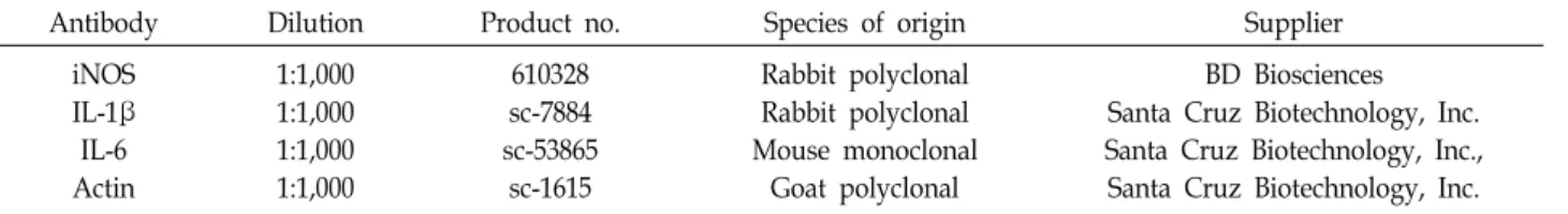
Table 2. List of antibodies used for western blot analysis in the present study
Antibody Dilution Product no. Species of origin Supplier
iNOS IL-1β IL-6 Actin
1:1,000 1:1,000 1:1,000 1:1,000
610328 sc-7884 sc-53865
sc-1615
Rabbit polyclonal Rabbit polyclonal Mouse monoclonal
Goat polyclonal
BD Biosciences Santa Cruz Biotechnology, Inc.
Santa Cruz Biotechnology, Inc., Santa Cruz Biotechnology, Inc.
단백질 발현 측정을 위한 Western blot analysis 전기영동을 위한 단백질 시료의 추출을 위해 배양이 끝난 세포를 수집하여 phosphate-buffered saline로 세척한 후, ly- sis buffer (0.5% Triton, 50 mM β-glycerophosphate (pH 7.2), 0.1 mM sodium vanadate, 2 mM MgCl
2, 1 mM ethyl- enediaminetetraacetic acid, 1 mM dithiothreitol, 2 μg/ml leupeptin, 0.1 mM phenylmethylsulfonylurea 및 4 μg/ml aprotinin, all from Sigma-Aldrich Chemical Co.)를 첨가하여 4℃에서 30분간 반응시켜 세포를 용해시키고 14,000 rpm에서 20분간 원심분리하였다. 원심분리하여 얻은 상층의 단백질은 Bradford protein assay kit (Bio-Rad Lab., Hercules, CA, USA)를 사용하여 595 nm에서 흡광도를 이용하여 정량하였 다. 동량의 단백질을 Laemmli sample buffer (Bio-Rad Lab.)와 혼합하여 sodium dodecyl sulfate-polyacrylamide gel로 전기 영동한 후, polyvinylidene difluoride membrane (Schleicher
& Schuell, Keene, NH, USA)에 전이시켰다. 항체의 비특이적 결합을 차단하기 위하여 전이시킨 membrane을 5% skim milk로 blocking하고, 분석하고자 하는 단백질에 해당되는 1 차 항체(BD Biosciences, San Jose, CA, USA 및 Santa Cruz Biotechnology, Inc. Santa Cruz, CA, USA, Table 2)를 가하여 4℃에서 overnight 반응시켰다. 이를 상온에서 2차 항체 (Amersham Corp., Arlington Heights, IL, USA)로 1시간 반응 후 enhanced chemiluminescence (ECL) detection kit (Amer- sham Corp.)를 적용시킨 후 단백질 발현 변화 여부를 가시화 하였다.
통계처리
본 연구의 물질 함량분석 결과는 반복 측정한 후 평균±표준 편차로 나타내었으며, SPSS Statistics (ver. 22, IBM, NY, USA) 를 사용하여 각 처리군 간의 유의성을 one-way analysis of variance (ANOVA) test를 실시한 후 Duncan’s multiple range test로 검증하였다. 세포실험 결과는 3회 실시한 독립적 인 실험을 통해 얻은 값을 평균±표준편차로 나타내었으며, Graph Pad Prism (Graph Pad Software Inc., San Diego, CA, USA)을 이용하여 one-way analysis of variance (ANOVA) test 실시한 후 Tukey test로 사후 검증하여 유의적 차이를 판 단하였다.
결과 및 고찰
Morroniside, loganin 및 cornin 함량
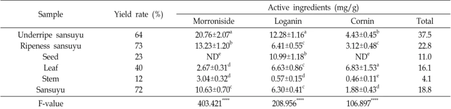
산수유 나무의 부위별 추출물의 기능성분 함량의 분석을 위하여 morroniside, loganin 및 cornin을 지표성분으로 선택 하였으며, 표준용액과 산수유 시료의 크로마토그램을 비교하 였다. 지표성분은 서로 간섭되지 않고 분리되었으며, 표준용 액과 시료의 peak 유지시간(retention time, RT) 또한 각각 morroniside (22.4 min), loganin (25.7 min) 및 cornin (26.2 min)과 일치하였다. 다음은 표준용액을 3.9, 7.8, 15.6, 31.3, 62.5, 125, 250, 500 μg/ml 농도에서 검량선을 작성한 결과, morroniside, loganin 및 cornin의 상관계수(R2) 값이 모두 0.99 이상으로 우수한 직선성을 보였다. 이를 바탕으로 산수유 나무의 열매(청산수유, 적산수유 및 건피), 씨앗, 잎, 줄기의 지표물질 함량을 분석한 결과, morroniside는 열매 중에서 청 산수유 및 적산수유에, loganin은 청산수유와 씨앗에 cornin은 잎과 청산수유에 높게 측정되었다(Table 3). 청산수유와 적산 수유의 지표물질 함량은 각각 37.5 mg/g 및 22.8 mg/g으로 가장 높으며, 열매의 성숙과정에서 지표물질 함량이 감소하는 것을 알 수 있었다. 건피의 경우 지표물질 함량이 18.8 mg/g으 로 적산수유에 비해 낮았으며, loganin은 유의적인 차이가 없 으나 morroniside와 cornin의 함량은 유의적으로 낮아졌다.
이는 데치기, 열풍건조 등으로 인해 감소하였으며, loganin은 상대적으로 열에 강한 것으로 판단된다. 이는 선행 연구의 결 과들과 유사하였다[5, 42]. 한편, 산수유 씨앗은 morroniside와 cornin은 검출되지 않았으며, loganin의 함량이 10.99 mg/g 청산수유 다음으로 높게 측정되었다. Cornin은 생과에서 씨앗 이 70% 이상을 차지하고 있으나, 기능성 소재의 원료로서 활 용도가 매우 낮은 편이다. 최근, 산수유 씨앗의 추출물을 이용 한 화장품의 방부 효능과 구성 성분에 관한 미백 효능 등과 같은 일부 효능이 보고되었으나[45] 다양한 기능성 및 안전성 평가에 대한 연구는 미비하다. Loganin과 cornin은 기억장애 개선 및 간 손상 보호[44] 등을 포함한 다양한 기능이 있는 것으로 보고되어 있으므로 다른 부위에 비해 loganin과 cornin 함량이 높은 잎을 활용한 기능성 연구는 향후 산업적 활용에 도움이 될 것으로 사료된다.
총 폴리페놀 함량 및 플라보노이드 함량
산수유 나무의 부위별 총 폴리페놀 함량을 측정한 결과는
Table 3. Active ingredients of morroniside, loganin and cornin of extracts from different parts of C. officinalis
Sample Yield rate (%) Active ingredients (mg/g)
Morroniside Loganin Cornin Total
Underripe sansuyu Ripeness sansuyu
Seed Leaf Stem Sansuyu
64 73 23 40 12 72
20.76±2.07a 13.23±1.20b
NDe 2.67±0.31d 3.04±0.32d 10.63±0.70c
12.28±1.16a 6.41±0.55c 10.99±1.18b
6.63±0.86c 0.57±0.15d 6.30±0.41c
4.43±0.45b 3.12±0.48c
NDe 6.83±1.53a 0.46±0.11e 1.88±0.43d
37.5 22.8 11.0 16.1 4.1 18.8
F-value 403.421**** 208.956**** 106.897****
Value are mean ± SD (n=8). Different letters within a column (a-f) indicate significant differences at p<0.05 by Duncan's multiple range test. ****p<0.0001.
ND, Not Detected.
Fig. 1. Total polyphenol contents of extract from different parts of C. officinalis. Value are mean ± SD (n=3). Different let- ters (a-f) above the bars are significant differences at p<0.05 by Duncan's multiple range test.
Fig. 3. DPPH free radical scavenging ability of extracts from different parts of C. fructus. Value are mean ± SD (n=3).
Different letters (a-f) above the bars are significant dif- ferences at p<0.05 by Duncan's multiple range test.
Fig. 1과 같으며, 추출 수율은 열매(64~73%)에서 가장 높았다 (Table 2). 적산수유와 청산수유가 각각 79.39 및 72.19 mg GAE/g으로 높게 측정되었다. 다음은 잎(64.84 mg GAE/g), 씨앗(56.91 mg GAE/g), 건피(38.49 mg GAE/g)의 순이었으 며, 줄기(14.32 mg GAE/g)가 가장 낮게 측정되었다. 비록 추 출 방법과 용매 및 시간에 따른 차이는 있으나, 선행연구 결과 [24, 25, 31]에 의하면, 산수유 열매, 꽃, 잎을 증류수와 99%
ethanol로 37℃에서 1~48시간 진탕 추출한 결과에서 추출 시 간이 길어질수록 추출 수율이 증가하였으며, 추출 부위는 열 매보다는 꽃과 잎이, 추출 용매는 ethanol 보다는 증류수에서 의 추출 수율이 높았다. 산수유 건피 추출 용매의 경우, Im 및 Lee [19]과 Kim 및 Son [22]의 100% 및 80% methanol 추출 물의 결과(각각 31.56 mg CAE/g 및 26.7 mg TAE/g)와 비슷 한 수준이었다. 산수유 나무의 부위별 총 플라보노이드 함량 을 측정한 결과는 Fig. 2와 같다. 제시된 결과에서처럼 잎이 20.45 mg QEU/g로 가장 높았으며, 열매는 청산수유(3.20 mg QEU/g)를 제외한 부위에서 거의 측정되지 않았다. 한편, Jeon et al. [20]은 총 플라보노이드가 5.67 mg QEU/g로 총 폴리페 놀 34.22 mg/g․hesperidin 함량보다 낮다고 보고한 바 있다.
Fig. 2. Total flavonoid contents of extract from different parts of C. officinalis. Value are mean ± SD (n=3). Different let- ters (a-d) above the bars are significant differences at p<0.05 by Duncan's multiple range test.
DPPH radical 소거능
산수유 나무의 부위별 DPPH radical 소거 활성으로 항산화
능을 측정한 결과는 Fig. 3과 같다. 산수유 전 부위가 양성 대조
A
B
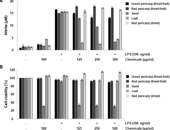
Fig. 4. Effects of the extracts from different parts of C. officinalis on NO production and cell viability in LPS-stimulated RAW 264.7 cells. The cells were pre-incubated with or without the indicated extracts from different parts of C. officinalis for 1 hr, and then treated with or without LPS (100 ng/ml) for 24 hr. (A) The nitrite concentration in culture media was evaluated by Greiss reaction. (B) Cell viability was determined by an MTT assay. Results are expressed as the percentage of surviving cells over control cells. Each data point represents mean ± SD of three independent experiments.
군 BHA 보다 항산화능이 낮은 것으로 나타났으나, 10 ppm 농도에서 BHA는 91.83%의 소거능을 보였으며, 잎과 씨앗이 각각 79.52%, 79.01%로 가장 높은 radical 소거 능을 보였다.
열매는 적산수유, 청산수유 순으로 각각 62.51, 35.22%의 radi- cal 소거 활성을 보였다. 건피와 줄기는 100 ppm 농도에서 각각 78.91, 25.48%의 radical 소거능을 보여 산수유 열매의 경우 건피보다 생과의 항산화 활성이 높은 것으로 확인되었 다. 유사한 선행 연구로서, Park et al. [31]은 100 ppm 농도에 서 산수유 성숙과와 완숙과가 각각 50.96%, 47.92%의 radical 소거 활성이 있었으며, Im 및 Lee [19]는 산수유 건피 meth- anol 추출물이 2.075 mg/ml로 본 실험결과가 더 낮은 농도에 서 radical 소거능을 보였다. 아울러 Lee et al. [25]은 대조구 0.1% 및 1% L-ascorbate의 91.13%와 92.21% 보다 산수유 건피 의 마이크로웨이브 추출물, 30, 60 및 90% 에탄올 추출물 및 물 추출 모두 93.54~97.20%의 높은 소거능이 있었음을 보고한 바 있다. 측정 조건의 차이는 있으나 산수유 부위별 모든 추출 물들은 모두 적절한 항산화능을 가지고 있음을 확인할 수 있 었다. 이러한 결과는 다양한 실험 모델에서 밝혀진 산수유 추 출물 및 유효 성분들의 항산화 효능을 잘 뒷받침하여 주는 결과이다[6, 18, 39, 40]. 이들 선행 연구에서 산수유 추출물
및 유효 성분들은 공통적으로 세포 내 다양한 신호 전달계의 조절을 통하여 항산화 효소의 활성을 증가시키거나 과도한 산화적 스트레스에 의한 세포사멸을 억제시켰다. 따라서 본 연구의 결과에서 얻은 항산화능이 높았던 추출물들을 대상으 로 그들의 항산화 효능과 연계된 기전 연구가 추가적으로 수 행되어야 할 것이다.
RAW 264.7 세포에서 NO 생성 및 세포 생존율에 미치는 각 추출물의 영향
염증 매개 인자인 NO는 NOS에 의하여 L-arginine으로부
터 생성되는 높은 반응성을 가진 생체 분자로서, 과도한 NO의
형성은 염증을 유도하며 산소와 결합하여 생성된 peroxyni-
trite (ONOO-)는 세포와 조직에 산화적 손상을 주고 유전자
변이, 패혈성 쇼크 및 신경 손상 등을 일으키기 때문에 생리적
수준의 NO 농도를 유지하는 것은 매우 중요하다[4, 30]. 본
연구에서는 RAW 264.7 세포에 LPS를 처리에 따라 유도되는
NO의 생성에 산수유 나무의 채취 부위별 에탄올 추출물이
미치는 영향에 과하여 조사하였다. Fig. 4에 나타낸 바와 같이,
LPS (100 ng/ml) 단독 처리에 의한 NO 생성은 대조군에 비하
여 약 14배 높게 나타났다. 산수유 나무의 채취 부위에 따른
A
B
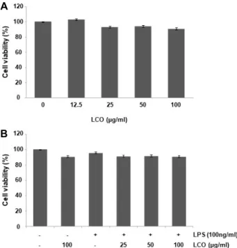
Fig. 5. Effects of leaf extracts of C. officinalis (LCO) on cell via- bility in RAW 264.7 cells. (A) The cells were treated with various concentrations of LCO extracts for 24 hr. (B) The cells were pretreated with the indicated concentrations of LCO for 1 hr prior to treatment with 100 ng/ml LPS for 24 hr. Cell viability was assessed by MTT assay. The results are expressed as means ± SD of three independent experiments duplicate in each run.
A
B
Fig. 6. Inhibition of NO production and iNOS expression by leaf extracts of C. officinalis (LCO) in RAW 264.7 cells.
The cells were exposed to the indicated concentrations of LCO 1 hr prior treatment of LPS (100 ng/ml) for 24 hr. (A) Amounts of NO production in culture media were determined by Greiss reaction. (B). Total proteins were isolated and subjected to Western blot analysis with specific antibody against iNOS. Experiments were repeated three times and similar results were obtained.
Actin was used as a control for equal loading. iNOS/ac- tin ratio was calculated and analyzed. All experiments were repeated three times (#p<0.05 in comparison with the control group; * p<0.05 in comparison with the LPS group).
각각의 에탄올 추출물을 다양한 농도(125, 250 및 500 μg/ml) 로 전처리한 후, LPS에 의한 NO 생성에 미치는 영향을 조사한 결과, 씨앗과 잎 에탄올 추출물에서 NO 생성이 유의적으로 억제되었음을 확인하였다. 동일 조건에서 MTT assay를 통해 세포 생존율의 변화를 확인해 본 결과, 산수유 나무 잎 추출물 (leaf extracts of C. officinalis, LCO)에서는 세포 생존에 유의적 인 차이가 나타나지 않았으나 125 μg/ml의 씨앗 추출물 처리 군에서는 대조군에 비하여 33.5%로 나타나 씨앗 추출물에 의 한 NO 생성의 억제는 세포독성에 기인한 것으로 사료된다.
따라서 이후 실험에서는 세포독성이 없으면서 NO의 생성을 효과적으로 억제시킨 잎 추출물을 대상으로 진행하였으며, 본 결과에서 가장 낮은 농도인 125 μg/ml에서도 강한 NO 생성 억제 효과를 나타내어 처리 농도 범위를 100 μg/ml 이하로 설정하였다.
산수유 나무 잎 추출물이 RAW 264.7 세포의 생존율과 LPS로 유도된 NO의 생성에 미치는 영향
RAW 264.7 세포에서 산수유 나무 잎 추출물 단독 처리 및 잎 추출물과 LPS의 동시 처리에 따른 세포 생존율의 변화를 조사하였다. 이를 위하여 LPS와 잎 추출물을 다양한 농도 (12.5, 25, 50 및 100 μg/ml)로 처리하고 24시간 동안 배양 후
MTT assay를 수행한 결과, 모든 실험 조건에서 세포 생존율이
90% 이상으로 나타났다(Fig. 5). 염증반응과 동반된 NO의 과
다 축적에는 iNOS의 전사 활성 증가가 관여하게 되며, iNOS
의 발현 수준은 NO 생성의 척도가 된다[1, 4]. 동일한 조건에
서 LPS로 자극된 RAW 264.7 세포의 NO 생성과 NO 생성에
관여하는 iNOS의 발현에 미치는 산수유 나무 잎 추출물의 효
과를 Griess 시약과 Western blotting을 이용하여 확인한 결과
는 Fig. 6에 나타냈다. 제시된 결과에서 알 수 있듯이, 잎 추출
물의 처리 농도 의존적으로 LPS에 의해 생성된 NO의 양이
유의적으로 감소하였으며, iNOS의 발현도 유사하게 LPS 단독
처리군에 비하여 감소되었다. 따라서 산수유 나무 잎 추출물
에 의한 NO의 생성 차단은 iNOS의 발현 억제에 의한 것임을
알 수 있다.
A
B
C
D
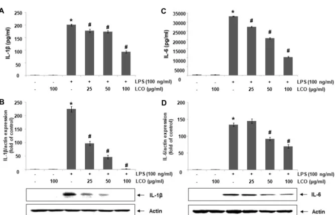
Fig. 7. Inhibition of IL-1β and IL-6 secretion and expression by leaf extracts of C. officinalis (LCO) in RAW 264.7 cells. The cells were exposed to the indicated concentrations of LCO 1 hr prior treatment of LPS (100 ng/ml) for 24 hr. (A and C) The IL-1β (A) and IL-6 (C) levels in medium were evaluated using commercial IL-1β and IL-6 cytokine ELISA kits. (B and D) Total proteins were isolated and subjected to Western blot analysis with specific antibodies against IL-1β and IL-6.
Experiments were repeated three times and similar results were obtained. Actin was used as a control for equal loading.
IL-1β and IL-6/actin ratio were calculated and analyzed. All experiments were repeated three times (#p<0.05 in comparison with the control group; * p<0.05 in comparison with the LPS group).
RAW 264.7 세포에서 LPS로 유도된 염증성 사이토카인 의 생성에 미치는 산수유 나무 잎 추출물의 영향
대식세포가 병원체 등의 감염에 의한 비정상적으로 활성화 되면 iNOS에 의한 NO 생성과 COX-2에 의한 PGE
2와 같은 염증 매개 인자의 생성 증가와 함께 염증성 사이토카인의 생 성 또한 증가한다[26, 34]. 따라서 본 연구에서는 LPS에 의한 IL-1β 및 IL-6와 같은 대표적인 염증성 사이토카인의 생성에 미치는 산수유 나무 잎 추출물의 영향을 조사하였다. 본 연구 의 결과에 의하면, LPS 단독 처리에 의해 IL-1β의 생성과 단백 질의 발현이 LPS를 처리하지 않은 대조군에 비하여 현저하게 증가되었다. 그러나 잎 추출물을 다양한 농도(25, 50 및 100 μg/ml)로 전처리한 군에서는 처리 농도 의존적으로 LPS에 의 해 증가된 IL-1β의 생성과 단백질의 발현이 억제되었다(Fig.
7A, Fig. 7B). 그리고 IL-6의 생성과 단백질 발현 또한 LPS에 의하여 증가되었으며 산수유 나무 잎 추출물을 전처리하였을 때 시료의 처리 농도가 높아질수록 IL-6의 생성 농도와 그 발 현이 점차적으로 감소하였다(Fig. 7C, Fig. 7D).
본 연구에서는 산수유 나무의 부위별 추출물에 함유된 유효 활성 성분 분석과 항산화 효능을 분석하였으며, 그중 가장 높
은 활성을 나타낸 잎 추출물을 이용하여 항염증 활성 실험을 수행하였다. 본 연구의 결과에 의하면, 잎 추출물은 강력한 항 산화 활성뿐만 아니라 항염증 활성을 지니고 있음을 확인하였 으며, 이는 산수유 추출물 및 구성 성분들의 효능에서 확인된 결과들과 잘 일치되었다[8, 10, 21, 37, 43]. 따라서 선행 연구들 의 결과를 토대로 산수유 나무 잎 추출물에 의한 항염증 효능 의 새로운 기전 연구가 추가로 수행되어야 할 것이며, 본 연구 에서 제시된 추출 방법은 향후 다양한 기능성 검증을 위한 추출법의 향상을 위한 조건 설정의 기준으로 활용될 것이다.
감사의 글
본 연구는 중소벤처기업부와 한국산업기술진흥원의 “지역 특화산업육성사업(R&D, S2874684)”와 구례군 야생화연구소 운영 연구개발비 지원으로 수행된 연구 결과임.
The Conflict of Interest Statement
The authors declare that they have no conflicts of interest
with the contents of this article.
References
1. Aktan, F. 2004. iNOS-mediated nitric oxide production and its regulation. Life Sci. 75, 639-653.
2. Apel, K. and Hirt, H. 2004. Reactive oxygen species: metabo- lism, oxidative stress, and signal transduction. Annu. Rev.
Plant Biol. 55, 373-399.
3. Blois, M. S. 1958. Antioxidant determination by the use of a stable free radical. Nature 181, 1199-1200.
4. Bogdan, C. 2015. Nitric oxide synthase in innate and adap- tive immunity: an update. Trends Immunol. 36, 161-178.
5. Cai, H., Cao, G. and Cai, B. 2013. Rapid simultaneous identi- fication and determination of the multiple compounds in crude Fructus Corni and its processed products by HPLC- MS/MS with multiple reaction monitoring mode. Pharm.
Biol. 51, 273-278.
6. Cao, G., Cai, H., Cai, B. and Tu, S. 2013. Effect of 5-hydrox- ymethylfurfural derived from processed Cornus officinalis on the prevention of high glucose-induced oxidative stress in human umbilical vein endothelial cells and its mechanism.
Food Chem. 140, 273-279.
7. Cao, G., Zhang, Y., Feng, J., Cai, H., Zhang, C., Ding, M., Cong, X. and Cai, B. 2011. A rapid and sensitive assay for determining the main components in processed Fructus cor- ni by UPLC–Q-TOF-MS. Chromatographia 73, 135-141.
8. Choi, Y. H., Jin, G. Y., Li, G. Z. and Yan, G. H. 2011. Cornuside suppresses lipopolysaccharide-induced inflammatory medi- ators by inhibiting nuclear factor-kappa B activation in RAW 264.7 macrophages. Biol. Pharm. Bull. 34, 959-966.
9. Chung, H. Y., Cesari, M., Anton, S., Marzetti, E., Giovannini, S., Seo, A. Y., Carter, C., Yu, B. P. and Leeuwenburgh, C.
2009. Molecular inflammation: Underpinnings of aging and age-related diseases. Ageing Res. Rev. 8, 18-30.
10. Cui, Y., Wang, Y., Zhao, D., Feng, X., Zhang, L. and Liu, C. 2018. Loganin prevents BV-2 microglia cells from Aβ (1-42)-induced inflammation via regulating TLR4/TRAF6/
NF-κB axis. Cell Biol. Int. 42, 1632-1642.
11. Deng, Y., Liu, Y., Tang, S., Zhou, C., Han, X., Xiao, W., Pastur-Romay, A. L., Vazquez-Naya, J. M., Loureiro, J. P., Munteanu, C. R. and Tan, Z. 2017. General machine learning model, review, and experimental-theoretic study of magno- lol activity in enterotoxigenic induced oxidative stress. Curr.
Top. Med. Chem. 17, 2977-2988.
12. Franceschi, C. and Campisi, J. 2014. Chronic inflammation (inflammaging) and its potential contribution to age-asso- ciated diseases. J. Gerontol. A Biol. Sci. Med. Sci. 69, S4-9.
13. Grune, T. 2000. Oxidative stress, aging and the proteasomal system. Biogerontology 1, 31-40.
14. Gülcin, I. 2012. Antioxidant activity of food constituents: an overview. Arch. Toxicol. 86, 345-91.
15. Han, Y., Jung, H. W. and Park, Y. K. 2014. Selective ther- apeutic effect of Cornus officinalis fruits on the damage of different organs in STZ-induced diabetic rats. Am. J. Chin.
Med. 42, 1169-1182.
16. Hanada, T. and Yoshimura, A. 2002. Regulation of cytokine signaling and inflammation. Cytokine Growth Factor Rev. 13, 413-421.
17. Huang, J., Zhang, Y., Dong, L., Gao, Q., Yin, L., Quan, H., Chen, R., Fu, X. and Lin, D. 2018. Ethnopharmacology, phy- tochemistry, and pharmacology of Cornus officinalis Sieb. et Zucc. J. Ethnopharmacol. 213, 280-301.
18. Hwang, K. A., Hwang, Y. J. and Song, J. 2016. Antioxidant activities and oxidative stress inhibitory effects of ethanol extracts from Cornus officinalis on raw 264.7 cells. BMC Complement. Altern. Med. 16, 196.
19. Im, D. Y. and Lee, K. I. 2017. Antioxidative activity and active compound analysis of the extract and fractions of Corni fructus. Kor. J. Pharmacogn. 48, 208-212.
20. Jeon, Y. H., Kim, M. H. and Kim, M. R. 2008. Antioxidative antimutagenic, and cytotoxic activities of ethanol extracts from Cornus officinalis. J. Kor. Soc. Food Sci. Nutr. 37, 1-7.
21. Kang, D. G., Moon, M. K., Lee, A. S., Kwon, T. O., Kim, J. S. and Lee, H. S. 2007. Cornuside suppresses cytokine-in- duced proinflammatory and adhesion molecules in the hu- man umbilical vein endothelial cells. Biol. Pharm. Bull. 30, 1796-1799.
22. Kim, Y. J. and Son, D. Y. 2016. Antioxidant activity and sup- pression of pro-inflammatory mediator of Corni fructus ex- tracts in activated RAW 264.7 macrophage. Kor. J. Food Preserv. 23, 876-882.
23. Lawrence, T., Bebien, M., Liu, G. Y., Nizet, V. and Karin, M. 2005. IKKα limits macrophage NF-κB activation and con- tributes to the resolution of inflammation. Nature 434, 1138.
24. Lee, M. H., Kim, J. M. and Park, E. J. 2011. Antioxidant and antigenotoxic effects of sansuyu fruit (Corni fructus) extracted with water at different temperatures. J. Kor. Soc.
Food Sci. Nutr. 40, 149-155.
25. Lee, N. H., Seo, C. S., Lee, H. Y., Jung, D. Y., Lee, J. K., Lee, J. A., Song, K. Y., Shin, H. K., Lee, M. Y., Seo, Y. B., Kim, H. and Ha, H. 2012. Hepatoprotective and anti- oxidative activities of Cornus officinalis against acetamino- phen-induced hepatotoxicity in mice. Evid. Based Comple- ment. Alternat. Med. 2012, 804924.
26. Lin, W. W. and Karin, M. 2007. A cytokine-mediated link between innate immunity, inflammation, and cancer. J. Clin.
Invest. 117, 1175-1183.
27. Lu, Y. C., Yeh, W. C. and Ohashi, P. S. 2008. LPS/TLR4 signal transduction pathway. Cytokine 42, 145-151.
28. Lushchak, V. I. 2014. Free radicals, reactive oxygen species, oxidative stress and its classification. Chem. Biol. Interact.
224, 164-175.
29. Ma, W., Wang, K. J., Cheng, C. S., Yan, G. Q., Lu, W. L., Ge, J. F., Cheng, Y. X. and Li, N. 2014. Bioactive compounds from Cornus officinalis fruits and their effects on diabetic nephropathy. J. Ethnopharmacol. 153, 840-845.
30. Moncada, S. and Higgs, A. 1993. The L-arginine-nitric oxide pathway. N. Engl. J. Med. 329, 2002-2012.
31. Park, S. J., Lee, G. E., Kim, Y. J. and Jeong, J. S. 2016.
Preparation and quality characterization of low sugar san-
suyu jam using fresh Corni fructus. J. Kor. Soc. Food Sci.
Nutr. 45, 222-229.
32. Ramos-Tovar, E. and Muriel, P. 2020. Free radicals, anti- oxidants, nuclear factor-E2-related factor-2 and liver damage.
J. Appl. Toxicol. 40, 151-168.
33. Rossol, M., Heine, H., Meusch, U., Quandt, D., Klein, C., Sweet, M. J. and Hauschildt, S. 2011. LPS-induced cytokine production in human monocytes and macrophages. Crit.
Rev. Immunol. 31, 379-446.
34. Schett, G., Elewaut, D., McInnes, I. B., Dayer, J. M. and Neurath, M. F. 2013. How cytokine networks fuel in- flammation: toward a cytokine-based disease taxonomy.
Nat. Med. 19, 822.
35. Schönbichler, S. A., Bittner, L. K. H., Pallua, J. D., Popp, M., Abel, G., Bonn, G. K. and Huck, C. W. 2013. Simultaneous quantification of verbenalin and verbascoside in Verbena offi- cinalis by ATR-IR and NIR spectroscopy. J. Pharm. Biomed.
Anal. 84, 97-102.
36. Singleton, V. L. and Rossi, Jr. J. A. 1965. Colorimetry of total phenolics with phosphomolybdic-phosphotungstic acid re- agents. Am. J. Enol. Vitic. 16, 144-158.
37. Sung, Y. H., Chang, H. K., Kim, S. E., Kim, Y. M., Seo, J.
H., Shin, M. C., Shin, M. S., Yi, J. W., Shin, D. H., Kim, H. and Kim, C. J. 2009. Anti-inflammatory and analgesic effects of the aqueous extract of corni fructus in murine RAW 264.7 macrophage cells. J. Med. Food. 12, 788-795.
38. Telang, N. T., Li, G., Sepkovic, D. W., Bradlow, H. L. and Wong, G. Y. C. 2012. Anti-proliferative effects of Chinese herb Cornus officinalis in a cell culture model for estrogen receptor-positive clinical breast cancer. Mol. Med. Rep. 5, 22-28.
39. Tian, W., Zhao, J., Lee, J. H., Akanda, M. R., Cho, J. H., Kim, S. K., Choi, Y. J. and Park, B. Y. 2019. Neuroprotective
effects of Cornus officinalis on stress-induced hippocampal deficits in rats and H2O2-induced neurotoxicity in SH-SY5Y neuroblastoma cells. Antioxidants (Basel) 9, 27.
40. Tseng, Y. T., Lin, W. J., Chang, W. H. and Lo, Y. C. 2019.
The novel protective effects of loganin against 1-meth- yl-4-phenylpyridinium-induced neurotoxicity: Enhance- ment of neurotrophic signaling, activation of IGF-1R/GLP- 1R, and inhibition of RhoA/ROCK pathway. Phytother. Res.
33, 690-701.
41. Victor, V. N. and Rocha, M. 2007. Targeting antioxidants to mitochondria: A potential new therapeutic strategy for cardiovascular diseases. Curr. Pharm. Des. 13, 845-863.
42. Wang, L., Chen, H., Jiang, Y., Liu, Z., Wang, Q. and Zheng, X. 2018. Simultaneous determination of 11 high-polarity components from Fructus Corni: A quantitative LC-MS/MS method for improved quality control. J. Chromatogr. Sci. 56, 56-64.
43. Wen, H., Xing, L., Sun, K., Xiao, C., Meng, X. and Yang, J. 2020. Loganin attenuates intestinal injury in severely burned rats by regulating the toll-like receptor 4/NF-κB sig- naling pathway. Exp. Ther. Med. 20, 591-598.
44. Yamabe, N., Noh, J. S., Park, C. H., Kang, K. S., Shibahara, N., Tanaka, T. and Yokozawa, T. 2010. Evaluation of loga- nin, iridoid glycoside from Corni Fructus, on hepatic and renal glucolipotoxicity and inflammation in type 2 diabetic db/db mice. Eur. J. Pharnacol. 648, 179-187.
45. Yang, J. C. 2016. A study on the cosmetic preservative effects of Cornus officinalis seed extracts. J. Kor. Oil Chemists. Soc.
33, 333-341.
46. You, Q., Yin, X. and Zhao, Y. 2013. Enzyme assisted ex- traction of polysaccharides from the fruit of Cornus officinalis. Carbohydr. Polym. 98, 607-610.
초록:산수유의 채취 부위에 따른 기능 성분 분석과 항산화 및 항염증 효과에 관한 연구
황보현
1,2․정지숙
3․김민영
1,2․지선영
1,2․윤선혜
4․김태희
5․김성옥
6․최영현
1,7*
(1동의대학교 항노화연구소, 2부산대학교 분자생물학과, 3구례군농업기술센터 야생화연구소, 4㈜네이처텍, 5함소아
제약 중앙연구소, 6경성대학교 식품영양학과, 7동의대학교 한의과대학 생화학교실)