ISSN 2288-1069 (Online)
http://dx.doi.org/10.12925/jkocs.2020.37.4.674
The Improvement of skin barrier function and anti-obesity effect of Codonopsis lanceolata by supercritical carbon
dioxide extraction
Bora Kim✝
Division of Chemistry and Cosmetics, Mokwon University, Daejeon, 35349, Republic of Korea (Received July 10, 2019; Revised August 17, 2019; Accepted August 18, 2020)
Abstract : The root of Codonopsis lanceolata has been used in traditional medicine. This study was conducted to confirm the comparative effect of ethanol solvent extraction (CLE) and supercritical carbon dioxide extraction (CLS) of C. lanceolata roots. CLS had higher antioxidant than CLE. For supercritical co-solvent modified carbon dioxide extraction (CLS), it were extracted at 250 bar 50℃ 150 min at a flow rate of ethyl alcohol 3 mL/min for 90min. In addition, CLS inhibited the adipocyte differentiation of 3T3-L1 cells. When treated with the extract at a concentration of 100 μg/mL, the Wnt/β-catenin pathway reporter luciferase activity of HEK 293-TOP cells increased approximately by 3-folds compared to that of the untreated control group. Also, the treatment by CLS (50 µg/mL) showed a significant increase of involucrin expression. These results indicate that supercritical carbon dioxide extract of C. lanceolatamay serve as a cosmeceutical agent for improving skin barrier function and the treatment of obesity.
Keywords : Codonopsis lanceolata, Supercritical carbon dioxide extraction, Involucrin, Wnt/β -catenin, Adipocyte differentiation
1. Introduction
Medicinal plants are currently the focus of a great deal of attention as functional materials, which are used for food, drug, and cosmetics.
Despite terrific therapeutic benefits of many biological compounds, most medicinal plants have been studied generality for the development of drugs and there are only some research on cosmeceutical materials.
Additionally, more effective and less toxic
✝Corresponding author
(E-mail; [email protected])
medicinal compounds are still required.
Supercritical carbon dioxide extraction (SCE) can provide several benefits compared to conventional solvent extraction: faster extraction time, improvement of the yield, a low environmental impact, and in the optimum process for obtaining extracts with high anti-oxidant quality[1].
The Wnt/β-catenin pathway plays a role in adipose cell communication and the inhibition of adipogenesis[2]. The understanding of molecular and cellular events regulating adipogenesis is crucial for designing rational therapies for prevention or treatment of obesity and for treatment of metabolic syndromes[3].
The skin barrier is located in the outermost layer of the epidermis, in the stratum corneum (SC), which consists of two major structural components, the corneocytes and intercorneocyte lipids[4]. Thus, the formation of the SC, layers of terminally differentiated cornified cells in the outermost epidermis, is responsible for the barrier properties of the skin[5].
Codonopsis lanceolata is a perennial vine plant belonging to the family Celadonaceae, predominantly found in Central, East, and South Asia. This plant has been widely used in traditional medicine and contains many biological compounds, including polyphenols, saponins, tannins, triterpene, alkaloids, and steroids[6]. Several studies have reported that the root of C. lanceolata shows positive biological effects, such as anti-inflammatory, anti-stress, antimicrobial, anti-oxidant, anti- cancer, and immunomodulatory activities[7].
However, the effects of its extract as a skin therapeutic agent for skin barrier function and as a nutraceutical product for obesity treatment have not been studied. This study aims to verify the effect of C. lanceolata extract on strengthening the skin barrier and suppressing obesity.
2. Experimental
2.1. Sample preparation
The C. lanceolata was purchased from the Jeonnam herbal farming cooperative (Hwasun- gun, Jeollanam-do, Korea). For supercritical co-solvent modified carbon dioxide extraction (CLS), the system and required components were acquired at 250 bar 50℃ 150 min at a flow rate of ethyl alcohol 3 mL/min for 90 min from Nano bio research center (Janseong- gun, Jeollanam-do, Korea). The extraction was performed by a method previously described[8]. In case of CLE, C. lanceolata was soaked in 99.9% ethyl alcohol for 7 days and then was dried by speed vacuum at 50℃.
2.2. Cell culture and materials
3T3-L1 preadipocytes, which were gained from Yonsei University, were cultured by a method previously described[10]. Human normal keratinocyte and HEK 293-TOP cells were purchased from American Type Culture Collection (ATCC, USA) and cultured by a method previously described[9].
2.3. Cell viability
Cell viability was measured using the 3-(4,5-dimethylthiazol-2-yl)-2,5-diphenyltetra zolium bromide (MTT) assay. Human normal keratinocytes were treated with different concentrations of C. lanceolata (CL) for 48 h at 37℃, followed by the addition of 50 μL of 2 mg/mL MTT (Sigma-Aldrich, St. Louis, MO, USA) solution to respective wells, and then incubation for 3 h at 37℃. The procedure was done according to the previous method[10].
2.4. Antioxidant assay
The 2,2-diphenyl-1-picrylhydrazyl (DPPH) assay was performed to determine the antioxidant capacity of CL. The SC50
(concentration required to obtain a 50%
antioxidant effect), a commonly used parameter to indicate the antioxidant capacity, was also measured[11].
2.5. Luciferase assay
HEK 293-TOP cells (3 x 10⁴) were seeded into 96-well plates and incubated in a medium with 10% FBS for one day. The procedure was done according to the previous method[12].
2.6. Adipocyte differentiation and Oil Red O staining
The 3T3-L1 preadipocytes were cultured in Dulbecco's modified Eagle's medium (DMEM) with high glucose, 110 mg/L pyruvate supplemented with heat inactivated 10% (v/v) calf serum (Gibco, CA, USA), 100 μg/mL penicillin, 100 μg/mL streptomycin in a CO2
incubator at 37 ℃. Adipocyte cell layers were washed with PBS, fixed with 4%
paraformaldehyde in PBS, stained with the Oil Red O dye solution for 1 hour, and then washed with distilled water. The procedure was done according to the previous method [12].
2.7. Western blot and Immunoblot
analysis of 3D skin equivalent model Normal human keratinocytes were washed once in PBS and lysed in a lysis buffer (Cell Signaling, MA, USA). Protein concentrations were determined using the Bradford protein assay kit (BioRad, CA, USA). The procedure was done according to the previous method[9].
The following primary antibodies were used:
rab bitanti-involucrin (Santa Cruz Biotechnology, Carlsbad, CA, USA), and mouse anti-β-actin (Sigma-Aldrich Co., St. Louis, MO, USA).
The band intensities were quantified using a Photo-Image System (Molecular Dynamics, Sweden). Skin models were prepared and cultured as described in Materials and methods and the experiments were performed by methods previously described[13]. The effect of CLS on involucrin synthesis was analyzed using EpiDermTM, a three-dimensional model of skin equivalents purchased from MatTek Corporation (Ashland, MA, USA). The prepared samples were observed by microscope (Carl Zeiss Inc., Oberkochen, Germany). The area was analyzed by involucrin with image quantitative analysis software (NIH ImageJ, version 1.61.).
2.8. Statistical analysis
All data are presented as the mean ± standard error of the mean (S.E.M). Statistical analyses were conducted with GraphPad Prism 5.0 (GraphPad Software, Inc. San Diego, CA, USA). Comparisons between multiple groups were performed using the one-way analysis of variance with Bonferroni post-hoc test.
P-value of less than 0.05 was considered significant.
3. Results and Discussion
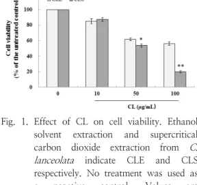
3.1. Effect of CL on cell viability
I performed the effect of CL on cell viability.
After 24 h treatment of Human normal keratinocytes with CL (10, 50, and 100 µg/mL). Cell viability was not affected by 100 µg/mL of CLE (Figure 1). In case of CLS, there was no cell toxicity up to 10 µg/mL.
Fig. 1. Effect of CL on cell viability. Ethanol solvent extraction and supercritical carbon dioxide extraction from C.
lanceolata indicate CLE and CLS respectively. No treatment was used as a negative control. Values are presented as means ± standard error of the mean (SEM). *p < 0.05, **p <
0.01 compared to the untreated control group.
3.2. Antioxidant effect of CL
The radical scavenging activities of CL increased in a concentration-dependent manner. The radical scavenging activity SC50
of CLE was 389.7 µg/ml and that of CLS was 217.1 µg/ml, therefore CLS has better antioxidant activity (Figure 2). The extraction efficiency as well as solvent power of carbon dioxide may be increased by the addition of a polar co-solvent such as ethanol. This supercritical co-solvent modified carbon dioxide extraction is more efficient method for antioxidants such as polyphenols.
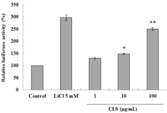
Fig. 3. Effect of CLS on Luciferase reporter activity. HEK293 cells containing pTOPFlash reporter gene in its chromosome were cultured and treated with different LC at 1, 10 or 100 μg/mL concentration, and cellular extract was prepared after 24 h as described in the Materials and methods. CLS indicate supercritical carbon dioxide extraction from C. lanceolata. *p < 0.05, **p < 0.01 compared to the untreated control group.
Fig. 2. Antioxidant effect on CL. Ethanol solvent extraction and supercritical carbon dioxide extraction from C.
lanceolata indicate CLE and CLS respectively. Values are presented as means ± standard error of the mean (SEM).
3.3. Effect of CLS on the adipocyte differentiation and Wnt/β-catenin activity
The reporter activity was increased approximately by 3-folds with the treatment of CLS at concentrations of 100 µg/mL, compared to that of the non-treated control group (Figure 3). Additionally, CLS inhibited adipocyte differentiation of 3T3-L1 cells in a
concentration dependent manner (Figure 4).
These results indicate that CLS induces differentiation of 3T3-L1 cells, showing the role of the Wnt/β-catenin activator in the adipocyte differentiation. Wnt/β-catenin signaling has been known to modulate additional developmental processes of adipocytes and to play a role in the suppression of differentiation of pre-adipocyte cells of 3T3-L1 cells[14, 15].
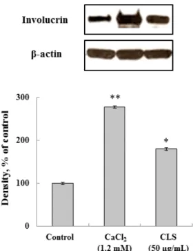
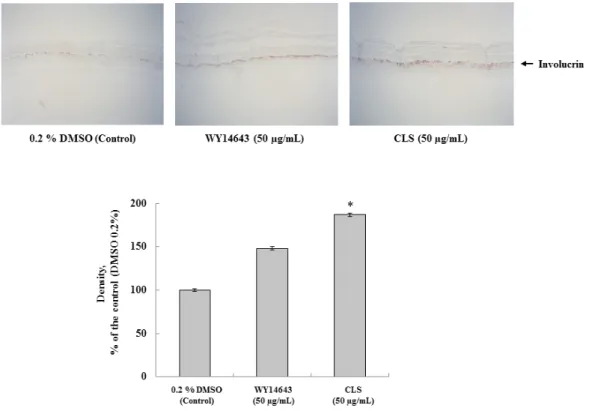
3.4. Effect of CLS on involucrin expression Involucrin is an essential cornified envelope component that can be used as biomarker for screening of new candidates for the improvement skin barrier function[16, 17]. As shown in Figure 5, the groups treated using CLS showed high levels on expression of involucrin compared to the untreated control group, but the levels were somewhat lower than that in the group treated with 1.2 mM CaCl2. Also, immunohistochemical analysis of involucrin related to keratinocyte differentiation was determined in 3D skin equivalent model.
As shown in Fig. 6, the treatment by CLS (50 µg/mL) showed a significant increase of involucrin expression compared with treatment by 0.2% DMSO as a negative control in skin equivalents, in addition, the group treated with
Fig. 4. Effect of CLS on the adipocyte differentiation of 3T3-L1 preadipocyte cells. 3T3-L1 cells were induced to differentiation for 7 days with or without 20 mM LiCl.
Intracellular lipids of 3T2-L1 cells were visualized by Oil Red O staining as described in the Materials and methods. Original magnification: x400. CLS indicate supercritical carbon dioxide extraction from C. lanceolata.
Fig. 5. Effect of C. lanceolata extract on expression of involucrin. Each signal was quantified by scanning densitometry. β-actin was used as an internal standard. 1.2 mM CaCl2 was used as a positive control and no treatment was used as a negative control. Values were nomalized to β-actin before calculating changes and presented as means ± standard error of the mean (SEM). *p < 0.05, **p < 0.01 compared to the untreated control.
Fig. 6. Immunohistochemical analysis of involucrin in 3D skin equivalent model. Each signal was quantified by ImageJ. Values are presented as means ± standard error of the mean (SEM). *p < 0.05 compared to the untreated control group. CLS indicate supercritical carbon dioxide extraction from C. lanceolata.
CLS showed higher level in the expression of involucrin compared to the group treated with the same concentration of WY14643. Our study shows that the expression of involucrin was increased in CLS. In conclusion, the effects of CLS treatment on skin barrier functions were evaluated. Therefore, supercritical extract from C. lanceolata may be an appropriate material for improving skin barrier function and inhibiting adipogenesis.
4. Conclusion
The carbon dioxide supercritical fluid extraction is environmental friendly technology and complementary and alternative method using organic solvent to solve problems such
as toxicity[18]. Supercritical fluid extraction is often used for the extraction of non-polar compounds. In case of C. lanceolata, the supercritical extract with modified carbon dioxide with ethanol (CLS) had the better physiological activity than the commonly used ethanol solvent extract (CLE). The extraction efficiency as well as solvent power of carbon dioxide may be increased by the addition of a polar co-solvent such as ethanol. It is presumed that this supercritical co-solvent modified carbon dioxide extraction is more efficient for bioactive substances including antioxidants such as polyphenols[19]. CLS showed the cytotoxicity of keratinocytes from concentrations above 50 µg/mL while CLE showed relatively low cytotoxicity. The radical scavenging activity SC50 of CLE was 389.7
µg/mL and that of CLS was 217.1 µg/mL, therefore CLS has better antioxidant activity.
CLS can increase the expression of involucrin and inhibited the adipocyte differentiation of 3T3-L1 cells. When treated with the extract at a concentration of 100 μg/mL, the Wnt/β -catenin pathway reporter luciferase activity of HEK 293-TOP cells was increased approximately by 3-folds compared to that of the untreated control group. Also, the treatment by CLS (50 µg/mL) showed a significant increase of involucrin expression.
This study suggests that CLS have potential as cosmeceutical agents for improving skin barrier functions and the treatment of obesity.
Acknowledgements
The author is grateful to Enprani Co. Ltd and Translational Research Center for Protein Function Control in Yonsei University for their support of this project and technical assistance.
References
1. K. Tyskiewicz, M. Konkol, E. Roj,
“Supercritical Carbon Dioxide (scCO2) Extraction of Phenolic Compounds from Lavender (Lavandula angustifolia) Flowers: A Box-Behnken Experimental Optimization”, Molecules, Vol.24, No.18. pp. 3354-3369, (2019).
2. J. Liu, S. R. Farmer, “Regulating the Balance between Peroxisome Proliferator- activated Receptor γ and β-Catenin Signaling during Adipogenesis”, Journal of Biological Chemistry, Vol.279, No.43 pp.
45020-45027, (2004).
3. G. Fruhbeck, J. Gomez-Ambrosi, F. J.
Muruzabal, M. A. Burrell, “The Adipocyte: a Model for Integration of Endocrine and Metabolic Signaling in Energy Metabolism Regulation”, American Journal of Physiology-Endocrinology and
Metabolism, Vol.280, No.6 pp. 827-847, (2001).
4. W. M. Holleran, Y. Takagi, G. K. Menon, S. M. Jackson, J. M. Lee, K. R. Feingold, P. M. Elias, “Permeability Barrier Requirements Regulate Epidermal beta- glucocerebrosidase”, Journal of Lipid Research, Vol.35, No.5 pp. 905-912, (1994).
5. D. T. Downing, “Lipid and Protein Structures in the Permeability Barrier of Mammalian Epidermis”, Journal of Lipid Research, Vol.33, No.3 pp. 301-313, (1992).
6. K. T. Lee, J. Choi, W. T. Jung, J. H.
Nam, H. J. Jung, H. J. Park, “Structure of a New Echinocystic acid Bisdesmoside isolated from Codonopsis lanceolata roots and the Cytotoxic Activity of Prosapogenins”, Journal of Agricultural and Food Chemistry, Vol.50, No.15 pp.
4190-4193, (2002).
7. S. E. Byeon, W. S. Choi, E. K. Hong, J.
Lee, M. H. Rhee, H. J. Park, J. Y. Cho,
“Inhibitory Effect of Saponin fraction from Codonopsis lanceolata on Immune Cell-mediated Inflammatory Responses”, Archives of Pharmacal Research, Vol.32.
No.6 pp. 813–822, (2009).
8. B. Kim, S. M. Lee, T. Y. Hwang, H. S.
Kim, “Anti-oxidative and Skin barrier effects of Natural Plants with a Supercritical Extract”, Korean Journal of Food Preservation, Vol.20, No.5 pp.
597-601, (2013).
9. B. Kim, Y. E. Choi, H. S. Kim, “Eruca sativa and its Flavonoid Components, Quercetin and Isorhamnetin, Improve Skin Barrier Function by Activation of Peroxisome proliferator-activated receptor (PPAR)-α and Suppression of Inflammatory Cytokines”, Phytotherapy Research, Vol.28, No.9 pp. 1359-1366, (2014).
10. B. Kim, H. S. Kim, “Novel Peptide Inhibits Inflammation by Suppressing of Protease activated receptor-2”, European
Journal of Pharmacology, Vol.832, No.1 pp. 25-32, (2018).
11. K. V. Sharma, R. Sisodia, “Evaluation of the Free Radical Scavenging Activity and Radioprotective Efficacy of Grewia asiatica fruit”, Journal of Radiological Protection, Vol.29, No.3 pp. 429-443, (2009).
12. S. H. Lee, B. Kim, M. J. Oh, J. Y. Yoon, H. Y. Kim, K. J. Lee, J. D. Lee, K. Y.
Choi, “Persicaria hydropiper (L.) spach and its Flavonoid Components, Isoquercitrin and Isorhamnetin, Activate the Wnt/-β catenin Pathway and Inhibit Adipocyte Differentiation of 3T3-L1 cells”, Phytotherapy Research, Vol.25, No.11 pp.
1629-1635, (2011).
13. B. Kim, “The Activation of PPAR-α and Wnt/β-catenin by Luffa cylindrica Supercritical Carbon Dioxide Extract”, Natural Product Sciences, Vol.25, No.4 pp.
341-347, (2019).
14. B. Kim, H. S. Kim, “Ricinus communis Extract Inhibits the Adipocyte Differentiation Through Activating the Wnt/β-catenin Signaling Pathway”, Korean Jourmal of Food Preservation, Vol.24, No.4 pp. 524-528, (2017).
15. J. Y. Ahn, H. J. Lee, S. N. Kim, T. Y.
Ha, “Curcumin‐Induced Suppression of Adipogenic Differentiation is Accompanied by Activation of Wnt/β‐catenin signaling”, American Journal of Physiology Cell Physiolology, Vol.298, No.6 pp. 1510–
1516, (2010).
16. Liu J, Farmer SR, Regulating the Balance between Peroxisome Proliferator-activated receptor γ and β-Catenin Signaling during Adipogenesis. Journal of Biological Chemistry, 279, pp. 45020-45027, (2004).
17. J. M. Jensen, R. Fölster-Holst, A.
Baranowsky, M. Schunck, S. Winoto- Morbach, C. Neumann, S. Schütze, E.
Proksch, “Impaired Spingomyelinase Activity and Epidermal Differentiation in Atopic Dermatitis”, Journal of Investigative Dermatology, Vol.122, No.6, pp.
1423-1431, (2004).
18. J. H. Seo, Y. J. Lee, Y. I. Jo, J. Y. Ko.
M. J. Mun, K. H. Park, S. E. Choi,
“Anti-fungal, Anti-oxidant, and Anti- inflammatory effects of Supercritical Fluid extracts from Ulmus davidiana”, Journal of the Korea Convergence Society, Vol..9.
No.8, pp. 225-233, (2018).
19. K. Tyskiewicz, M. Konkol, E. Roj, “The Application of Supercritical Fluid Extraction in Phenolic Compounds Isolation from Natural Plant Materials”, Molecules, 23, pp. 2625-2652, (2018).