Baicalein이 Lipopolysaccharide에 의한 생쥐의 Neuroinflammation에 미치는 영향
하경운#, 김연섭*
가천대학교 한의과대학 해부경혈학교실
Effects of Baicalein on Neuroinflammation in Lipopolysaccharide-treated Mice
Gyung-Woon Ha
#, Youn-Sub Kim
*Department of Anatomy-Pointology, College of Oriental Medicine, Gachon University
ABSTRACT
Objects : Baicalein is a major bioactive flavonoid component of Scutellaria baicalensis Georgi that shows a wide range of biological activities, including neuroprotections and anti-inflammatory actions. Hence it is a potential therapeutic material for the treatment of neuroinflammation. In this study, we investigated the modulatory effect of baicalein on neuroinflammation.
Method : Pro-inflammatory cytokines (TNF-α, IL-1β and IL-6 mRNA), COX-2 mRNA expression and microglial activation in the brain tissue is induced by systemic lipopolysaccharide (LPS) treatment in C57BL/6 mice.
Baicalein was treated orally with 10, 20, and 30 mg/kg 1 hour prior to the LPS (3 ㎎/㎏, i.p.) injection.
TNF-α, IL-1β, IL-6 and COX-2 mRNA expression in the brain tissue was measured by the quantitative real-time polymerase chain reaction(PCR) method. Iba1 expression in the brain was measured by western blotting method. Microglia was observed with immunohistochemistry.
Results : Baicalein 30 mg/kg significantly attenuated the expression of TNF-α, IL-1β, IL-6 and COX-2 mRNA in the brain tissue. Baicalein 20 mg/kg significantly attenuated the expression of IL-6 mRNA in the brain tissue.
Baicalein 30 mg/kg significantly attenuated the expression of Iba1 protein expression in the brain tissue. Baicalein 30 mg/kg significantly decreased the number and cell size of microglia in the cerebral cortex and hypothalamic region and the area percentage of Iba1-expressed microglia in the hippocampus.
Conclusion : These results demonstrated that baicalein attenuates LPS induced neuroinflammation in the mice via reduction of pro-inflammatory cytokines (TNF-α, IL-1β, IL-6), COX-2 mRNA expression and microglial activation.
Key words : Baicalein, TNF-α, IL-1β, IL-6, COX-2, Microglia
서 론
1)2)
중추신경계 염증은 항 염증성 cytokine, 케모카인, 신경 전달물질 및 활성산소종이 과다 분비됨으로써 중추신경계 에 상주하는 microglia, 별아교세포 및 침윤된 주변 식세포 가 활성화되고 이로 인하여 염증을 가속시킨다
1).
Microglia는 중추신경계의 면역세포로 중추신경계 염증 유발에 관여하며, 뇌조직에서 염증유발 cytokine을 분비하 고
2)부상이나 질병 시 중추신경계의 숙주방어 및 조직손 상에 관여하는 것으로 알려져 있다
3). 또한 뇌실질과 면역 계를 연결시키며, 뇌조직 염증, 면역반응, 퇴행성 변화 등 에서 중추적인 역할을 한다. 병리적인 변화 시 가장 먼저
* 교신저자 : 김연섭, 경기도 성남시 수정구 복정동 산 65, 가천대학교 한의과대학 해부경혈학교실 ·Tel : 031-750-5420 ·E-mail : [email protected]
# 제1저자 : 하경운, 경기도 성남시 수정구 복정동 산 65, 가천대학교 한의과대학 해부경혈학교실 ·Tel : 031-750-5427 ·E-mail : [email protected]
·접수:2013년 2월 6일 ·수정:2013년 3월 21일 ·채택:2013년 3월 21일
활성화되어 방어기전의 중심이 되며, 활성화된 microglia는 손상부위로 이주하고 증식하여 phagocytic cell로 변하여 nitric oxide(NO), tumor necrosis factor-α(TNF-α), interleukin(IL)과 같은 세포독성 물질들을 분비한다. 그리 고 microglia는 조직을 수복하는 역할이 있으므로 병리적 인 변화로 활성화되었을 경우 감시, 방어, 신경보호 역할을 하지만, 과도하게 활성화되거나 방어기전에서 실패하게 되 면 뇌손상에 영향을 주게 된다
4,5).
黃芩(Scutellaria baicalensis GEORGI)은 꿀풀과 (Labiatae)에 속하는 속썩은 풀의 주피를 벗긴 뿌리로
6)항 염증, 항바이러스 및 항박테리아 작용과 같은 다양한 약리 작용이 있다고 알려져 있다
7).
Baicalein((5,6,7-trihydroxy-2-phenyl-4H-1-benzop yran-4-one)은 黃芩의 뿌리에 함유되어 있는 flavonoid 성 분중 하나로써 lipoxygenase의 저해
8), 마이크로솜(microsome) 의 지질과산화반응 억제
9), 산소라디칼(oxygen radicals) 생성 의 저해
10), 그리고 hydrogen peroxide(H
2O
2), hydroxyl radical(OH) 및 superoxide anions(O
2-)과 같은 활성산소종 (free radical species)으로 유도된 fibroblast의 손상을 억제 하는 것으로 보고되고 있다
11,12). 그리고 세포사망을 유도하는 작용과 12-lipoxygenase 억제 작용에 의한 알츠하이머병 (Alzheimer's, AD)에 대한 보호 작용이 있으며
13), 파킨슨병 (Parkinson's disease, PD)의 in vitro와 in vivo 실험 모델 에서는 신경보호에 효과가 있다고 하였다
14). 그리고 피부손상 에 대한 염증을 억제시키며 cyclooxynase-2 (COX-2)의 발 현을 억제한다고 하였다
15). 또한 baicalein은 강력한 항염증 및 항산화 작용을 가지고 있어서 다양한 염증성 질환을 치료 하고 예방하는데 사용되고 있다
16).
이에 저자는 baicalein이 중추신경계염증(neuroinflammation) 에 대한 보호 작용이 있을 것이라 추측하고, lipopolysaccharide (LPS)를 mouse의 복강에 투여하여 중추신경계염증을 유발시 켰다. 뇌조직에서 대표적인 염증유발 cytokine인 TNF-α, IL-1β 및 IL-6 mRNA 발현과 COX-2 mRNA 발현을 살 펴보았다. 그리고 뇌조직의 microglia 활성에 미치는 영향을 확인하기 위해 Iba1 단백질 발현, 대뇌피질과 시상하부의 microglia 수와 크기 및 해마에서 microglia 면적비율의 변 화를 관찰한바 유의한 결과를 얻었기에 보고하는 바이다.
재료 및 방법
1. 실험동물
실험동물은 나라바이오텍 (Nara Biotechnology, Korea) 에서 구입한 25-28 g의 수컷 C57BL/6 생쥐를 사용하였다.
실험에 사용한 생쥐는 온도 (21-23℃), 습도 (40-60%)와 조 명 (12시간 명/암)이 자동적으로 유지되는 사육실에서 무균음 수와 사료를 자유롭게 공급하며 사육되었고, 실험실 환경에 1 주 이상 적응시킨 후 사용하였다.
2. 약물 및 시약
본 실험에 사용한 baicalein (C15H10O5; Molar mass, 270.24; #465119)과 lipopolysaccharide (LPS; from
Escherichia coli O55:B5)는 Sigma-Aldrich (St. Louis, MO, USA)에서 구입하였다. Rabbit anti-ionized calcium binding adaptor molecule 1 (Iba1) antibody (#016-20001, #019-19741)는 Wako Pure Chemical Industries (Osaka, Japan)에서, mouse anti-β-actin antibody는 Chemicon International (Temecula, CA, USA) 에서, goat anti-rabbit와 goat anti-mouse IgG horseradish peroxidase conjugated secondary antibody는 Santa Cruz Biotechnology (Santa Cruz, CA, USA)에서 구입하여 사 용하였다. 그 이외의 시약들은 전문제조회사의 실험실용 최고 품질의 시약을 구입하여 사용하였다.
Fig. 1. Chemical structure of baicalein
3. 실험군의 구분 및 처치
생쥐를 무작위로 5개 군으로 구분하였다. 정상군 (Normal)은 약물처치 없이 통상적으로 사육되었으며, 대조 군 (Control)은 LPS 3 ㎎/㎏을 복강주사 하였다. Baicalein 을 투여한 군은 투여용량에 따라 10 ㎎/㎏ 투여군 (Baicalein 10), 20 ㎎/㎏ 투여군 (Baicalein 20) 및 30 ㎎ /㎏ 투여군 (Baicalein 30)으로 구분하였으며, 각각의 용량 을 생리식염수에 녹여 LPS의 복강주사 1시간 전에 1회 경구 투여 하였다. 실험동물은 real-time polymerase chain reaction (PCR) 측정에 30마리, western blotting 측정에 18마리, 면역조직화학염색에 18마리, 총 66마리의 생쥐가 사용되었다.
4. 염증유발 cytokine과 COX-2 mRNA의 real-time PCR 측정
염증유발 cytokine 중 TNF-α, IL-1β, IL-6 및 COX-2 mRNA 발현을 뇌조직에서 정량적인 real-time PCR 방법으로 측정하였다. Baicalein 처치와 LPS 복강주사 4시간 후에 생쥐를 단두로 희생시키고 뇌를 적출한 다음, 뇌조직으로 부터 Trizol (Qiagen, Germany)을 사용하여 total RNA을 추출하였다. 이후 1 µg의 total RNA와 Script cDNA synthesis Kit (Bio-Rad, USA)를 사용하여 DNA로 역전사 하였으며, 정량적 real-time PCR은 CFX 96 REAL-TIME PCR Detection System (Bio-Rad, USA)에서 iQ SYBR Green Supermix kit (Bio-Rad, USA)와 preoptimized primer/probe mixture를 사용하여 측정하였다.
Housekeeping gene으로는 β-actin을 사용하였으며, 측정에
사용된 gene의 primer sequence는 아래 Table 1과 같다. 이
외 측정방법은 제조사의 사용설명서에 따랐으며, 각 시료로부
터 측정된 mRNA 발현량은 Normal군의 측정치를 1로 하여
그 배수 (fold change)를 자료로 사용하였다.
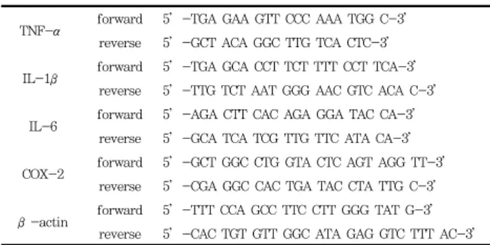
Table 1. Primer Sequences
TNF-α forward 5’-TGA GAA GTT CCC AAA TGG C-3’
reverse 5’-GCT ACA GGC TTG TCA CTC-3’
IL-1β forward 5’-TGA GCA CCT TCT TTT CCT TCA-3’
reverse 5’-TTG TCT AAT GGG AAC GTC ACA C-3’
IL-6 forward 5’-AGA CTT CAC AGA GGA TAC CA-3’
reverse 5’-GCA TCA TCG TTG TTC ATA CA-3’
COX-2 forward 5’-GCT GGC CTG GTA CTC AGT AGG TT-3’
reverse 5’-CGA GGC CAC TGA TAC CTA TTG C-3’
β-actin forward 5’-TTT CCA GCC TTC CTT GGG TAT G-3’
reverse 5’-CAC TGT GTT GGC ATA GAG GTC TTT AC-3’
5. Iba1 발현의 western blotting 측정
염증유발 cytokine mRNA의 real-time PCR 측정용과는 별도의 생쥐를 baicalein 처치와 LPS 복강주사 4시간 후에 생쥐를 단두로 희생시키고 뇌를 적출한 다음, 뇌조직을 lysis buffer (50 mM Tris–HCl, pH 8.0, 150 mM NaCl, 1% Triton X-100, 0.5% sodium deoxycholate, 0.1%
sodium dodecyl sulfate (SDS), 1 mM EDTA, 1%
protease inhibitor cocktail; Sigma, USA)에 넣고 얼음으로 차갑게 한 상태에서 균질화한 다음 원심분리하여 상층액을 분리하였다. 단백질 총량은 Bradford 방법을 사용하여 정량 하였으며, 50 µg의 단백질을 포함한 용해물을 SDS–10%
polyacrylamide gel electrophoresis에 의해 세분하고, western blotting을 시행하였다. 1차항체는 rabbit anti-Iba1 antibody (#016-20001, Wako, Japan)와 mouse anti-β -actin antibody (MAB1501, Chemicon, USA)를 사용하였 다.
6. 뇌조직의 처리
Real-time PCR 및 western blotting 측정용과는 별도의 생쥐를 baicalein 처치와 LPS 복강주사 4시간 후에 tiletamine 과 zolazepam (50:50, 18 ㎎/㎏, Zoletil, Virbac Laboratoris, Carros, France)으로 깊게 마취한 다음 개흉 하고, 심장을 통하여 0.05 M phosphate buffered saline (PBS)과 4% paraformaldehyde로 충분히 관류하였다. 이 후 뇌를 적출하여 24시간 정도 post-fixation하고, sucrose 용액에 담궈 침전시킨 다음 뇌조직을 -40℃의 dry ice-isophentane 용액으로 동결시켰다. 동결조직은 cryocut으로 30 µm 두께의 횡단절편으로 제작하여 조직염색 에 사용하였다.
7. Microglia의 면역조직화학염색
Microglia를 면역조직화학염색하기 위해, 뇌조직 절편을 0.05 M PBS로 5분간 3회 씻어내고, 1% H2O2에서 10분 간 반응시킨 다음 다시 3회 씻어낸 뒤 10% normal horse serum (Vectastain, USA)과 bovine serum albumin (Sigma-Aldrich, USA)를 PBS에 섞은 blocking solution에 한 시간 정도 반응시켰다. 이후 PBS로 3회 씻어 낸 후, microglia 표식자인 anti-Iba1 (1:500, #019-19741, Wako, Japan)을 1차항체로 사용하여 PBS와 Triton
X-100을 섞은 용액으로 희석한 후 4℃에서 반응시켰다.
이후 2차항체에 반응시키고, avidin–biotin complex (Vector Laboratories, USA) 방법에 따라 0.05% DAB (Sigma-Aldrich, USA)로 발색한 다음 통상적인 면역조직 화학염색법에 따라 조직표본을 제작하였다. Iba1에 면역염 색된 microglia는 CCD camera (DP70, Olympus, Japan)가 장착된 광학현미경 (BX51, Olympus, Japan)으로 관찰하였 다.
8. Microglia에 대한 영상분석
Iba1에 면역염색된 microglia의 변화를 정량적으로 관찰 하기 위해 뇌 부위별로 Iba1 양성반응의 microglia 수와 크기 및 면적비율을 ImageJ software (Ver. 1.44p, NIH, USA)를 사용하여 측정하였다. 간단히 설명하면, 광학현미 경으로부터 컬러영상을 얻고 (step-1), 그 영상을 흑백이 치영상 (black-white binary image)로 변환시키고 (step-2), 다음 원본 영상과 대조하여 gray value를 결정 하고 (step-3), 결정된 gray value보다 높은 값의 pixel들 을 선정하여 그 수와 크기 및 면적비율을 측정하였다 (step-4) (Fig. 2). 면역염색된 microglia의 특성에 따라 대뇌피질 부위와 시상하부 부위에서는 일정면적 (100,000 µm
2) 당 microglia의 수와 microglia의 크기를, 해마의 치상 회 부위에서는 Iba1이 발현된 microglia의 면적비율을 측정 하였다. 측정한 뇌부위는 Fig. 3과 같으며, 각 실험동물 당 4 장의 조직표본, 각 조직표본 당 3개의 부위에 대해 영상분석 을 실시하고 그 평균값을 각 실험동물의 측정치로 사용하였 다.
Fig. 2. Representative process of image analysis. Capture of original image (step-1), inverting to black-white binary image (step-2), determination of threshold gray value and pixels definition, refer to original image (step-3), measurement of all pixels which gray value is greater than threshold (step-4).
Fig. 3. Brain areas to process the image analysis.
9. 통계처리
본 연구에서 제시된 자료들은 Mean ± Standard error 로 표시되었으며, 각 자료의 통계적 유의성 검증은 student's t-test와 ANOVA 분산분석을 사용하여 Control 군과 Sample군들 사이에서 p<0.05 이상의 유의수준으로 검정하였다.
결 과
1. 뇌조직의 염증유발 cytokine과 COX-2 mRNA 발현에 미치는 영향
뇌조직의 염증유발 cytokine 및 염증 관련인자로 TNF- α와 IL-1β, IL-6 및 COX-2 mRNA 발현에 미치는 영향 을 baicalein의 용량별 10, 20 및 30 ㎎/㎏의 처리군 (각각 Baicalein 10, 20 및 30)에서 관찰하였다.
1) TNF-α mRNA 발현의 변화
염증유발 cytokine의 일종인 TNF-α mRNA 발현을 real-time PCR 방법으로 측정한 결과, Normal군은 5.2±0.8 배 이었으며, Control군은 22.2±3.6 배로 증가하였 다. Baicalein 10군은 16.0±2.2 배, Baicalein 20군은 15.5±1.0 배, Baicalein 30군은 12.6±1.0 배로 Control군에 비하여 모든 군에서 증가를 억제하였으나, Baicalein 30군에서 만 p<0.05의 유의성 있는 TNF-α mRNA 발현의 감소를 나 타내었다 (Fig. 4).
Fig. 4. Effect of baicalein on TNF-α mRNA in the LPS-treated mouse brain tissue. Normal; no-treated group.
Control; LPS-treated group. Baicalein 10; baicalein 10 ㎎/㎏
and LPS 3 ㎎/㎏-treated group. Baicalein 20; baicalein 20 ㎎/
㎏ and LPS 3 ㎎/㎏-treated group. Baicalein 30; baicalein 30
㎎/㎏ and LPS 3 ㎎/㎏-treated group. LPS increases brain TNF-α mRNA, while baicalein treatment attenuated brain TNF-α mRNA at 30 ㎎/㎏ administration. Data are represented by mean ± SEM (n=6 in each group). Statistical significances are based on comparison to the Control group (*, p<0.05).
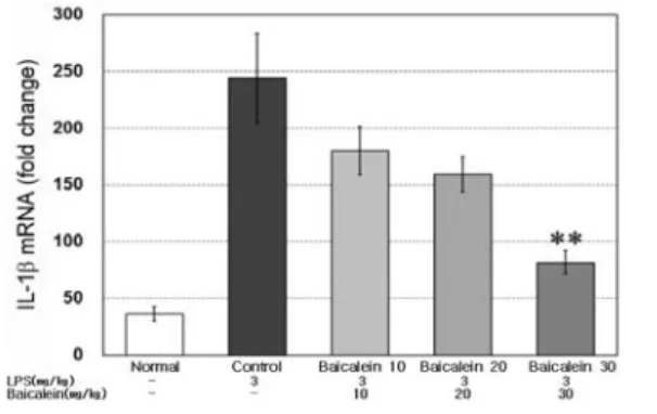
2) IL-1β mRNA 발현의 변화
염증유발 cytokine의 일종인 IL-1β mRNA 발현을 real-time PCR 방법으로 측정한 결과, Normal군은 36.4±6.7 배 이었으며, Control군은 발현이 244.0±39.5 배 이였다. Baicalein 10군은 217.9±21.3 배, Baicalein 20군
은 159.5±15.3 배, Baicalein 30군은 81.7±10.2 배로 Control군에 비하여 모든 군에서 증가를 억제하였으나, Baicalein 30군에서만 p<0.01의 유의성 있는 IL-1β mRNA 발현의 감소를 나타내었다 (Fig. 5).
Fig. 5. Effect of baicalein on IL-1β mRNA in the LPS-treated mouse brain tissue. LPS increases brain IL-1β mRNA, while baicalein treatment attenuated brain IL-1β mRNA at 30 ㎎/㎏ administration. Data are represented by mean ± SEM (n=6 in each group). Statistical significances are based on comparison to the Control group (**, p<0.01).
3) IL-6 mRNA 발현의 변화
염증유발 cytokine의 일종인 IL-6 mRNA 발현을 real-time PCR 방법으로 측정한 결과, Normal군은 39.7±5.0 배 이었으며, Control군은 139.1±18.1 배 이었 다. Baicalein 10군은 100.5±9.6 배, Baicalein 20군은 88.6±11.8 배, Baicalein 30군은 62.2±12.6 배로 Control 군에 비하여 Baicalein 20군은 p<0.05, Baicalein 30군은 p<0.01의 유의성 있는 IL-6 mRNA 발현의 감소를 나타 내었다(Fig. 6).
Fig. 6. Effect of baicalein on IL-6 mRNA in the LPS-treated mouse brain tissue. LPS increases brain IL-6 mRNA, while baicalein treatment attenuated brain IL-6 mRNA at 20 and 30
㎎/㎏ administration. Data are represented by mean ± SEM (n=6 in each group). Statistical significances are based on comparison to the Control group (*, p<0.05; **, p<0.01).
4) COX-2 mRNA 발현의 변화
뇌조직에서 염증 COX-2 mRNA 발현을 real-time PCR
방법으로 측정한 결과, Normal군은 1.26±0.19 배 이었으며,
Control군은 4.86±0.71 배 이였다. Baicalein 10군은
3.44±0.40 배, Baicalein 20군은 3.05±0.58 배, Baicalein
30군은 2.49±0.35 배로 Control군에 비하여 모든 군에서 증
가를 억제하였으나, Baicalein 30군에서만 p<0.05의 유의성 있는 COX-2 mRNA 발현의 감소를 나타내었다 (Fig. 7).
Fig. 7. Effect of baicalein on COX-2 mRNA in the LPS-treated mouse brain tissue. LPS increases brain COX-2 mRNA, while baicalein treatment attenuated brain COX-2 mRNA at 30 ㎎/㎏ administration. Data are represented by mean ± SEM (n=6 in each group). Statistical significances are based on comparison to the Control group (*, p<0.05).
2. 뇌조직의 microglia 활성화에 미치는 영향
1) Iba1 단백질 발현의 변화뇌조직에서 microglia 활성의 표지단백질인 Iba1 발현을 western blotting 방법으로 측정한 결과, Normal군의 100%에 대하여, Control군은 LPS처리에 의하여 Iba1 단 백질 발현이 142.1±5.5% 로 증가하였다. Baicalein을 30
㎎/㎏ 처리한 군은 121.2±5.4%로 Control군에 비하여 p<0.05의 유의성 있는 Iba1 단백질 발현의 억제가 관찰되 었다 (Fig. 8).
Fig. 8. Effect of baicalein on Iba1 expression in the LPS-treated mouse brain. Representative Western blots illustrate differences in the bands of Iba1 (A). LPS up-regulates Iba1 expression in brain tissue, while baicalein treatment attenuated up-regulation of Iba1 expression at dose of 30 ㎎/㎏ administration (B). Data are represented by mean
± SEM (n=6 in each group). Statistical significance is based on comparison to the Control group (*, p<0.05).
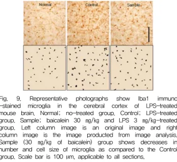
2) 대뇌피질에서 microglia 수와 크기의 변화
대뇌피질 부위에서 microglia의 표지단백질인 Iba1에 양 성반응을 나타낸 microglia의 수를 측정한 결과, Normal군
은 53.7±4.6 개/10
5µm
2이었으며, Control군은 82.3±6.3 개/10
5µm
2로 증가하였다. Baicalein을 30 ㎎/㎏ 처리한 Sample군은 61.3±6.2 개/10
5µm
2로 Control군에 비하여 p<0.05의 유의성 있는 microglia 수의 감소가 관찰되었다.
또한 microglia의 크기를 관찰한 결과 Normal군은 51.09±0.63 µm
2에 대하여 Control군은 61.15±0.89 µm
2로 증가하였다. Baicalein을 30 ㎎/㎏ 처리한 Sample군은 57.63±0.86 µm
2로 Control군에 비하여 p<0.05의 유의성 있는 microglia 크기의 감소가 관찰되었다 (Fig. 9).
Fig. 9. Representative photographs show Iba1 immuno -stained microglia in the cerebral cortex of LPS-treated mouse brain. Normal; no-treated group. Control; LPS-treated group. Sample; baicalein 30 ㎎/㎏ and LPS 3 ㎎/㎏-treated group. Left column image is an original image and right column image is the image producted from image analysis.
Sample (30 ㎎/㎏ of baicalein) group shows decreases in number and cell size of microglia as compared to the Control group. Scale bar is 100 µm, applicable to all sections.
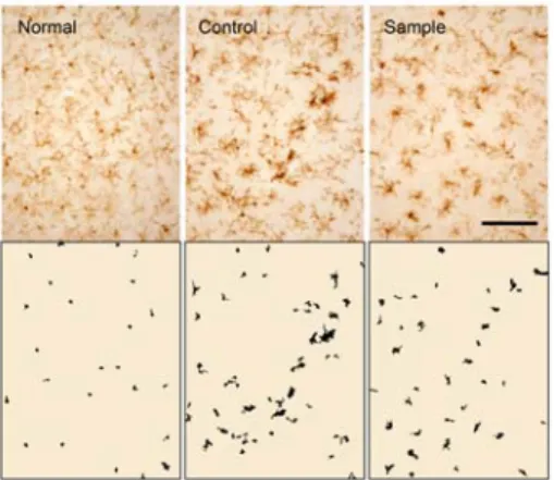
3) 해마에서 microglia 면적비율의 변화
치상회 부위를 중심으로 한 해마에서는 조직 구조와 Iba1에 대한 면역염색 결과의 특성상 microglia의 수와 크 기를 측정하지 못하고 microglia의 면적비율을 측정한 결 과, Normal군의 2.12±0.14%이었으며, Control군은 LPS처 리에 의하여 microglia의 면적비율이 4.91±0.38%로 증가 하였다. Baicalein을 30 ㎎/㎏ 처리한 Sample군은 3.34±0.43%로 Control군에 비하여 p<0.05의 유의성 있는 microglia 면적비율의 감소가 관찰되었다 (Fig. 10).
Fig. 10. Representative photographs show Iba1 immuno -stained microglia in the hippocampus of LPS-treated mouse brain. Upper column image is a representative original image and lower column image is the image producted from image analysis. Sample (30 ㎎/㎏ of baicalein) group shows a decrease in microglia activation as compared to the Control group. Scale bar is 100 µm, applicable to all sections.
4) 시상하부에서 microglia 수와 크기의 변화
시상하부 에서 microglia의 표지단백질인 Iba1에 양성반응 을 나타낸 microglia의 수를 측정한 결과, Normal군은 26.0±1.6 개/10
5µm
2이었으며, Control군은 46.3±2.4 개 /10
5µm
2로 증가하였다. Baicalein을 30 ㎎/㎏ 처리한 Sample군은 36.5±3.2 개/10
5µm
2로 Control군에 비하여 p<0.05의 유의성 있는 microglia 수의 감소가 관찰되었다.
또한 microglia의 크기를 관찰한 결과 Normal군은 44.20±1.88 µm
2이었으며, Control군은 96.47±4.14 µm
2로 크게 증가하였다. Baicalein을 30 ㎎/㎏ 처리한 Sample 군은 79.09±2.81 µm
2로 Control군에 비하여 p<0.01의 유 의성 있는 microglia 크기의 감소가 관찰되었다 (Fig. 11).
Fig. 11. Representative photographs show Iba1 immuno -stained microglia in the hypothalamic region of LPS-treated mouse brain.
Upper column image is a representative original image and lower column image is the image producted from image analysis.
Sample (30 ㎎/㎏ of baicalein) group shows decreases in number and cell size of microglia as compared to the Control group.
Scale bar is 100 µm, applicable to all sections.
고 찰
凊熱燥濕과 瀉火解毒을 하는 黃芩
6)의 주요성분은 염증, 암, 바이러스관련 질병치료 사용하는 flavonoid계 화합물들이 며, 그 중 대표적인 성분인 baicalein은 H
2O
2에 의한 신경세 포 손상을 보호하며
11), O
2와 glucose의 박탈로 유발되는 해 마신경세포의 손상을 보호하고
17), 1 - Methyl - 4 - phenyl - 1,2,3,6 - tetra - hydropyridine (MPTP) 이나 6 - hydroxydopamine (6-OHDA)로 유발되는 dopamine 신경 세포의 손상을 보호한다고 하였다
18,19). 또한 산화적 손상과 중추신경계 염증을 억제하며 기억력을 개선한다고 하였다
20).
LPS는 Gram-negative 박테리아의 세포벽 구성성분으로 endotoxin으로 작용하며
21), 중추신경계 조직 내의 microglia, 성상아교세포, 신경세포 및 뇌혈관세포 등 다양한 세포들에 위치한 tolk-like receptors (TLRs)에 의해 인지된 다
22). TLRs는 세포내 신호전달체계인 nuclear factor-kappa B (NF-kB) 활동을 증강시키는 기전에 의해 microglia를 활성화시키고, 이어서 염증유발 cytokine과 adhesion molecules, nitric oxide, prostaglandin 등의 분 비에 의해 뇌조직의 염증을 확산시키고 신경세포를 손상시킨 다
22,23).
TNF-α는 LPS 자극으로 염증반응을 일으키는 중요한 매
개체로 미생물 감염이나 조직손상 시에 나타나는
pro-inflammatory cytokine이다
24). Cytokines 중 TNF- α, IL-1, IL-6은 염증반응에 직접적으로 관여하며, 특히 TNF-α는 뇌조직의 신경세포 괴사, 염증반응 확산 등 다양 한 병리기전에 개입 한다
25). 따라서 본 실험은 뇌조직에서 TNF-α mRNA 발현을 real-time PCR을 통해 관찰한 결 과, baicalein 30 ㎎/㎏ Sample군에서 유의성 있는 감소를 나타내었다.
IL-1β는 LPS 독성의 염증매개인자로 알려진 염증유발 cytokine으로써
26), 혈액뇌장벽(Blood-Brain Barrier, BBB) 의 손상에 영향을 미치고 외상 후 세포사멸 및 괴사성세포사 멸을 유도하는 것으로 알려져 있다
27). 따라서 본 실험에서 IL-1β mRNA 발현을 real-time PCR을 통해 관찰한 결과, baicalein 30 ㎎/㎏ Sample군에서 유의성 있는 감소를 나타 내었다.
IL-6은 염증유발 및 염증억제의 모든 역할을 하는 cytokine으로 염증 시에는 과잉생산 된다
28). 또한 높은 농도 의 IL-6와 같은 cytokine의 생성과 확산은 microglia를 활 성화시켜 신경세포 사망을 이르게 하는 궁극적인 촉매자이기 도 하다
27). 따라서 본 실험에서 IL-6 mRNA 발현을 real-time PCR을 통해 관찰한 결과, baicalein 20, 30 ㎎/
㎏ Sample군에서 유의성 있는 감소를 나타내었다.
COX는 COX-1과 COX-2로 분류되는데, COX-1은 정상 적인 상황에서 발현하여 신체의 항상성 유지에 관여한다. 반 면 COX-2는 염증반응의 진행과정에 중요한 역할을 하며 면 역반응 시 유사분열 촉진물질(mitogen), cytokine, 및 염증 매개인자에 의해 손상부위에서 발현되며
29), 중추신경계 손상 시 과다 생성되는 것으로 보고되고 있다
30). 따라서 본 실험에 서 COX-2 mRNA 발현을 real-time PCR을 통해 관찰한 결과, baicalein 30 ㎎/㎏ Sample군에서 유의성 있는 감소 를 나타내었다.
Baicalein의 항염증효능 작용기전과 관련된 이전의 연구보 고에 의하면, baicalein은 NF-κB의 불활성화 및 IκB-α 인산화/붕괴 억제에 의해 염증 cytokine인 IL-1β, IL-6의 발현을 억제하였으며
31), C/EBPβ DNA 바인딩 활동을 차단 함으로써 COX-2 유전자 발현을 억제하였고
32), FOXO를 활성 화 시켜 방사선으로 유도된 염증 반응을 억제한다고 하였다
33). 또한 baicalein은 외상성 뇌손상 대뇌피질에서 염증 cytokine 인 TNF-α, IL-1β, IL-6의 발현을 억제하였으며 상해 후 조직학적 및 기능적으로도 개선된 결과가 있었다고 보고되었다
27)
. 이러한 연구결과들을 종합적으로 볼 때, 본 연구의 결과는 baicalein이 염증유발 cytokine인 TNF-α, IL-1β, IL-6 및 COX-2 mRNA 발현을 억제하는 기전을 통해 중추신경계 염증을 억제하는 작용이 있는 것으로 생각된다.
Microglia는 정상적인 상태에서 가늘고 긴 돌기들을 갖는 형태로 뇌와 척수의 넓은 지역을 커버 하지만 뇌조직 손상이 나 면역성 자극 등으로 세포체가 커지고 굵고 짧은 돌기들을 갖는 형태로 변하며, 더 활성화되면 운동성과 포식 작용을 가 진 원형의 세포로 변형 되어
23)집중적으로 손상지역의 사멸세 포와 일차구심성말초신경의 퇴화 잔해를 제거하여
34)뇌의 항 상성을 유지하는데 기여한다. 그러나 microglia가 뇌손상과 감염 및 염증반응 등의 자극에 의해 손상된 BBB를 통하여
면역계 세포들이 중추신경계로 침입하면, 활성화된
microglial cell들이 TNF-α, IL-1 및 IL-6 등의 염증유발 cytokines를 과다하게 생성하게 되고 이로 인하여 뇌에서는 cytokine homeostasis가 파괴되어 중추신경계의 염증 반응 을 일으킨다
35,36). 따라서 본 실험에서 baicalein이 염증유발 cytokine의 공급원인 microglia의 활성화에 미치는 영향을 알아보기 위해 microglia 활성화 표지 단백질인 Iba1을 western blot 방법과 면역조직화학염색을 통해 살펴본 바, 30 ㎎/㎏를 처리한 Sample군에서 Iba1 단백질의 발현이 유 의성 있게 억제되었으며, 대뇌피질과 시상하부에서 microglia 의 수와 크기가 모두 유의성 있게 감소되었고, 해마에서도 유 의성 있는 microglia 면적비율의 감소가 관찰되었다.
이상의 결과들을 종합해보면 baicalein은 TNF-α, IL-1 β, IL-6 같은 염증매개성 cytokine 및 COX-2 mRNA 발 현을 억제하고 microglia의 활성을 억제시켜 뇌신경보호에 효과가 있으므로 중추신경계염증 치료제로 개발 될 수 있는 가능성을 가지고 있다고 생각된다.
결 론
Mouse의 뇌조직에서 염증유발 cytokine과 microglia의 활성화를 통해 중추신경계염증에 대한 baicalein의 신경보호 작용을 살펴보았다. LPS 3 ㎎/㎏을 복강주사 1시간 전에 C57BL/6 생쥐에 baicalein 10, 20, 30 ㎎/㎏을 용량별로 경구투여 하였다. LPS 복강주사 4시간 후 뇌조직에서 염증유 발 cytokine인 TNF-α, IL-1β, IL-6과 COX-2 mRNA 발현 및 대뇌피질, 시상하부 및 해마에서 microglia의 활성에 미치는 영향을 관찰한 결과 다음과 같다.
1. Baicalein 30 ㎎/㎏을 투여한 군은 대조군에 비해 유의 성 있는 TNF-α mRNA 발현의 감소를 나타내었다.
2 Baicalein 30 ㎎/㎏을 투여한 군은 대조군에 비해 유의 성 있는 IL-1β mRNA 발현의 감소를 나타내었다.
3. Baicalein 20과 30 ㎎/㎏을 투여한 군은 대조군에 비 해 유의성 있는 IL-6 mRNA 발현의 감소가 관찰되었 다.
4. Baicalein 30 ㎎/㎏을 투여한 군은 대조군에 비해 유의 한 COX-2 mRNA 발현의 감소를 나타내었다.
5. Baicalein 30 ㎎/㎏ Sample군은 대조군에 비해 유의 성 있는 Iba1 단백질 발현의 억제가 관찰되었다.
6. 대뇌피질과 시상하부에서 baicalein 30 ㎎/㎏ Sample 군은 대조군에 비해 유의성 있는 microglia 수와 크기 의 감소가 관찰되었다.
7. 해마에서 baicalein 30 ㎎/㎏ 처리한 Sample군은 대조 군에 비해 유의성 있는 microglia 면적비율의 감소가 관찰되었다.
이러한 결과들로 보아 baicalein이 중추신경계염증 억제에 효능이 있다고 생각된다.
감사의 글
이 연구는 2013년도 가천대학교 지원(GCU-2013-R080) 에 의한 결과임.
참고문헌