One-visit Apexification Using MTA and Reattachment of a Crown-root Fractured Tooth with Severe Coronal Damage: A Case Report
Youngjun Park
1, Jewoo Lee
2, Jiyoung Ra
21
Department of Pediatric Dentistry, College of Dentistry, Wonkwang University
2
Department of Pediatric Dentistry and Dental Research Institute, College of Dentistry, Wonkwang University
In dental trauma, reattachment of the original tooth fragment improves the reproduction of original tooth shape, texture, color, and radiolucency; thus, it provides good aesthetics.
A 9-year-old boy was referred due to complicated crown-root fracture of the maxillary right central incisor. Although it had poor prognosis due to severe coronal damage and subcrestal fracture, reattachment of the tooth fragment was chosen due to the patient’s age. One-visit apexification with mineral trioxide aggregate (MTA) was performed, followed by osteotomy and reattachment of the tooth fragment with post placement.
Regular observation revealed no clinical signs or symptoms and no radiologic complications.
Key words : Crown-root fracture, Dental trauma, Fragment reattachment, Fiber post Abstract
Corresponding author : Jiyoung Ra
Department of Pediatric Dentistry, College of Dentistry, Wonkwang University, 895 Muwang-ro, Iksan, Jeollabuk-do, 54538, Korea Tel: +82-63-580-6633 / Fax: +82-63-858-2957 / E-mail: [email protected]
Received July 24, 2018 / Revised October 8, 2018 / Accepted August 13, 2018
Ⅰ. Introduction
Crown-root fractures, defined as fractures involving enamel, dentin, and cementum, are classified as uncomplicated or complicated depending on pulpal involvement. These crown- root fractures comprise 5% of injuries affecting permanent dentition[1]. Complicated crown-root fractures present both endodontic and restorative problems due to pulpal exposure and fracture lines extending subgingivally[2].
Restorative treatment options for crown-root fractures are determined by the degree of subgingival extension; this includes restoration after surgical exposure of the fracture surface, restoration after orthodontic extrusion, or surgical ex- trusion of apical fragment and intentional replantation[1,3-6].
However, extraction or decoronation is indicated if the fracture line follows the long axis of the tooth, or if the coronal frag- ment comprises more than one third of the root[1].
Reattachment of the tooth fragment serves as a transitional treatment alternative until an age when gingival margin con- tours are relatively stable[1]. This technique can improve the reproduction of original tooth shape, texture, radiolucency, and function; further, it can shorten the treatment time[5,7].
Although this procedure requires strict moisture control, suc- cessful cases have been reported in crown-root fracture[7-10].
In extensive tooth fractures, reattachment of the tooth frag-
ment is difficult to perform[3], but reattachment with a post
for inner reinforcement has been reported[11]. In this situation,
the space provided by the pulp chamber is used as the space
for the post.
Apexification is needed to place the post when trauma oc- curs in immature permanent teeth. Traditional apexification with calcium hydroxide has a disadvantage in that this proce- dure demands time and periodic material changes. In recent years, mineral trioxide aggregate (MTA) has been introduced.
Importantly, MTA has a fast setting time, good biological and physical properties, and is less sensitive to moisture[12].
This report describes the treatment procedure and results of reattachment of a crown-root fractured tooth with severe coronal damage, including one-visit apexification with MTA and intracanal-post reinforcement.
Ⅱ. Case Report
A 9-year-old boy visited the Department of Pediatric Den- tistry, Wonkwang University, with the chief complaint of injury to the upper front tooth after trauma from a baseball bat. The maxillary right central incisor was fractured, and the maxil- lary right lateral incisor was completely avulsed (Fig. 1). The avulsed tooth could not be found in the injured area. The pa- tient’s medical history was insignificant and extra-oral exami- nation was normal.
The intra-oral examination revealed a complicated crown- root fracture in the maxillary right central incisor. The radio- graphic examination revealed an incomplete root apex without alveolar bone fracture, root fracture, or dislocation. The frac- tured coronal fragment of the maxillary right central incisor was gently removed. The tooth remnant exhibited no mobility.
The maxillary right central incisor had a poor prognosis due
to severe coronal damage, and the parents were notified of the possibility of extraction or decoronation. However, consid- ering the age of the patient and avulsion of the maxillary right lateral incisor, reattachment of the tooth fragment was chosen despite potential limitations of the treatment.
On the day of the trauma, pulp extirpation and working- length determination were performed under 2% lidocaine buccal infiltration (Fig. 2A). Subsequently, apexification was performed with MTA (ProRoot MTA, Dentsply Tulsa Dental, Johnson City, TN, USA). MTA was filled to a level of apical 5.0 mm. Temporary filling with cotton and caviton was performed to wait for the final setting of the MTA (Fig. 2B). The coronal fragment was stored in saline solution for reattachment. CT images were taken for extraction of palatally impacted me- siodens (Fig. 3).
Fig. 1. Complicated crown-root fracture of the maxillary central incisor detected during initial radiographic exam.
Fig. 2. (A) Working-length determination. (B) Apexification was performed with MTA.
Fig. 3. The fracture line and MTA plug of the maxillary right
central incisor were observed on CT images.
After 10 days, the tooth remnant was buried in the gingiva.
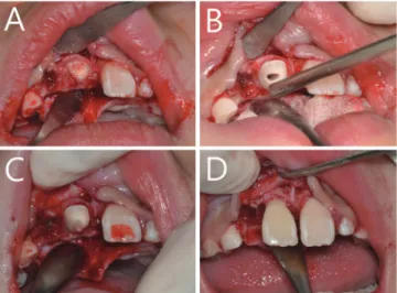
Under 2% lidocaine nasopalatine nerve block and buccal in- filtration, buccal and palatal flaps were elevated, and the root portion was exposed (Fig. 4A). Osteotomy was performed to completely expose the fracture line under the alveolar bone (Fig. 4B). The caviton was removed and the setting of the MTA was confirmed. The fiber post (FRC Postec Plus, Ivoclar-Viva- dent AG, Schaan, Liechtenstein) was then placed in the root canal and confirmed to fit with the coronal fragment.
In order to eliminate moisture contamination, rubber dams or gingival cord packing were recommended in the bonding process. However, in this case, there was no undercut to main- tain the rubber dam clamp, so cotton and suction tips were used to prevent blood contamination.
The tooth remnant was acid-etched for 15 seconds with 37% phosphoric acid gel (DenFil™ Etchant-37, Vericom, Chun- cheon, Korea), then washed with air-water spray and dried.
Next, a conventional 2-bottle adhesive system (Scotchbond MultiPurpose Plus, 3M ESPE, St. Paul, MN, USA) was applied and light cured for 20 seconds. The root canal was filled with dual-cured resin (LuxaCore Z-dual, DMG, Hamburg, Germany), the post was placed, and the remnant was polymerized (Fig.
4C).
The crown fragment was treated in the same manner as the root, and reattachment was performed with a flowable resin (Filtek Z350 XT, 3M ESPE, St. Paul, MN, USA). Excess cement was removed prior to polymerization. Because the attachment parts were not completely matched, they were reinforced with resin and polished with an ET bur to achieve a smooth surface (Fig. 4D). Sutures were then applied. During the operation, the palatal impacted supernumerary tooth was extracted. A peri- apical radiograph was taken after surgery (Fig. 5A).
One week later, sutures were removed and follow-up was performed every 3 months. Pocket depth measurement and periapical radiography were performed at admission. A pocket depth of 3.0 - 4.0 mm was observed on the palatal side;
Fig. 4. The reattachment procedure. (A) Flaps were elevated and the root portion was exposed. (B) Osteotomy was performed. (C) FRC post was placed with dual-cured resin.
(D) Reattachment of coronal fragment and polishing were performed.
Fig. 5. (A) Radiograph following reattachment. (B, C, D, E) Radiographs obtained at 2-, 5-, 11-, and 23-month follow-ups. (F, G,
H) Clinical view at 5-, 11-, and 23-month follow-ups.
bleeding on probing was not observed. Plaque management and tooth brushing instruction were performed as needed.
Follow-up at 23 months showed acceptable periodontal health, no signs of ankylosis or poor aesthetics, and normal function.
Notably, there were no pathological findings in the apical root (Fig. 5, 6).
Ⅲ. Discussion
Crown-root fracture is a relatively common trauma to the maxillary anterior teeth in children[13]. Because trauma to oral structures has a significant psychological impact on children, careful treatment approaches are needed[8].
In subgingival crown-root fractures, treatment is often chal- lenging because the fracture line invades the biologic width.
The biologic width is defined as the junctional epithelium and supracrestal connective tissue attachment surrounding each tooth. The mean dimensions of the biologic width range from 1.5 to 2.7 mm. If the margins of the restoration invade this bi- ologic width, increased plaque accumulation occurs, potentially causing more severe gingival inflammation. This inflammation may be followed by periodontal destruction with increased pocket depth, loss of attachment, and gingival recession[14].
Therefore, root recontouring, gingivectomy and osteotomy, orthodontic extrusion, or surgical extrusion are proposed to secure the biologic width. If the decision is made to reattach the tooth fragment, gingivectomy and osteotomy are indi- cated[10].
The gingivectomy and osteotomy procedure decreases the crown-root ratio, which is not indicated in the aesthetic area.
However, this gingivectomy and osteotomy procedure is ad- vantageous in that it is simple and immediate restoration is possible[15]. In this case, reattachment of the tooth fragment was selected because of the young age of the patient, as well as because the maxillary lateral incisor was avulsed and the tooth fragment was well-preserved. Thus, flap surgery with osteotomy was performed to secure complete isolation and biologic width of the operative field.
The reattachment of a tooth fragment can be performed quickly, requiring less cooperation of the patient. Such reat- tachment can improve the reproduction of tooth color, shape, and contour, increase the patient’s self-esteem, and provide a better biological surface for periodontal attachment[2,3,7].
Adhesive agents can also provide sufficient bonding strength to withstand the slow loading from masticatory stresses[16].
Moreover, when comparing treatment satisfaction of parents of child patients, satisfaction with reattachment of the tooth fragment is higher than that of resin restoration[11]. Therefore, if an intact fragment is available, its reattachment will be the most functional and aesthetic treatment option[8,17].
When extensive tooth fractures are present, intracanal rein- forcement with a post may be needed to reattach the tooth fragment. Posts provide greater retention and stability of the tooth fragment and distribute the stress along the root. A va- riety of posts have been proposed to support the fragments.
Fiber posts are often used because of the advantage of a modulus of elasticity similar to that of dentin[13].
Although pulpotomy is reportedly successful in completing
the root formation in trauma with pulp involvement[2], apexi-
fication is required for post placement and coronal restora-
Fig. 6. Because cystic change of both maxillary canines was observed at 19 months, CT images were taken. These showed
acceptable periodontal health and no pathological findings in the apical root of the maxillary right central incisor.
tion. In the past, apexification was performed by using calcium hydroxide in the root canal to induce formation of hard tissue and apical closure, followed by filling with gutta-percha cone.
This procedure demands time, periodic material replacement, and can create problems with the quality of the induced hard tissue, particularly with respect to material weakening the den- tin[12,18].
One-visit apexification was proposed to place the MTA at the apical 5.0 mm to avoid root fracture due to dentin weak- ness. The fast setting time of the MTA enabled immediate crown restoration. A study by Simon et al .[19] concluded that one-visit apexification with MTA is a predictable and reproduc- ible clinical procedure through adequate follow-up. The au- thors of the prior study suggested that this technique may be an alternative to the use of calcium hydroxide.
Lin et al .[20] reported that MTA has a similar success rate to that of calcium hydroxide in the apexification of immature per- manent teeth. Moreover, MTA can achieve higher success rates, because it is easy to obtain patient cooperation with such a short treatment time. MTA has been used as a promising suc- cessor to replace calcium hydroxide for pulpal and periodontal healing complications after trauma[18].
In this case, one-visit apexification with MTA was performed on the day of trauma. Then, using the space of the pulp cham- ber, a post was placed in the root canal and reattachment of the tooth fragment was possible in a short period of time.
This case was limited because the biologic width on the palatal side was not fully established. In principle, in a case of subcrestal crown-root fracture, restoration after orthodontic extrusion or surgical extrusion of the apical fragment is in- dicated. To compensate for this limitation, pocket depth was measured at each visit to assess periodontal health; rigorous plaque management and tooth brushing instruction were also performed. Long-term prognostic evaluation of periodontal health will be needed.
In addition, ankylosis is infrequently reported when MTA is placed in the root perforation area in animal studies[21]. The occurrence of ankylosis of the maxillary right central incisor should be evaluated periodically.
Ⅳ. Summary
In dental trauma, reattachment of the tooth fragment im- proves the reproduction of original tooth shape, texture, color, and radiolucency; thus, it provides good aesthetics. However,
this technique cannot be considered a durable procedure for the management of extensive fractures.
The findings from this case suggest that good preservation of a tooth fragment, combined with the ability to perform flap surgery with osteotomy, may enable reattachment of the tooth fragment with post placement. Moreover, this approach can be a viable treatment option for complicated crown-root frac- tures, despite the occurrence of severe coronal damage. If the tooth has an incomplete root, one-visit apexification with MTA can be considered.
References
1. Andreasen JO, Andreasen FM, Andreasen L : Textbook and Color Atlas of Traumatic Injuries to the Teeth, 4th ed.
Munksgaard, 314-336, 2013.
2. Eden E, Yanar SC, Sönmez S : Reattachment of subgingivally fractured central incisor with an open apex. Dent Trauma- tol , 23:184-189, 2007.
3. Saito CT, Guskuma MH, Panzarini SR, et al . : Management of a complicated crown-root fracture using adhesive frag- ment reattachment and orthodontic extrusion. Dent Trau- matol , 25:541-544, 2009.
4. Calişkan MK, Türkün M, Gomel M : Surgical extrusion of crown-root-fractured teeth: a clinical review. Int Endod J , 32:146-151, 1999.
5. Dogan MC, Akgun EO, Yoldas HO : Adhesive tooth frag- ment reattachment with intentional replantation: 36-month follow-up. Dent Traumatol , 29:238-242, 2013.
6. Wang Z, Heffernan M, Vann WF Jr : Management of a com- plicated crown-root fracture in a young permanent incisor using intentional replantation. Dent Traumatol , 24:100-103, 2008.
7. Oz IA, Haytaç MC, Toroglu MS : Multidisciplinary approach to the rehabilitation of a crown-root fracture with original fragment for immediate esthetics: a case report with 4-year follow-up. Dent Traumatol , 22:48-52, 2006.
8. Chaugule V, Bhat C, Patil V, H Mithiborwala S : Reattach- ment of a vertical complicated subgingival crown root frac- ture in a 10-year old child: a case report. Int J Clin Pediatr Dent , 2:53-59, 2009.
9. Grossmann Y, Araúz-Dutari J, Sadan A, et al . : A conserva- tive approach for the management of a crown-root frac- ture. Quintessence Int , 37:753-759, 2006.
10. Baratieri LN, Monteiro Júnior S, Cardoso AC, de Melo Filho
JC : Coronal fracture with invasion of the biologic width: a case report. Quintessence Int , 24:85-91, 1993.
11. de Alcântara CE, Corrêa-Faria P, Vasconcellos WA, Ramos- Jorge ML : Combined technique with dentin post reinforce- ment and original fragment reattachment for the esthetic recovery of a fractured anterior tooth: a case report. Dent Traumatol , 26:447-450, 2010.
12. Bramante CM, Menezes R, Letra A, et al . : Use of MTA and intracanal post reinforcement in a horizontally fractured tooth: a case report. Dent Traumatol , 22:275-278, 2006.
13. Wang J, Li M : Multidisciplinary treatment of a complicated crown-root fracture. Pediatr Dent , 32:250-254, 2010.
14. Schmidt JC, Sahrmann P, Walter C, et al . : Biologic width dimensions-a systematic review. J Clin Periodontol , 40:493- 504, 2013.
15. Fidel SR, Fidel-Junior RA, Fidel RA, et al . : Clinical manage- ment of a complicated crown-root fracture: a case report.
Braz Dent J , 22:258-262, 2011.
16. Murchison DF, Burke FJ, Worthington RB : Incisal edge re- attachment: indications for use and clinical technique. Br Dent J , 186:614-619, 1999.
17. Martos J, Pinto KVA, Miguelis TMF, Xavier CB : Manage- ment of an uncomplicated crown fracture by reattach- ing the fractured fragment-Case report. Dent Traumatol, 33:485-489, 2017.
18. Bakland LK, Andreasen JO : Will mineral trioxide aggregate replace calcium hydroxide in treating pulpal and periodon- tal healing complications subsequent to dental trauma? A review. Dent Traumatol, 28:25-32, 2012.
19. Simon S, Rilliard F, Berdal A, Machtou P : The use of min- eral trioxide aggregate in one-visit apexification treatment:
a prospective study. Int Endod J , 40:186-197, 2007.
20. Lin JC, Lu JX, Ling JQ, et al . : Comparison of mineral triox- ide aggregate and calcium hydroxide for apexification of immature permanent teeth: A systematic review and meta- analysis. J Formos Med Assoc , 115:523-530, 2016.
21. Katsamakis S, Slot DE, Van der Sluis LW, Van der Weijden
F : Histological responses of the periodontium to MTA: a
systematic review. J Clin Periodontol , 40:334-344, 2013.
국문초록
심한 치관 손상이 발생한 치관-치근 파절 치아의 일회 내원 치근관형성술 및 파절편 재부착 : 증례 보고
박영준
1전공의 ㆍ이제우
2교수 ㆍ라지영
2교수
1
원광대학교 부속치과병원 소아치과
2
원광대학교 치과대학 소아치과학교실 및 치의학연구소
치아의 외상에서 파절편의 재부착은 본래 치아의 형태, 질감, 색, 방사선투과성을 재현하여 우수한 심미성을 제공한다.
9세의 남자 환자가 상악 우측 중절치의 복잡 치관-치근 파절로 내원하였다. 심한 치관 손상과 골 연하로 연장된 파절로 인해 예후가 불량했지만 환자의 나이를 고려하여 치아 파절편을 재부착하기로 결정하였다. Mineral trioxide aggregate(MTA)를 이용한 일회 내원 치근관형성술 후에 골절제술과 포스트 식립을 동반하여 치아 파절편을 재부착하였다.
정기적인 관찰결과 임상 및 방사선학적으로 양호한 결과를 나타냈기에 보고하고자 한다.