• 검색 결과가 없습니다.

Neuroendocrine Differentiation in Acquired Resistance to Epidermal Growth Factor Receptor Tyrosine Kinase Inhibitor

N/A
N/A
Protected

Academic year: 2021

Share "Neuroendocrine Differentiation in Acquired Resistance to Epidermal Growth Factor Receptor Tyrosine Kinase Inhibitor"

Copied!
9
0
0

로드 중.... (전체 텍스트 보기)

전체 글

(1)

Neuroendocrine Differentiation in Acquired Resistance to Epidermal Growth Factor

Receptor Tyrosine Kinase Inhibitor

Youjin Chang, M.D.

1,

*, Seon Ye Kim, M.Sc.

1,

*, Yun Jung Choi, Ph.D.

1

, Kwang Sup So, M.Sc.

1

, Jin Kyung Rho, Ph.D.

1,2

, Woo Sung Kim, M.D., Ph.D.

1

, Jae Cheol Lee, M.D., Ph.D.

3

, Jin-Haeng Chung, M.D., Ph.D.

4

and Chang-Min Choi, M.D., Ph.D.

1,3

1

Department of Pulmonary and Critical Care Medicine,

2

Asan Institute for Life Sciences,

3

Department of Oncology, Asan Medical Center, University of Ulsan College of Medicine, Seoul,

4

Department of Pathology, Seoul National University Bundang Hospital, Seoul National University College of Medicine, Seongnam, Korea

Background: Small cell lung cancer (SCLC) transformation during epidermal growth factor receptor tyrosine kinase inhibitor (EGFR-TKI) treatment in lung cancer has been suggested as one of possible resistance mechanisms.

Methods: We evaluated whether SCLC transformation or neuroendocrine (NE) differentiation can be found in the cell line model. In addition, we also investigated its effect on responses to conventional chemotherapeutic drugs of the SCLC treatment.

Results: Resistant cell lines to various kinds of EGFR-TKIs such as gefitinib, erlotinib, CL-387,785 and ZD6474 with A549, PC-9 and HCC827 lung adenocarcinoma cell lines were established. Among them, two resistant cell lines, A549/GR (resistant to gefitinib) and PC-9/ZDR (resistant to ZD6474) showed increased expressions of CD56 while increased synaptophysin, Rb, p16 and poly(ADP-ribose) polymerase were found only in A549/GR in western blotting, suggesting that NE differentiation occurred in A549/GR. A549/GR cells were more sensitive to etoposide and cisplatin, chemotherapeutic drugs for SCLC, compared to parental cells. Treatment with cAMP and IBMX induced synaptophysin and chromogranin A expression in A549 cells, which also made them more sensitive to etoposide and cisplatin than parental cells. Furthermore, we found a tissue sample from a patient which showed increased expressions of CD56 and synaptophysin after development of resistance to erlotinib.

Conclusion: NE differentiation can occur during acquisition of resistance to EGFR-TKI, leading to increased chemosensitivity.

Keywords: Small Cell Lung Carcinoma; Cell Transformation, Neoplastic; Drug Resistance, Neoplasm

Copyright © 2013 The Korean Academy of Tuberculosis and Respiratory Diseases. All rights reserved.

Address for correspondence: Chang-Min Choi, M.D., Ph.D.

Department of Pulmonary and Critical Care Medicine, Asan Medical Center, College of Medicine, University of Ulsan, 88 Olympic-ro 43-gil, Songpa-gu, Seoul 138-736, Korea

Phone: 82-2-3010-5902, Fax: 82-2-3010-6968, E-mail: [email protected]

*Youjin Chang and Seon Ye Kim contributed equally to this work.

Received: May 6, 2013 Revised: May 31, 2013 Accepted: Jun. 5, 2013

cc

It is identical to the Creative Commons Attribution Non-Commercial License (http://creativecommons.org/licenses/by-nc/3.0/).

(2)

Introduction

Sensitizing epidermal growth factor receptor (EGFR) mu- tation such as G719X, deletion on exon 19, L858R or L861X in non-small cell lung cancer (NSCLC) is a strong predictive marker for EGFR-tyrosine kinase inhibitors (EGFR-TKIs)

1,2

. However, despite initial dramatic response in patients har- boring such EGFR mutations, acquired resistance eventu- ally develops in most of them

3,4

. Through enormous efforts to identify and understand resistance mechanisms, several causes and overcoming strategies have been revealed. Among them, secondary threonine-to-methionine mutation at co- don 790 in EGFR (T790M) is the most common mechanism representing almost 50% of them

5,6

. More potent irreversible EGFR-TKIs and mutant selective EGFR inhibitors have been actively investigated for the possibility of therapeutic options to overcome T790M-mediated resistance

7-9

. Bypass signals by met proto-oncogene (MET) amplification or AXL receptor tyrosine kinase (AXL) overexpression also have been sug- gested as other possible causes leading to resistance. Several MET and AXL inhibitors are currently under clinical trials or development because combined treatment with EGFR-TKIs showed excellent response in experimental models

10,11

.

In 2011, Sequist et al.

12

reported the frequency of observed EGFR-TKI resistance mechanisms by analysis of patients undergoing post-resistance biopsy at Massachusetts General Hospital. The detection rate of T790M (49%) was not differ- ent, while that of MET amplification (5%) was much lower in this cohort, compared to those of previous studies

10,13

. Inter- estingly, small cell lung cancer (SCLC) transformation from NSCLC was found in 5 patients (14%) although its original EGFR mutation was maintained in resistant tumors, and 3 of them showed the response to chemotherapy for SCLC. Con- sidering that there have been several similar case reports

14

, SCLC transformation would be one of the resistance mecha- nisms for EGFR-TKI therapy. However, it is not clear whether combined SCLC cells selected by EGFR-TKI treatment were initially present in un-biopsied parts of tumor or real SCLC transformation from NSCLC occurred in tumors of those patients. In this study, we investigated whether it would be possible to find the phenomenon of SCLC transformation or neuroendocrine (NE) differentiation in EGFR-TKI-resistant cell line models, and that could affect the response to conven- tional chemotherapeutic drugs for SCLC treatment.

Materials and Methods

1. Cell culture and reagents

The A549 and HCC827 cells were purchased from the American Type Culture Collection (Rockville, MD, USA). The PC-9 cell line was a kind gift from Dr. Kazuto Nishio (National

Cancer Center Hospital, Tokyo, Japan). All cells were cultured in RPMI-1640 (Invitrogen, Carlsbad, CA, USA) containing 10%

fetal bovine serum, 100 U/mL penicillin and 100 μg/mL strep- tomycin (Invitrogen) at 37

o

C in an atmosphere of 5% CO

2

. Gefitinib, erlotinib (reversible EGFR-TKIs) and ZD6474 (an EGFR and vascular endothelial growth factor receptor inhibi- tor) were purchased from Selleck Chemicals (Houston, TX, USA). CL-387,785 (an irreversible EGFR-TKI) was purchased from Calbiochem (San Diego, CA, USA). The 3-(4,5-dimeth- ylthiazol-2-yl)-2,5-diphenyltetrazolium bromide (MTT) solu- tion, cAMP, 3-isobutyl-1-methylxanthine (IBMX), cisplatin and etoposide were all purchased from Sigma (St. Louis, MO, USA).

2. Establishment of the EGFR-TKIs-resistant cell lines The A549/GR, PC-9/GR, PC-9/ER, and HCC827/CLR were established in our previous researches

15-17

. The PC-9/CLR and PC-9/ZDR cells were established by using the previously de- scribed method

15

. Briefly, PC-9 cells were exposed to 10 nmol/

L of CL-387,785 or ZD6474 for 72 hours in media containing 10% fetal bovine serum. Then, they were washed and cultured in drug-free media until surviving cells were 80% confluent.

These cells were re-exposed to increasing concentrations of CL-387,785 or ZD6474. Cells which could grow in the 100 nmol/L CL-387,785 or ZD6474 were obtained 6 months after initial exposure. The surviving cells were cloned, then, the CL- 387,785 and ZD6474-resistant cell lines were designated as PC-9/CLR and PC-9/ZDR, respectively. For all in vitro studies, resistant cells were cultured in drug-free media for at least 1 week to eliminate CL-387,785 and ZD6474.

3. MTT assay

Briefly, the cells were seeded onto 96-well plates overnight.

Each of drugs was added to them in a dose-dependent man- ner, then, the cells were incubated for 72 hours. The viability of cells was determined by using the MTT assay in accordance with the method described by Carmichael et al.

18

.

4. Western blot analysis

The cell lysates were prepared by using the previously de-

scribed method

19

. The membrane was probed with antibod-

ies against CD56, synaptophysin (SYP), chromogranin (Chr-A)

(Santa Cruz Biotechnology, Santa Cruz, CA, USA), p-Rb, Rb,

p16, poly(ADP-ribose) polymerase (PARP) and ß-actin (Cell

Signaling Technology, Beverly, MA, USA) as primary antibod-

ies, and then the membrane was treated with horseradish-

peroxidase-conjugated secondary antibody. The membrane

was developed using an ECL kit (Amersham Biosciences,

Piscataway, NJ, USA).

(3)

5. Immunohistochemistry analysis

The cells were grown in a chamber slide (Nalge Nunc International, Naperville, IL, USA). They stained with H&E visualized with a Nikon light microscope (Nikon, Inc., Melville, NY, USA) that had the capability of digital photography. For immunohistochemical analysis, the paraffin sections (4 μm thick) were deparaffinized with xylene, rinsed thoroughly with ethanol and then soaked in 0.03% hydrogen peroxide in methanol to inactivate the endogenous peroxidase activity.

The sections were incubated with either 10% goat serum or

10% rabbit serum, and then they were covered with the pri- mary antibodies, washed with phosphate-buffered saline and processed further with using a DAKO EnVision kit (DAKO, Los Angeles, CA, USA), as directed by the manufacturer. The color was developed with 3,3’-diaminobenzindine (DAB) that contained 0.3% H

2

O

2

. Primary antibodies against CD56 and SYP were used.

6. Induction of NE differentiation by cAMP/IBMX To induce NE differentiation by the previously described

Figure 1. The basal level of neuroendocrine marker proteins was evaluated by western blotting (A, B) and immunocytochemistry (C). The

chemosensitivity of gefitinib-resistant cells was determined by a MTT assay (D). The cells were exposed to the indicated concentrations of

etoposide or cisplatin for 72 hours (*p<0.01 compared with A549 cells, **p<0.001 compared with A549 cells). Cells stained with H&E showed

morphological changes between A549 cells and A549/GR cells (E). SYP: synaptophysin; Chr-A: chromogranin A.

(4)

method

20

, A549 cells were seeded at 50% confluency in a chamber slide. The cells were treated with 0.5 mmol/L cAMP/

IBMX combinations for 72 hours, and then they were fixed for 1 hour with methanol. Immunocytochemical staining of the cells was performed by the same procedure used for the par- affin sections.

Results

1. NE differentiation only in A549/GR cells leading to increased chemosensitivity

We investigated the change of NE markers in lung cancer cells which acquired resistance to EGFR-TKIs by performing western blotting and immunocytochemistry. PC-9/CLR and PC-9/ZDR cells were cloned with continuously exposed to increasing concentrations of CL-387,785 (IC

50

: <1 nM in the PC-9 cells and >100 nM in the PC-9/CLR; data not shown) or ZD6474 (IC

50

: 50 nM in the PC-9 cells and 500 nM in the PC-9/ZDR; data not shown), and other EGFR-TKI-resistant

cells were established in previous studies

15-17

. The expression of CD56 was increased in both the A549/GR and PC-9/ZDR cells (Figure 1A). However, the enhanced expression of SYP, Rb and p16 were found only in the A549/GR cells (Figure 1B, C). As morphological changes, the cluster of A549/GR cells became more compact and their nuclear/cytoplasmic ratios were increased. These cells were also more sensitive to etopo- side or cisplatin than parental A549 cells (Figure 1D, E). These results suggest that NE differentiation occurred in A549/GR cells.

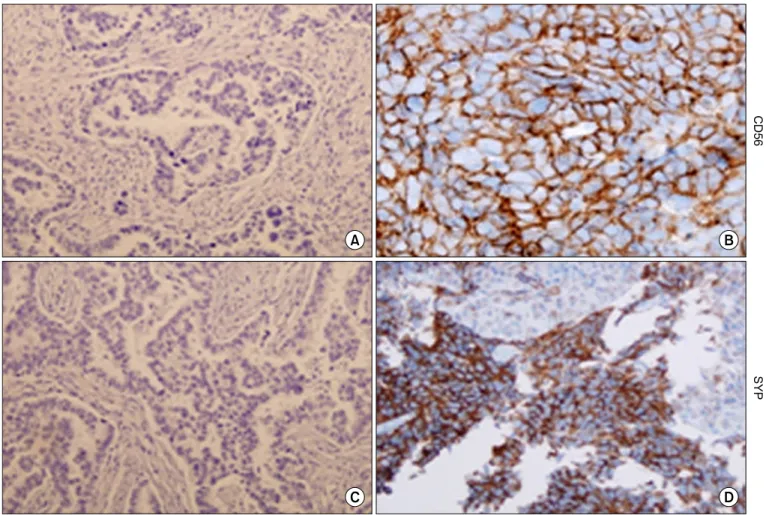
2. Increased CD56 and SYP expression in a rebiopsied tumor after development of resistance to erlotinib Rebiopsy of regrowing tumor during treatment with erlo- tinib was performed in a patient with adenocarcinoma who had initially responded well to erlotinib. A deletion muta- tion on exon 19 of EGFR gene was noted in the tissues of the regrowing tumor as well as in those of the original tumor.

Interestingly, the expression of CD56 and SYP was increased after acquiring resistance (Figure 2), which suggests that NE

Figure 2. The expression of synaptophysin (SYP) and CD56 was increased in re-biopsied tumor tissues procured after development of erlo-

tinib-resistance in a patient. (A, C) Tumor obtained before treatment with erlotinib. (B, D) Tumor after acquisition of resistance.

(5)

differentiation occurred in this patient.

3. Induced NE differentiation by cAMP and IBMX with increasing chemosensitivity

A previous study demonstrated that combined cAMP and IBMX treatment led to NE differentiation in NSCLC cells

20

. We induced NE differentiation of A549 cells by treating them with the same method. When the cells were co-treated with cAMP and IBMX, they became scattered and extended with a slen- der shape (Figure 3A). The expression of SYP was increased on the western blots and immunocytochemistry while that of Chr-A was slightly increased. However, CD56 expression was not observed (Figure 3B, C). Transformed cells obtained the increased sensitivity to etoposide and cisplatin compared to parental cells (Figure 3D).

4. Increased PARP expression in A549/GR cells without affecting to the response to chemotherapeutic drugs by its inhibition

Recently, overexpression of PARP1 and enhancer of zeste homolog 2 (EZH2) has been indentified in an integrated analysis of multiple proteins involved in intracellular signaling pathway in SCLC cell lines

21

. They showed that PARP1 was highly expressed at the mRNA and protein levels in SCLCs, and then SCLC was significantly more sensitive to PARP inhibitors than NSCLCs. Thus, they suggested that PARP in- hibition downregulated the key components of DNA repair machinery and enhanced the efficacy of chemotherapy. Con- sistent with this, PARP1 expression was increased in A549/GR cells (Figure 4A). However, the response to AZD2281, a PARP inhibitor, was not different between A549 and A549/GR cells (Figure 4B). In addition, PARP inhibition by AZD2281 did not affect the response to etoposide and cisplatin (Figure 4C).

Figure 3. Neuroendocrine (NE) differentiation was induced by treatment with 0.5 mmol/L cAMP and IBMX for 72 hours. Morphological

changes by light microscopy (A), immunocytochemistry (B) and western blots (C) indicated NE differentiation. (D) MTT assay was per-

formed to confirm whether small cell lung cancer can enhance the sensitivity against etoposide or cisplatin. The cells were treated with the

indicated doses of etoposide or cisplatin in the presence or absence of cAMP and IBMX for 72 hours. The p-values were calculated to assess

the effect of combined treatment of cAMP and IBMX (*p<0.01, **p<0.001). SYP: synaptophysin; Chr-A: chromogranin.

(6)

Discussion

This is the first study showing that NE differentiation from

NSCLC cells occurred during acquisition of resistance to

EGFR-TKI to date. In general, EGFR-TKIs don’t seem to be

effective in SCLC although some early experimental studies

Figure 4. The poly (ADP-ribose) polymerase (PARP) expression was increased in A549/GR cells, which did not affect chemosensitivity. (A)

The basal level of PARP protein was evaluated by western blotting. (B) Response to AZD2281, a PARP inhibitor, was determined by a MTT

assay. The cells were exposed to the indicated concentrations of AZD2281 for 72 hours. (C) Cells were treated with indicated doses of etopo-

side or cisplatin for 72 hours alone, AZD2281 and two drugs in combination. The viability of the cells was determined using the MTT assay.

(7)

suggest their efficacy in cell line models

22

. Only 2 of 19 pa- tients with SCLC showed stable disease while 17 patients had progressive disease by gefitinib in a phase II study

23

. However, the benefit from gefitinib therapy could be obtained if SCLC would have a sensitizing EGFR mutation although it is very rare. A Japanese never-smoker patient with SCLC responded to gefitinib which was initiated at patient’s request

24

. Deletion mutation on exon 19 (delE746-A750) was found by direct se- quencing in her tumor tissue. Zakowski et al.

25

also reported a female American case of SCLC with deletion mutation on exon 19. Erlotinib led to partial response and the response du- ration was 18 months.

However, it is too early to conclude that the EGFR mutation in SCLC is a predictive marker for EGFR-TKIs because SCLC transformation harboring EGFR mutation was found in resis- tant tumor samples after EGFR-TKI therapy. If the assertion that SCLC transformation is one of the resistant mechanisms to EGFR-TKI would be true, what’s the difference of EGFR mu- tations between EGFR-TKI-responsive and -resistant SCLC?

This question leads us to consider the possibility that SCLC transformation or NE differentiation could be an accompany- ing phenomenon occurring during acquisition of resistance, not the main cause of resistance. We expect it could be eluci- dated through further studies in near future.

Approximately 15% of NSCLCs have NE features

26

and they have been suggested to exhibit the biological characteristics similar to SCLC such as early metastasis, initial responsive- ness to chemotherapeutic drugs and expression of NE mark- ers including neuron-specific enolase, L-dopa decarboxylase, Chr-A, SYP, and cytoplasmic dense core granules. However, there have been conflicting results regarding chemosensitivity of NSCLC with NE features. Some revealed that NE differen- tiation led to chemosensitivity

27,28

while others failed to dem- onstrate any correlation with response to drugs

29-31

. Probably, this might be caused by discrepancies on how to define NE differentiation, how to interpret the results of immunohis- tochemical staining and what kinds of antibodies should be used. In our study, regardless of whether it exerts a significant role in resistance to EGFR-TKI or not, NE differentiation made transformed cells more susceptible to chemotherapeutic drugs such as etoposide and cisplatin which are currently be- ing used for treatment of SCLC, suggesting that these drugs could be the next therapeutic option.

The case reported by Morinaga raised another question whether SCLC found in resistant samples arose from the transformation of adenocarcinoma or the selection of pre- existing minor clone

14

. The first and third biopsies after treat- ment of irinotecan and cisplatin revealed adenocarcinoma, while the second biopsy at the same site in the mass in prog- ress during EGFR-TKI therapy showed SCLC in that report.

Although the biopsies were done at the same site, it seems to be very hard to prove the reality of transformation because small pieces of tissues procured by needle biopsy cannot

represent the whole nature of cancer. However, our study per- formed with cell lines firstly demonstrated that NE differentia- tion can occur during EGFR-TKI treatment.

Recently, Byers et al. investigated molecular characteristics of SCLC using an integrative proteomic and transcriptomic analysis

21

. They found that the expression of PARP1, a DNA repair protein and E2F1, co-activator, was highly increased both at the mRNA and protein levels in SCLC, and SCLC growth was inhibited by PARP1 knockdown. In our study, PARP1 expression was also increased in A549 cells with NE differentiation compared to parental cells. However, there was no difference in the response of A549/GR cells to a PARP1 inhibitor compared to parental cells. Further, the combination of AZD2281 with etoposide or cisplatin could not enhance the growth-inhibitory effect in A549/GR cells. These are clearly contrasted with results from Byers et al. suggesting that although A549/GR cells were differentiated to possess some NE features, they would not acquire the whole characteristics of SCLC. However, further studies would be required for the response to EGFR-TKI treatment of other lung cancers with NE differentiation such as large cell carcinoma or carcinoid tumor.

Although we proved that NE differentiation can occur dur- ing EGFR-TKI treatment, not by selection of pre-existing cells, established A549/GR does not seem to be clinically appropri- ate model because A549 cells do not harbor EGFR mutations.

Nevertheless, we had a similar experience in epithelial to mes- enchymal transition (EMT) by EGFR-TKI. This phenomenon was also firstly found in A549 cells with acquired resistance to gefitinib

15

, which was also not appropriate, it led to the discov- ery of EMT in EGFR-mutant lung cancer cells

16

and clinical samples

12,17

later.

In summary, NE differentiation occurred during acquisition of resistance to EGFR-TKI leading to increased chemosensi- tivity suggesting therapeutic drugs for SCLC would be useful.

However, it should be more clarified whether NE differentia- tion is the real main cause of EGFR-TKI resistance.

Acknowledgements

This work is supported by the 2012 grant from “The Korean Academy of Tuberculosis and Respiratory Diseases.”

References

1. Kris MG, Natale RB, Herbst RS, Lynch TJ Jr, Prager D, Belani CP, et al. Efficacy of gefitinib, an inhibitor of the epidermal growth factor receptor tyrosine kinase, in symptomatic pa- tients with non-small cell lung cancer: a randomized trial.

Jama 2003;290:2149-58.

2. Perez-Soler R. Phase II clinical trial data with the epidermal

(8)

growth factor receptor tyrosine kinase inhibitor erlotinib (OSI-774) in non-small-cell lung cancer. Clin Lung Cancer 2004;6 Suppl 1:S20-3.

3. Jackman D, Pao W, Riely GJ, Engelman JA, Kris MG, Janne PA, et al. Clinical definition of acquired resistance to epidermal growth factor receptor tyrosine kinase inhibitors in non- small-cell lung cancer. J Clin Oncol 2010;28:357-60.

4. Oxnard GR, Arcila ME, Chmielecki J, Ladanyi M, Miller VA, Pao W. New strategies in overcoming acquired resistance to epidermal growth factor receptor tyrosine kinase inhibitors in lung cancer. Clin Cancer Res 2011;17:5530-7.

5. Kobayashi S, Boggon TJ, Dayaram T, Janne PA, Kocher O, Meyerson M, et al. EGFR mutation and resistance of non- small-cell lung cancer to gefitinib. N Engl J Med 2005;352:786- 92.

6. Pao W, Miller VA, Politi KA, Riely GJ, Somwar R, Zakowski MF, et al. Acquired resistance of lung adenocarcinomas to ge- fitinib or erlotinib is associated with a second mutation in the EGFR kinase domain. PLoS Med 2005;2:e73.

7. Kwak EL, Sordella R, Bell DW, Godin-Heymann N, Okimoto RA, Brannigan BW, et al. Irreversible inhibitors of the EGF re- ceptor may circumvent acquired resistance to gefitinib. Proc Natl Acad Sci U S A 2005;102:7665-70.

8. Miller VA, Hirsh V, Cadranel J, Chen YM, Park K, Kim SW, et al. Afatinib versus placebo for patients with advanced, metastatic non-small-cell lung cancer after failure of erlo- tinib, gefitinib, or both, and one or two lines of chemotherapy (LUX-Lung 1): a phase 2b/3 randomised trial. Lancet Oncol 2012;13:528-38.

9. Zhou W, Ercan D, Chen L, Yun CH, Li D, Capelletti M, et al.

Novel mutant-selective EGFR kinase inhibitors against EGFR T790M. Nature 2009;462:1070-4.

10. Engelman JA, Zejnullahu K, Mitsudomi T, Song Y, Hyland C, Park JO, et al. MET amplification leads to gefitinib resis- tance in lung cancer by activating ERBB3 signaling. Science 2007;316:1039-43.

11. Zhang Z, Lee JC, Lin L, Olivas V, Au V, LaFramboise T, et al.

Activation of the AXL kinase causes resistance to EGFR- targeted therapy in lung cancer. Nat Genet 2012;44:852-60.

12. Sequist LV, Waltman BA, Dias-Santagata D, Digumarthy S, Turke AB, Fidias P, et al. Genotypic and histological evolution of lung cancers acquiring resistance to EGFR inhibitors. Sci Transl Med 2011;3:75ra26.

13. Bean J, Brennan C, Shih JY, Riely G, Viale A, Wang L, et al.

MET amplification occurs with or without T790M mutations in EGFR mutant lung tumors with acquired resistance to gefi- tinib or erlotinib. Proc Natl Acad Sci U S A 2007;104:20932-7.

14. Morinaga R, Okamoto I, Furuta K, Kawano Y, Sekijima M, Dote K, et al. Sequential occurrence of non-small cell and small cell lung cancer with the same EGFR mutation. Lung Cancer 2007;58:411-3.

15. Rho JK, Choi YJ, Lee JK, Ryoo BY, Na, II, Yang SH, et al. Epithe- lial to mesenchymal transition derived from repeated expo-

sure to gefitinib determines the sensitivity to EGFR inhibitors in A549, a non-small cell lung cancer cell line. Lung Cancer 2009;63:219-26.

16. Chung JH, Rho JK, Xu X, Lee JS, Yoon HI, Lee CT, et al. Clini- cal and molecular evidences of epithelial to mesenchymal transition in acquired resistance to EGFR-TKIs. Lung Cancer 2011;73:176-82.

17. Rho JK, Choi YJ, Lee JK, Ryoo BY, Na II, Yang SH, et al. The role of MET activation in determining the sensitivity to epidermal growth factor receptor tyrosine kinase inhibitors. Mol Cancer Res 2009;7:1736-43.

18. Carmichael J, DeGraff WG, Gazdar AF, Minna JD, Mitchell JB.

Evaluation of a tetrazolium-based semiautomated colorimet- ric assay: assessment of chemosensitivity testing. Cancer Res 1987;47:936-42.

19. Rho JK, Choi YJ, Ryoo BY, Na II, Yang SH, Kim CH, et al. p53 enhances gefitinib-induced growth inhibition and apoptosis by regulation of Fas in non-small cell lung cancer. Cancer Res 2007;67:1163-9.

20. Walker GE, Antoniono RJ, Ross HJ, Paisley TE, Oh Y. Neuroen- docrine-like differentiation of non-small cell lung carcinoma cells: regulation by cAMP and the interaction of mac25/

IGFBP-rP1 and 25.1. Oncogene 2006;25:1943-54.

21. Byers LA, Wang J, Nilsson MB, Fujimoto J, Saintigny P, Yordy J, et al. Proteomic profiling identifies dysregulated pathways in small cell lung cancer and novel therapeutic targets including PARP1. Cancer Discov 2012;2:798-811.

22. Tanno S, Ohsaki Y, Nakanishi K, Toyoshima E, Kikuchi K.

Small cell lung cancer cells express EGFR and tyrosine phosphorylation of EGFR is inhibited by gefitinib (“Iressa”, ZD1839). Oncol Rep 2004;12:1053-7.

23. Moore AM, Einhorn LH, Estes D, Govindan R, Axelson J, Vinson J, et al. Gefitinib in patients with chemo-sensitive and chemo-refractory relapsed small cell cancers: a Hoosier On- cology Group phase II trial. Lung Cancer 2006;52:93-7.

24. Okamoto I, Araki J, Suto R, Shimada M, Nakagawa K, Fukuoka M. EGFR mutation in gefitinib-responsive small-cell lung cancer. Ann Oncol 2006;17:1028-9.

25. Zakowski MF, Ladanyi M, Kris MG; Memorial Sloan-Kettering Cancer Center Lung Cancer OncoGenome Group. EGFR mu- tations in small-cell lung cancers in patients who have never smoked. N Engl J Med 2006;355:213-5.

26. Sterlacci W, Fiegl M, Hilbe W, Auberger J, Mikuz G, Tzankov A.

Clinical relevance of neuroendocrine differentiation in non- small cell lung cancer assessed by immunohistochemistry:

a retrospective study on 405 surgically resected cases. Vir- chows Arch 2009;455:125-32.

27. Carles J, Rosell R, Ariza A, Pellicer I, Sanchez JJ, Fernandez- Vasalo G, et al. Neuroendocrine differentiation as a prog- nostic factor in non-small cell lung cancer. Lung Cancer 1993;10:209-19.

28. Graziano SL, Mazid R, Newman N, Tatum A, Oler A, Mortim-

er JA, et al. The use of neuroendocrine immunoperoxidase

(9)

markers to predict chemotherapy response in patients with non-small-cell lung cancer. J Clin Oncol 1989;7:1398-406.

29. Gajra A, Tatum AH, Newman N, Gamble GP, Lichtenstein S, Rooney MT, et al. The predictive value of neuroendocrine markers and p53 for response to chemotherapy and survival in patients with advanced non-small cell lung cancer. Lung Cancer 2002;36:159-65.

30. Graziano SL, Kern JA, Herndon JE, Tatum A, Brisson ML,

Memoli V, et al. Analysis of neuroendocrine markers, HER2 and CEA before and after chemotherapy in patients with stage IIIA non-small cell lung cancer: a Cancer and Leukemia Group B study. Lung Cancer 1998;21:203-11.

31. Schleusener JT, Tazelaar HD, Jung SH, Cha SS, Cera PJ, Myers

JL, et al. Neuroendocrine differentiation is an independent

prognostic factor in chemotherapy-treated nonsmall cell lung

carcinoma. Cancer 1996;77:1284-91.

수치

Figure 1. The basal level of neuroendocrine marker proteins was evaluated by western blotting (A, B) and immunocytochemistry (C)
Figure 2. The expression of synaptophysin (SYP) and CD56 was increased in re-biopsied tumor tissues procured after development of erlo- erlo-tinib-resistance in a patient
Figure 3. Neuroendocrine (NE) differentiation was induced by treatment with 0.5 mmol/L cAMP and IBMX for 72 hours

참조

관련 문서

However, recent reports suggested that dynamin has been implicated in altering cell membrane shape during cell migration associated with cytoskeleton

It might also act in conjunction with a growth factor, such as bone morphogenic protein (BMP), insulin-like growth factor, or platelet-derived growth factor.

Background : Janus tyrosine kinase 2 (JAK2) V617F mutation has been described in a high proportion of patients with Philadelphia

In this study, platelet-derived growth factors-BB (PDGF-BB), vascular endothelial growth factor (VEGF), and insulin-like growth factor-1 (IGF-1) were quantified in the PRF

Activating mutation in the tyrosine kinase JAK2 in polycythemia vera, essential thrombocythemia and myeloid metaplasia with myelofibrosis.. A gain-of-function

Stimulates secretion of growth hormone Resistance to a wide variety of viruses Stimulates cell immune system. A bone-resorbing factor Sulfate

In the present study, consistent to that report, up-regulation of VEGF mRNA was observed in the colon cancer cell with the acquired resistance to 5-FU and this

In this paper, it has been investigated that how to determine the growth conditions such as the growth temperature, the ratio of Ⅵ/Ⅱ and the growth rate