R E S E A R C H A R T I C L E Open Access
Risk factors of uncontrolled periprosthetic knee joint infection after two-stage
reimplantation
Du-Han Kim, Ki-Cheor Bae
*, Dong-Wan Kim and Byung-Chan Choi
Abstract
Background: Periprosthetic infection after total knee arthroplasty is a challenging problem, and physicians should identify risk factors to decrease recurrence. However, risk factors for reinfection with two-stage reimplantation have not yet been well established. The purpose of this study was to assess treatment outcomes of subsequent two- stage knee reimplantation and identify risk factors for uncontrolled periprosthetic knee joint infections.
Methods: We retrospectively reviewed 70 knees diagnosed with a periprosthetic knee joint infection treated with two-stage reimplantation between September 2011 and October 2016 at our institution. Patients in the controlled infection group (group C) required no further medication or surgical treatment within 2 years after reimplantation.
Patients in the uncontrolled infection group (group U) displayed symptoms of active infection after resection arthroplasty or were reinfected after two-stage reimplantation. We compared group C and group U, and analyzed potential risk factors for uncontrolled prosthetic joint infection (PJI).
Results: Of 70 knees included in this analysis, 53 (75.7%) were clinically deemed free from infection at the latest follow-up. The remaining 17 knees (24.3%) required additional surgical procedures after two-stage reimplantation.
Demographics were not statistically significantly different between the two groups. Wound complications were statistically more frequent in group U (p = 0.030). Pre-reimplantation C-reactive protein (CRP) was statistically different between groups C and U (0.44 and 1.70, respectively, p = 0.025). Among the cultured microorganisms, fungus species were statistically more frequently detected in group U compared with group C (p = 0.031).
Conclusions: The reinfection rate of our two-stage reimplantation protocol was 24.3% in the included cases.
Wound complications, higher pre-reimplantation CRP levels, and fungus species were statistically more common in group U compared with group C. Our findings will help in counseling patients and physicians to understand that additional caution may be required when treating PJI if the aforementioned risk factors are present.
Level of evidence: IV, case series.
Keywords: Knee, Arthroplasty, Infection, Complications, Reimplantation
© The Author(s). 2020 Open Access This article is licensed under a Creative Commons Attribution 4.0 International License, which permits use, sharing, adaptation, distribution and reproduction in any medium or format, as long as you give appropriate credit to the original author(s) and the source, provide a link to the Creative Commons licence, and indicate if changes were made. The images or other third party material in this article are included in the article's Creative Commons licence, unless indicated otherwise in a credit line to the material. If material is not included in the article's Creative Commons licence and your intended use is not permitted by statutory regulation or exceeds the permitted use, you will need to obtain permission directly from the copyright holder. To view a copy of this licence, visithttp://creativecommons.org/licenses/by/4.0/.
The Creative Commons Public Domain Dedication waiver (http://creativecommons.org/publicdomain/zero/1.0/) applies to the data made available in this article, unless otherwise stated in a credit line to the data.
* Correspondence:
[email protected]Department of Orthopedic Surgery, Keimyung University School of Medicine,
1035, Dalgubeol-daero, Dalseo-gu, Daegu 42601, South Korea
Background
Remarkable outcomes have been achieved in many pa- tients who underwent total knee arthroplasty (TKA), in- cluding improved quality of life [1]. Importantly, however, some patients treated with primary TKA do not achieve optimal outcomes, and total failure requiring revision arthroplasty may occur. Prosthetic joint infec- tion (PJI) is one of the most common causes of TKA failure, occurring in approximately 2% of patients [2]. As the mean age of those who undergo TKA decreases and people live longer, the number of people suffering from PJI after TKA is rapidly increasing.
For cases of acute infections with a stable implant and adequate soft tissue mass, treatments designed to retain the existing implant are recommended; however, treat- ments are more complicated in chronic infections [3].
For patients with a chronic, peri-knee implant infection, two-stage reimplantation is preferred as this approach is associated with the highest chance to both eradicate the infection and provide patients with a functional and pain-free TKA [4]. However, numerous two-stage reim- plantation protocols have been reported and treatment results vary by surgeon [3, 5].
Risk factors associated with the occurrence of infection after primary TKA have been extensively studied and re- ported in the literature [6–10]. Risk factors include patient-associated factors (e.g., young age, male sex, high body mass index (BMI), diabetes mellitus (DM), pre- operative comorbidities, previous knee surgeries, rheumatoid arthritis) and surgical factors (e.g., operative time, surgeon experience and patient volume, hospital experience and patient volume) [11, 12]. However, risk factors for reinfection with two-stage reimplantation have not yet been well established. Therefore, the
purpose of this study was to assess outcomes of two- stage reimplantation for PJI and to identify risk factors associated with failure after two-stage reimplantation knee arthroplasty.
Methods
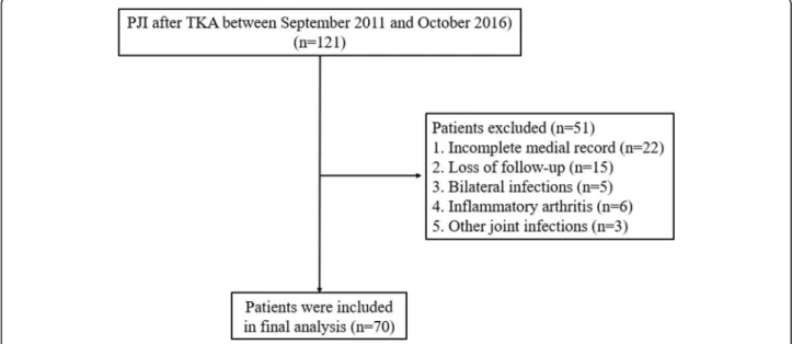
We retrospectively reviewed 121 knees diagnosed with a PJI after TKA between September 2011 and October 2016 at our institution. This study was approved by our institutional review board (IRB No. 2019-05-001). PJI was treated with two-stage reimplantation by a single surgeon and a uniform protocol. Inclusion criteria follow the Musculoskeletal Infection Society (MSIS) guidelines for PJI [13]. Patients treated with alternative approaches or those who refused reimplantation for personal issues were excluded. Patients with incomplete medical re- cords, less than 2 years of follow-up, or bilateral cases were also excluded (Fig. 1).
A total of 70 knees met the inclusion criteria and were retrospectively reviewed. Patients were placed in the controlled infection group (group C) if they required no further medication or surgical treatment within 2 years after reimplantation. Patients were placed in the uncon- trolled infection group (group U) if they presented with symptoms of active infection after resection arthroplasty or were reinfected after two-stage reimplantation.
Data collection
Multiple potential predictive variables were collected from the medical record, including demographic data (e.g., age, gender, BMI, affective side, symptom dur- ation), comorbidities (e.g., hypertension, DM, liver cir- rhosis, chronic kidney disease, cancer history, previous infection history, anticoagulant abuse), and operation-
Fig. 1 Patient flow chart. PJI prosthetic joint infection, TKA total knee arthroplasty
related factors (wound complications, hemarthrosis, transfusion, deep vein thrombosis). Clinical outcomes were assessed using the Korean Knee Score (KKS). C- reactive protein (CRP) and the erythrocyte sedimenta- tion rate (ESR) were retrospectively evaluated pre- resection (first stage) and pre-reimplantation (second stage). Cultured microorganisms were classified into one of five groups and analyzed: methicillin-sensitive organ- isms; methicillin-resistant organisms; fungus species;
Pseudomonas species; and other species.
First stage procedure: resection arthroplasty
The first stage involved resection arthroplasty with antibiotic-loaded cement spacer (Fig. 2). All patients with PJI underwent resection arthroplasty with the re- moval of all prosthetic components and cement as well as debridement of necrotic tissue. Bone cement and vancomycin were mixed at a mass ratio of 10:1.
Antibiotic-mixed cement was placed on each articular side for two reasons: to cover the bone defect and pre- vent soft tissue contracture; and to provide direct local delivery of high doses of antibiotics, avoiding systemic toxicity [14]. We reuse the femoral implant after the autoclaving process, and the femoral implant with gentamicin-mixed cement (CMW; DePuy Synthes, Warsaw, IN, USA) is placed on the articular side of the femur. A high cross-linked polyethylene liner (TC3 knee system; DePuy Synthes) was placed between the femoral component and bone cement on the tibial side, which acted like a bearing. A drain was left in the knee joint and aided in evaluating the knee joint status (Fig. 3).
Management between first stage and second stage All patients underwent 6 weeks of parenteral drugs, selected based on sensitivity to intraoperative-cultured pathogens. At postoperative week 6, if the patient still showed no sign of infection, parenteral drugs were changed to oral medicines for 4 weeks and patients were
closely monitored in outpatient clinics. The timing of the second-stage reimplantation was based on clinical condition and laboratory data. Reimplantation was per- formed after 2 weeks of the antibiotic holiday without elevation of the ESR and CRP. In patients who did not confirm normal laboratory data, we performed reimplan- tation according to the clinical condition combined with a trend of decreased ESR and CRP levels after dis- continuing oral medicines.
Second stage procedure: reimplantation
At postoperative week 12, reimplantation was per- formed. Meticulous debridement was conducted again.
If intraoperative findings revealed suspicion of infection (e.g., purulent exudates), the protocol dictated a return to the first stage with the replacement of the cement spacer. If intraoperative findings suggested eradication of infection, revision arthroplasty was planned. Any debris or soft tissue that showed loss of viability was debrided.
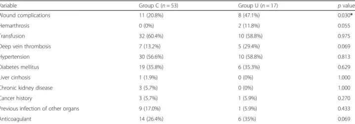
The femoral prosthesis, polyethylene liner, and cement were removed and the tibial and femoral medulla were curetted. All patients were treated with a standard rotat- ing hinge prosthesis (TC3; DePuy Synthes). Metal aug- mentation and cement were used to cover bone defects and a stem was used to provide stability to the prosthesis (Fig. 4). As the previous infection was considered con- trolled, first-generation cephalosporins were adminis- tered for 6 days postoperatively. Patients were followed in the outpatient clinic every 3 months.
Statistical analysis
Analyses were performed using SPSS version 21.0 (SPSS Incorporated, Chicago, IL, USA). Categorical and continu- ous variables were expressed as the count and the mean ± standard deviation (range), respectively. Proportional haz- ard regression univariate analysis was performed to assess the association of clinical covariates with the risk of
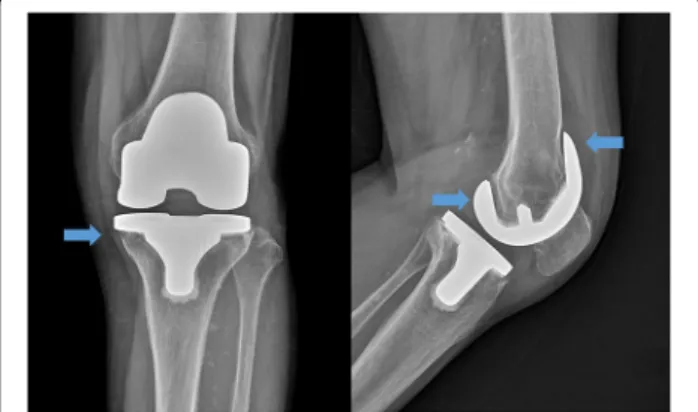
Fig. 2 Plain radiograph from a 76-year-old female 3 years after primary total knee arthroplasty. Blue arrows indicate bone resorption around the femoral prosthesis and medial condyle of tibia
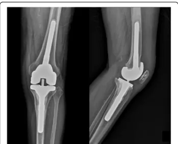
Fig. 3 Postoperative plain radiograph of resection arthroplasty. The
infected prosthesis was removed, and antibiotic-mixed cement was
placed on the articular side of the femur and tibia
uncontrolled infection. The Mann–Whitney test or chi- square/Fisher’s exact test was used to compare patient characteristics, comorbidities, type of cultured pathogens, and laboratory results between groups C and U. p < 0.05 was considered statistically significant.
Results
Of the 70 knees included in this analysis, 67 were deemed clinically stable after resection arthroplasty; the remaining three required additional surgical treatment due to remaining infection and were classified as the first-stage failure group. Of the 67 clinically stable knees, 53 were clinically deemed free from infection after
reimplantation and assigned to group C. The remaining 14 knees were reinfected after two-stage reimplantation;
each retreated with two-stage reimplantation (Fig. 5).
There were no statistically significant differences in demographics (i.e., mean age, male-to-female sex ratio, height, weight, direction of affected site, duration of follow-up) between groups C and U. Clinical outcome at final follow-up assessed using the KKS was 68.0 in group C and 59.5 in group U, a difference that was not statisti- cally significant (p = 0.180) (Table 1).
Univariate analysis for risk factors revealed that wound complications (e.g., dehiscence, discharge) occurred sig- nificantly more frequently in group U (n = 8) compared with group C (n = 11) (p = 0.03). However, the preva- lence of other comorbidities was not significantly differ- ent between group C and group U (Table 2).
Pre-reimplantation CRP was significantly higher in group U than group C (1.70 ± 2.85 and 0.44 ± 0.47, re- spectively; p = 0.025). Pre-resection CRP, pre-resection ESR, and pre-implantation ESR were not significantly different between group C and group U (p = 0.205, p = 0.593, and p = 0.509, respectively). The presence of fun- gus species using culture tests was statistically more fre- quent in group U compared with group C (p = 0.031).
The presence of methicillin-resistant or methicillin- sensitive organisms, Pseudomonas, and other species was not significantly different between groups (p = 0.882, p = 0.517, p = 0.327, and p = 0.572, respectively) (Table 3).
Discussion
In this study, we found that 17 out of 70 patients (24.3%) developed recurrent infections after our two-stage
Fig. 5 Patient flow chart for the controlled infection group (group C) and the uncontrolled infection group (group U)
Fig. 4 Post-reimplantation plain radiograph
reimplantation. These outcomes are similar to findings of other studies in the literature (i.e., incidence of infection re- currence ranges from 10% to 28%) [4, 15, 16]. Additionally, the univariate analysis identified pre-reimplant CRP, wound complications, and fungal species as risk factors for uncontrolled PJI following two-stage reimplantation.
Several risk factors for PJI have been published; how- ever, little is known about potential risk factors for PJI fol- lowing failed two-stage reimplantation arthroplasty.
Sakellariou et al. identified patients with a background of inflammatory arthritis or those who were immunocom- promised to be at an increased risk for reinfection [16].
Also, in an analysis of 368 patients by Kubista et al., it was noted that the strongest positive predictors of treatment failure included chronic lymphoedema, and revision be- tween resection and definitive reimplantation and patients treated with intravenously administered cefazolin had a
significant reduction in recurrent infection rates [4]. Watts et al. reported that morbidly obese patients (i.e., BMI ≥ 40 kg/m
2) had significantly higher rates of subsequent revi- sion (hazard ratio, 4.45), reinfection (hazard ratio, 4.86), and reoperation (hazard ratio, 4.37) following two-stage TKA revision for PJI when compared with a matched co- hort of nonobese patients (i.e., BMI < 30 kg/m
2) [17]. Simi- lar studies were also conducted in joints other than the knee. Jhan et al. evaluated 62 patients with chronic PJI of the hip joint treated with two-stage reimplantation and found that obesity, liver cirrhosis, Gram-negative organ- isms, and presence of a sinus tract were significantly re- lated to the risks of failure [5]. In our study, the prevalence of DM and obesity, and the use of anticoagu- lant (also a known risk factor of PJI), were higher in group U compared with group C; however, this difference was not statistically significant.
Table 1 Demographic data for patients in group C and group U
Variable Group C (n = 53) Group U (n = 17) p value
Age 69.72 ± 6.46 66.88 ± 6.67 0.121
Sex 1.000
Male 9 (17.0%) 3 (17.6%)
Female 44 (83.0%) 14 (82.4%)
Body mass index 25.5 ± 8.4 24.4 ± 9.2 0.438
Affected side 0.525
Right 27 (50.9%) 7 (41.2%)
Left 26 (49.1%) 10 (58.8%)
Symptom duration (months) 7.3 ± 11.6 7.8 ± 11.0 0.715
Interval between TKA and resection arthroplasty (months) 24.2 ± 21.4 16.9 ± 11.8 0.854
Duration of follow-up (months) 32.6 ± 8.0 33.7 ± 7.7 0.629
Previous history of knee surgery 9 (17.0%) 1 (5.9%) 1.000
Final Korean Knee Score 68.0 ± 12.1 59.5 ± 10.3 0.180
Data presented as mean ± standard deviation or n (%). Group C controlled infection group, Group U uncontrolled infection group, TKA total knee arthroplasty
Table 2 Univariate analysis of selected variables for patients in group C and group U
Variable Group C (n = 53) Group U (n = 17) p value
Wound complications 11 (20.8%) 8 (47.1%) 0.030*
Hemarthrosis 0 (0%) 2 (11.8%) 0.055
Transfusion 32 (60.4%) 10 (58.8%) 0.975
Deep vein thrombosis 7 (13.2%) 5 (29.4%) 0.069
Hypertension 30 (56.6%) 10 (58.8%) 0.813
Diabetes mellitus 19 (35.8%) 6 (35.3%) 0.629
Liver cirrhosis 1 (1.9%) 0 (0%) 1.000
Chronic kidney disease 3 (5.7%) 0 (0%) 1.000
Cancer history 3 (5.7%) 1 (5.9%) 0.270
Previous infection of other organs 9 (17.0%) 1 (5.9%) 0.433
Anticoagulant 14 (26.4%) 6 (35%) 0.069
Data presented as n (%). Group C controlled infection group, Group U uncontrolled infection group
*p < 0.05 considered statistically significant
Although interpreting laboratory tests is crucial, there is ongoing debate and controversy relating to the role of laboratory results in PJI. Ghanem et al. studied 109 patients with infected TKAs who underwent two-stage revision and were unable to define an absolute CRP or ESR threshold for infection eradication despite a 21%
(23 of 109) reinfection rate at an average of 2 years [18].
Stambough et al. analyzed 291 cases of PJI and suggested that the percent change in serum ESR and CRP inflam- matory markers before and after two-stage reimplanta- tion for PJI was not associated with the reinfection risk when controlling for ASA class [19]. Furthermore, this group concluded that inflammatory markers provide no additional diagnostic accuracy for determining the tim- ing of reimplantation. Lee and Chen reported in a sys- tematic review and meta-analysis that no single marker was superior to all of the others. Because none is likely sufficient to confirm control of infection after the first stage of the two-stage protocol for PJI, they suggested that multiple tools are needed for ensuring infection eradication. [20]. In this present study, there was no sig- nificant difference in pre-resection ESR results between both groups; however, the pre-resection CRP level was lower in group U than in group C. Similar to our study, Petrikkos et al. noted that both serum procalcitonin and CRP levels were lower in patients with fungal infections than in those with bacterial infection [21].
Several studies of uncontrolled PJI and microorgan- isms have been published. Earlier studies reported higher failure rates in periprosthetic infection when methicillin- resistant bacteria are present [22–24]. Importantly, we note that these studies included patients with hip PJI treated with several different strategies. Kubista et al.
were unable to detect a statistically significant difference in recurrence rates between patients with confirmed in- fection with methicillin-sensitive and methicillin- resistant organisms [4 ]. The presence of Enterococcus or
Streptococcus species was also associated with a higher risk of failure in a study by Citak et al. [25] Furthermore, several authors reported that polymicrobial peripros- thetic infections were at an increased risk for recurrence infection [16, 26, 27]. Here, we report no significant dif- ferences between methicillin-sensitive and methicillin- resistant species, an observation consistent with studies by Kubista et al. [4] This observation is thought to be the result of vancomycin usage, which spreads from ce- ment to kill methicillin-resistant or methicillin-sensitive species. Recently, attempts have been made to mix amphotericin B or voriconazole into cement, drugs which are effective against fungal species leading to favorable outcomes [28]. Further studies on the heat stability and local spread of antimicrobial agents are needed.
The strengths of this study include that it was per- formed in a homogeneous group of patients who underwent surgery by a single surgeon. Also, this was the first report of risk factors for uncontrolled infec- tion after two-stage reimplantation in Korean patients.
However, our study has several limitations: a retro- spective design and relatively small cohort; our insti- tution is a tertiary referral medical center, so primary arthroplasty was performed by various surgeons and methods—prostheses were also from different com- panies and biology of the knee can be ruined in vari- ous ways, which may affect the final treatment result;
intraoperative frozen section histopathology, which yields a high diagnostic accuracy matched with MSIS criteria, was not routinely checked; and we did not check the comorbidity scores. Further well-designed prospective clinical studies are needed to confirm our results.
Conclusion
The reinfection rate of our two-stage reimplantation protocol showed 24.3% in the included cases. Wound Table 3 Univariate analysis of laboratory results and identification of pathogens between group C and group U
Variables Group C (n = 53) Group U (n = 17) p value
Pre-resection CRP 5.87 ± 6.74 3.11 ± 3.33 0.205
Pre-resection ESR 80.48 ± 32.88 75.53 ± 29.36 0.593
Pre-reimplantation CRP 0.44 ± 0.47 1.70 ± 2.85 0.025*
Pre-reimplantation ESR 42.93 ± 22.59 50.71 ± 33.72 0.509
Identification of pathogen 34 (64.2%) 12 (70.6%) 0.111
Methicillin-resistant organisms 19 (35.8%) 6 (35.3%) 0.882
Methicillin-sensitive organisms 11 (20.8%) 2 (11.8%) 0.517
Fungal species 2 (3.8%) 3 (17.6%) 0.031*
Pseudomonal species 1 (1.9%) 1 (5.9%) 0.327
Other organisms 1 (1.9%) 0 (0%) 0.572
Data presented as mean ± standard deviation or n (%). CRP C-reactive protein, ESR erythrocyte sedimentation rate, Group C controlled infection group, Group U uncontrolled infection group
*p < 0.05 considered statistically significant