https://doi.org/10.3350/cmh.2021.0178 Clinical and Molecular Hepatology 2021;27:363-401
Guideline
Corresponding author : Yong Kyun Cho
Division of Gastroenterology and Hepatology, Department of Internal Medicine, Kangbuk Samsung Hospital, Sungkyunkwan University School of Medicine, 29 Saemunan-ro, Jongno-gu, Seoul 03181, Korea
Tel: +82-2-2001-8557, Fax: +82-2-2001-2610
E-mail: [email protected], [email protected] https://orcid.org/0000-0002-9231-006X
* Seong Hee Kang, Hye Won Lee, Jeong-Ju Yoo ,Yuri Cho, and Seung Up Kim equally contributed to this work as co-first authors.
Abbreviations:
AGREE II, Appraisal of Guidelines for Research and Evaluation II; ALT, alanine aminotransferase; AST, aspartate aminotransferase; AUC, area under the receiver operating characteristic curve; BMI, body mass index; CAP, controlled attenuation parameter; CKD, chronic kidney disease; CT, computed tomography;
CVD, cardiovascular disease; ELF, enhanced liver fibrosis; FIB-4, fibrosis-4 index; FLI, fatty liver index; GLP-1, glucagon-like peptide-1; GRADE, Grading of Recommendations, Assessment, Development and Evaluation; GREACE, GREek Atorvastatin and Coronary-heart-disease Evaluation; HCC, hepatocellular carcinoma; HIS, hepatic steatosis index; KASL, the Korean Association for the Study of the Liver; LDL-C, low density lipoprotein cholesterol; LT, liver transplantation; MAFLD, metabolic (dysfunction)-associated fatty liver disease;
METs, metabolic equivalents of task; MRE, magnetic resonance elastography;
MRI-PDFF, MRI proton density fat fraction; MRI, magnetic resonance imaging;
MRS, MR spectroscopy; NAFL, nonalcoholic fatty liver; NAFLD, nonalcoholic fatty liver disease; NAS, NAFLD activity score; NASH, nonalcoholic steatohepatitis;
NFS, NAFLD fibrosis score; NLFS, NAFLD liver fat score; PIVENS, Pioglitazone versus Vitamin E versus Placebo for the Treatment of Nondiabetic Patients with Nonalcoholic Steatohepatitis; PNPLA3, the patatin-like phospholipase domain–containing 3; PPAR-γ, peroxisome proliferator activated receptor gamma; SAMM50, sorting and assembly machinery component 50; SWE, shear wave elastography; T2DM, type 2 diabetes mellitus; TM6SF2, transmembrane 6 superfamily, member 2
Received : Jun. 22, 2021 / Accepted : Jun. 22, 2021 Editor: Seung Up Kim, Yonsei University College of Medicine, Korea
KASL clinical practice guidelines: Management of non- alcoholic fatty liver disease
Seong Hee Kang
1,*, Hye Won Lee
2,*, Jeong-Ju Yoo
3,*, Yuri Cho
4,*, Seung Up Kim
2,*, Tae Hee Lee
5, Byoung Kuk Jang
6, Sang Gyune Kim
3, Sang Bong Ahn
7, Haeryoung Kim
8, Dae Won Jun
9, Joon-Il Choi
10, Do Seon Song
11, Won Kim
12, Soung Won Jeong
13, Moon Young Kim
1, Hong Koh
14, Sujin Jeong
15, Jin-Woo Lee
16, and Yong Kyun Cho
17on behalf of The Korean Association for the Study of the Liver (KASL)
1
Department of Internal Medicine, Yonsei University Wonju College of Medicine, Wonju;
2Department of Internal Medicine, Yonsei University College of Medicine, Seoul;
3Department of Internal Medicine, Soonchunhyang University Bucheon Hospital, Bucheon;
4
Center for Liver and Pancreatobiliary Cancer, National Cancer Center, Goyang;
5Department of Internal Medicine, Konyang University College of Medicine, Daejeon;
6Department of Internal Medicine, Keimyung University School of Medicine, Daegu;
7Department of Inter- nal Medicine, Nowon Eulji Medical Center, Eulji University School of Medicine, Seoul;
8Department of Pathology, Seoul National Univer- sity Hospital, Seoul National University College of Medicine, Seoul;
9Department of Internal Medicine, Hanyang University College of Medicine, Seoul;
10Department of Radiology, Seoul St. Mary’s Hospital, College of Medicine, The Catholic University of Korea, Seoul;
11
Department of Internal Medicine, St. Vincent’s Hospital, College of Medicine, The Catholic University of Korea, Seoul;
12Department of Internal Medicine, Seoul Metropolitan Government Boramae Medical Center, Seoul National University College of Medicine, Seoul;
13
Department of Internal Medicine, Soonchunhyang University Seoul Hospital, Soonchunhyang University College of Medicine, Seoul;
14
Division of Gastroenterology, Hepatology and Nutrition, Department of Pediatrics, Severance Children’s Hospital, Yonsei University College of Medicine, Seoul;
15Division of Pediatric Gastroenterology Hepatology and Nutrition, CHA Bundang Medical Center, CHA University, Seongnam;
16Department of Internal Medicine, Inha University Hospital, Inha University School of Medicine, Incheon;
17
Division of Gastroenterology and Hepatology, Department of Internal Medicine, Kangbuk Samsung Hospital, Sungkyunkwan Univer-
sity School of Medicine, Seoul, Korea
INTRODUCTION Preamble
The prevalence of nonalcoholic fatty liver disease (NAFLD) is in- creasing rapidly worldwide as the obese and diabetic populations increase, and it has been estimated to be 20–30% in Korea. Con- sidering the increased popularity of a westernized diet and life- style, lack of exercise, and the resulting increase in obesity and di- abetes, NAFLD is predicted to become more prevalent in the future and to become a major cause of chronic liver disease. In some patients, NAFLD progresses to end-stage liver diseases such as cirrhosis and hepatocellular carcinoma (HCC), and it is an inde- pendent cardiovascular risk factor.
In 2013, the Korean Association for the Study of the Liver (KASL) enacted a clinical practice guideline for the diagnosis and treatment of NAFLD to improve understanding of the disease and provide useful clinical information and direction for healthcare providers. The research results that have accumulated since then necessitate a revision. Accordingly, the clinical practice guidelines committee began revising the guidelines to reflect the results of Korean and international research and develop new recommenda- tions based on a systematic approach that reflects evidence- based medicine and expert opinions. However, evidence remains insufficient, and many studies are currently being conducted. As medical evidence and new findings accumulate in the future, these guidelines will require ongoing supplementation and revision.
Target population
Patients diagnosed with NAFLD based on clinical, biochemical, radiological, or pathological findings in the absence of significant alcohol consumption and liver diseases such as viral hepatitis were the primary research population involved in the development of these guidelines. These guidelines are also based on data from pediatric and adolescent NAFLD patients, whose unique findings distinguish them from adult NAFLD patients.
Intended users
The aim of these guidelines is to provide useful clinical informa- tion and direction to healthcare providers involved in the diagno- sis and treatment of NAFLD patients. Moreover, these guidelines are intended to provide definite and practical information to resi- dent physicians, practitioners, and trainers.
Developer and funding information
The Clinical Practice Guideline Committee for the Management of NAFLD (Committee) was organized in accordance with propos- als by the approval of the KASL Board of Executives and consists of 16 gastroenterologists, one pathologist, one radiologist, and two pediatricians specializing in hepatology. All expenses were paid by KASL. Each committee member collected and analyzed the source data in his or her own field, and the members then wrote the manuscript together.
Evidence collection
The committee systematically collected and reviewed the inter- national and domestic literature published in PubMed, MEDLINE, KoreaMed, and other databases. The literature was limited to re- search papers published in the English and Korean languages. The keywords used were ‘nonalcoholic fatty liver disease,’ ‘nonalco- holic fatty liver,’ ‘nonalcoholic steatohepatitis,’ ‘fatty liver,’ ‘hepat- ic steatosis,’ and ‘steatohepatitis.’ In addition, keywords related to specific clinical questions were included.
Levels of evidence and grades of recommendations
The literature gathered for data collection was analyzed in a
systematic review, and the quality of evidence was classified
based on the modified Grading of Recommendations, Assess-
ment, Development and Evaluation (GRADE) system (Table 1). Ac-
cording to the types of studies, randomized, controlled studies
were approached from a high level of evidence, while observa-
tional studies were approached from a low level of evidence. Sub-
sequently, the level of evidence basis sets in corresponding stud-
Keywords: Non-alcoholic fatty liver disease; Epidemiology; Diagnosis; Treatment; Guideline
ies was elevated or lowered by accounting for the factors influencing the quality of the studies. Through follow-up studies, the level of evidence was defined as follows: A, indicating the highest level of evidence with the smallest possibility of any changes in the conclusion; B, indicating a moderate level of po- tential changes; and C, indicating the lowest level of evidence with the greatest possibility of any changes.
The strength of each recommendation was suggested according to the GRADE system. In addition to the level of evidence, the re- sults of studies were considered based on aspects of clinical mul- tipliers and socio-economic factors, such as cost. Grading of the recommendations was performed as follows: 1, strong recom- mendation or 2, weak recommendation. A strong recommenda- tion indicated, for example, that the interventions could be ap- plied in most patients with strong certainty, there was a greater possibility of desirable effects, and there was high-quality evi- dence, as well as presumed patient-important outcomes, cost-ef- fectiveness, preference, and compliance. A weak recommendation indicated a suggestion made with less certainty but that could be considered favorable for many patients. Alternative interventions could be chosen for “weak recommendations”, according to cost and the preferences of the patients or medical practitioners.
These Clinical Practice Guidelines for the Management of NAFLD have been developed through the reviews of medical ex- perts to be used practically for treatment, research, and educa- tion. These recommendations are not absolute standards for treatment, and adoption of these guidelines in clinical practice can differ for individual patients.
List of key questions
The committee considered the following clinical questions as key components to be covered in the guidelines.
1. What are the definition and categories?
2. What are the incidence rate and prevalence rate?
3. How does NAFLD progress?
4. What causes NAFLD-related deaths?
5. What are the risk factors of NAFLD?
6. What are NAFLD’s comorbidities?
7. Who should be targeted for NAFLD screening, and how is screening conducted?
8. Which non-invasive surrogates are available to diagnose he- patic steatosis?
9. Which non-invasive surrogates are available to diagnose NASH?
10. Which non-invasive surrogates are available to assess liver fibrosis?
11. Which differential tests are available for advanced fibrosis?
12. What are the indications for liver biopsy?
13. What are the histopathological features of NAFLD?
14. Is surveillance for HCC necessary?
15. How can HCC be prevented?
16. Who should be treated, and what is the aim of the treat- ment?
17. What do lifestyle modifications include?
18. What is the effect of moderate or less alcohol consumption?
19. What are the types, indications, effects, and side effects of medications for NAFLD?
20. What are the indications for and post-operative manage-
Table 1. The grading of recommendations, assessment, development, and evaluation (GRADE) systemCriteria
Quality of evidence
High quality Further research is very unlikely to change our confidence in the estimate of the effect. A Moderate quality Further research is likely to have an important impact on our confidence in the estimate of the effect
and may change the estimate. B
Low quality Further research is very likely to have an important impact on our confidence in the estimate of the effect and is likely to change the estimate. Any change of estimate is uncertain.
C Strength of
recommendation
Strong Factors influencing the strength of the recommendation include the quality of the evidence, presumed patient-important outcomes, and cost.
1 Weak Variability in preference and values or greater uncertainty. The recommendation is made with less
certainty, higher cost or resource consumption. 2
ment of bariatric surgery?
21. What are the indications for liver transplantation (LT) and post-LT management?
Regarding NAFLD in children and adolescents:
1. What is the prevalence rate?
2. How does NAFLD progress?
3. What genetic diseases are associated with risk factors?
4. Is related NAFLD related to family history and genetic predis- position?
5. Who should be targeted for NAFLD screening, and how is screening conducted?
6. What are the diagnostic methods?
7. Who should be treated, and how?
Review of the manuscript and approval process
The manuscripts written by the committee members were re- viewed and approved through meetings of the committee. The quality of the manuscripts was evaluated based on the integrity of the contents and the standards of Appraisal of Guidelines for Re- search and Evaluation II (AGREE II). The guidelines were also re- viewed at a meeting of an external review board consisting of 11 specialists in the field of hepatology and at a symposium open to all KASL members, and they were then further modified prior to publication. The final manuscript was endorsed by the Board of Executives of KASL.
Release of the guidelines and plan for updates
The Korean version of the KASL Clinical Practice Guideline for the Management of NAFLD was released at Liver Week 2021 (May 15, 2021), and published in the Clinical and Molecular Hepatology (July 2021). This guideline in Korean is available on the KASL website (http://www.kasl.org). Updates are planned when new
reliable evidence has accumulated. Detailed plans for updates will be posted on the KASL website.
DEFINITION
What are the definition and categories?
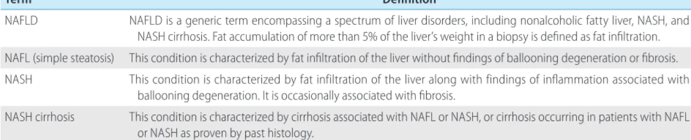
NAFLD is a condition characterized by finding fat infiltration of the liver on radiological exams or biopsy without significant alco- hol intake, viral hepatitis, medication intake that would cause a fatty liver, or other obvious cause. NAFLD is defined as a disease with findings suitable for clinical, biochemical, imaging, and path- ological examinations. NAFLD is a generic term that encompasses the spectrum of nonalcoholic fatty liver (NAFL), nonalcoholic ste- atohepatitis (NASH), and NASH cirrhosis (Table 2). The significant safe limits of daily alcohol intake that distinguish NAFLD from al- coholic fatty liver disease range from 10–40 g (pure alcohol), though that range varies between studies. Therefore, definite cri- teria are difficult to recommend. The Clinical Practice Guideline of the European Association for the Study of the Liver agreed to de- fine the amount of significant alcohol consumption as weekly al- cohol consumption ≥210 g in men and ≥140 g in women.
1No ethnic differences have been reported regarding safe alcohol lim- its that do not produce liver damage. The KASL Clinical Practice Guideline for NAFLD uses the amount of significant alcohol con- sumption stated above in clinical treatment and in studies for in- ternational comparison with the results of future studies.
The term NAFLD was introduced by Schaffner in 1986.
2Howev- er, as information about the causes and mechanisms of the dis- ease accumulated, an opinion emerged that the term NAFLD does not reflect the condition’s heterogeneous pathogenesis and vari- ous courses. Furthermore, the overestimation of the exclusion of alcohol has sparked heated debate about the threshold of “signif-
Table 2. Definition of nonalcoholic fatty liver disease-related terms
Term Definition
NAFLD NAFLD is a generic term encompassing a spectrum of liver disorders, including nonalcoholic fatty liver, NASH, and NASH cirrhosis. Fat accumulation of more than 5% of the liver’s weight in a biopsy is defined as fat infiltration.
NAFL (simple steatosis) This condition is characterized by fat infiltration of the liver without findings of ballooning degeneration or fibrosis.
NASH This condition is characterized by fat infiltration of the liver along with findings of inflammation associated with ballooning degeneration. It is occasionally associated with fibrosis.
NASH cirrhosis This condition is characterized by cirrhosis associated with NAFL or NASH, or cirrhosis occurring in patients with NAFL or NASH as proven by past histology.
NAFLD, nonalcoholic fatty liver disease; NASH, nonalcoholic steatohepatitis; NAFL, nonalcoholic fatty liver.
icant” alcohol consumption required for the diagnosis of NAFLD, and the potential (contradictory) role of alcohol consumption in these “non-alcohol” diseases has been repeatedly raised. In 2019, a consensus by 32 experts suggested alternative terminology, metabolic (dysfunction)-associated fatty liver disease (MAFLD), to more accurately reflect the pathogenesis of this disease.
3The di- agnosis of MAFLD is based on evidence of fat accumulation in the liver in the presence of one of the following three criteria: over- weight/obesity, type 2 diabetes mellitus (T2DM), and evidence of metabolic dysregulation. Further research results are needed to verify the validity of this term.
EPIDEMIOLOGY
What are the incidence rate and prevalence rate?
Incidence
In 2007, researchers published results from tracking 5,237 men for more than 4 years and reported for the first time that the an- nual incidence of NAFLD was 74.1 cases per 1,000 persons.
4The annual incidence rate of NAFLD diagnosed by abdominal ultraso- nography in health screening examinees was about 48.2 cases per 1,000 persons (range, 13.4–77.7).
5-11As diagnosed by the he- patic steatosis index (HSI), the annual incidence rate was 21.1 cases per 1,000 persons.
12According to a meta-analysis in Asia, the annual incidence rate in Korea was 45.1 cases per 1,000 per- sons.
13,14Prevalence
The prevalence of NAFLD varies depending on the study popu- lation, definition of NAFLD, and diagnostic modality. In 2002, the prevalence among 1,074 people receiving health checkups and di- agnosed by abdominal ultrasound was 48.6%.
15The prevalence among 141,610 people receiving health checkups in the Seoul and Gyeonggi area and diagnosed by abdominal ultrasound was 25.2% (male, 34.4%; female, 12.2%),
16and the prevalence in other studies varied from 21% to 44%.
17-23In a meta-analysis, the prevalence in Korea, as diagnosed by ultrasonography, was 32.9%.
13The prevalence diagnosed using the fatty liver index (FLI) was 12.6–16.1%,
22,24and the prevalence diagnosed with transient elastography was 42.9%.
25The prevalence diagnosed by liver bi- opsy among 589 living liver donors in Korea was 51%.
26Lean/non-obese NAFLD
People with normal body weight (body mass index [BMI; kg/m
2] of less than 23 kg/m
2for Asians, less than 25 kg/m
2for Western- ers) or non-obese weight (BMI of less than 25 kg/m
2for Asians, less than 30 kg/m
2for Westerners) can also be diagnosed with NAFLD, but data on the incidence rate among those people in Ko- rea are limited. In 2004, the prevalence of NAFLD was 16.1%
among 460 people with a BMI between 18.5 kg/m
2and 25 kg/m
2who received domestic health checkups.
27In several domestic studies, the average prevalence of non-obese NAFLD was 18.8%
(12.4–27.1%).
28-34Summary
1. The annual incidence of NAFLD in Korea is about 45 cases per 1,000 persons.
2. The prevalence of NAFLD in Korea is approximately 30%.
3. The prevalence of nonalcoholic fatty liver disease in the domestic non-obese population is about 19%.
Natural course and cause of death How does NAFLD progress?
Because the natural course of NAFLD can only be confirmed through repeated liver biopsy, it has been reported only in small studies. The incidence of NASH was reported to vary from 8.5%
to 64.0% over a 3.0–6.6 years follow-up period, a large variation that appears to result from differences in the number of patients, various follow-up periods, and diagnostic criteria.
35-37In a meta-analysis comparing NAFL and NASH, the percentage of patients who progressed to more than one stage of liver fibro- sis were similar at 39.1% and 34.5%, respectively, but the time it took those patients to progress more than one stage was 14.3 years and 7.1 years, respectively. NASH thus showed faster pro- gression.
38In a cohort study of NASH patients, the incidence of cirrhosis varied by race and region, with an average of 21–26% of patients progressing to cirrhosis in eight years.
39,40According to a study of an LT waitlist, the number of patients with end-stage liver disease because of NAFLD had tripled from 10 years previously, making NAFLD the second most common reason for needing LT.
41Recently, HCC caused by NAFLD has rapidly increased, becom-
ing the third most common cause of HCC development in United
States, and that number is expected to increase by 9% per
year.
42,43This trend is believed to be due to the rapid increase in
the prevalence of obesity, which is a risk factor for NAFLD. NAFLD is associated with a higher incidence of HCC than other etiologies of liver disease in the absence of advanced fibrosis or cirrhosis.
44-46What causes NAFLD-related deaths?
According to data from the U.S. National Vital Statistics System, the mortality rate of NAFLD has increased in the past decade.
47In a meta-analysis, patients with NAFLD had a 1.6 times higher mor- tality rate than the general population. The major causes of death were cardiovascular disease (CVD), malignancy, and liver disease, and in the presence of steatohepatitis, liver disease-related deaths increased.
48-50In several cohort studies, the prognosis of patients with NAFLD was most closely related to the degree of liver fibro- sis.
51,52In a large-scale cohort study based on liver biopsy, the mortality rate of NAFL patients was 1.7 times that of the normal control group; the mortality rate of patients who had steatohepa- titis without hepatic fibrosis was 2.1 times that of the normal con- trol group; patients with liver fibrosis had a mortality rate 2.4 times that of the normal control group; patients with cirrhosis had a mortality rate that was 3.8 times that of the normal control group. Thus, the mortality rate increased with the degree of liver fibrosis.
53Summary
1. Liver fibrosis can progress faster in NASH than in NAFL.
2. The incidence of liver cirrhosis and HCC associated with NAFLD is increasing, and HCC can develop even in the absence of liver cirrhosis.
3. The main causes of death among patients with NAFLD are CVD, malignant tumors, and liver diseases, and liver disease- related mortality increases when steatohepatitis and liver fibrosis are present.
What are the risk factors of NAFLD?
NAFLD is closely related to obesity, diabetes, dyslipidemia, and metabolic syndrome (Fig. 1). Obesity is a well-known risk factor for NAFLD, and the prevalence of NAFLD increases as BMI in- creases.
54Among obese patients who underwent bariatric sur- gery, the prevalence of NAFL, NASH, and significant liver fibrosis were 61–91%, 30–37%, and 29.3%, respectively.
55,56Metabolic syndrome, which consists of abdominal obesity, impaired fasting blood sugar, hypertriglyceridemia, low high-density cholesterol- emia, and hypertension, is a major risk factor for NAFLD, just like
obesity. In the presence of metabolic syndrome, the prevalence of NAFLD was 50%.
57,58The prevalence of NAFLD was also high in diabetic patients, 60–75%.
14In patients with dyslipidemia, anoth- er risk factor, the prevalence of NAFLD was 50%.
59,60In the presence of hypothyroidism, the prevalence of NAFLD in- creases by 1.6 times,
61and in the presence of polycystic ovary syndrome, the incidence increases by about 2.2 times.
62In addi- tion, obstructive sleep apnea,
63hypopituitarism,
64hypogonad- ism,
65pancreatoduodenal resection,
66and psoriasis
67increase the prevalence of NAFLD.
Decreased physical activity and sarcopenia increase the risk of NAFLD.
68,69The prevalence and incidence of NAFL increased in the group with reduced physical activity.
70-72In the presence of sarcopenia, the risk of NAFLD increased by about four times re- gardless of obesity or metabolic syndrome.
68,69When sarcopenia was accompanied by NAFLD, the risk of advanced liver fibrosis in- creased by 1.8 times.
69Genetic factors play a major role in the occurrence of NAFLD.
Typically, the patatin-like phospholipase domain-containing 3 ( PNPLA3) and transmembrane 6 superfamily, member 2 (TM6SF2) single nucleotide polymorphisms affect the development and pro- gression of the disease. In Korea, PNPLA3 and sorting and assem- bly machinery component 50 (SAMM50) were associated with the prevalence and severity of NAFLD.
73Summary
1. Major risk factors for NAFLD include obesity, diabetes, dyslipidemia, and metabolic syndrome.
What are NAFLD’s comorbidities?
NAFLD is closely related to systemic metabolic diseases,
74and it
is an independent risk factor for the occurrence of various non-
Figure 1. Risk factors for nonalcoholic fatty liver disease.hepatic diseases, including CVD, T2DM, metabolic syndrome, chronic kidney disease (CKD), and malignant tumors.
75-77CVD
In a retrospective cohort study of 1,804 patients with NAFLD in Denmark in the 2000s, the mortality rate from CVD was higher than that in the normal control group.
50Several cohort studies that followed also found NAFLD to be an independent risk factor for CVD.
78,79In a study that followed patients with histologically diagnosed NAFLD for an average of 26.4 years, the mortality rate increased by 1.3 times, and the risk of CVD increased by 1.6 times compared with the normal control group, with both increases as- sociated with the degree of liver fibrosis.
78A meta-analysis of pa- tients with NAFLD diagnosed histologically or radiologically con- cluded that the risk of CVD was increased by about 1.6 times compared with the normal control group. The risk of CVD also in- creased when liver fibrosis was assessed using the NAFLD fibrosis score (NFS) or liver biopsy.
80According to two Korean cohort studies, NAFLD was independently associated with the incidence of coronary artery calcification and atherosclerosis, which are di- rect causes of CVD.
18,81T2DM
T2DM and NAFLD influence each other.
82In a large-scale cohort study of healthy Koreans, the prevalence and incidence of T2DM in patients with NAFLD (assessed using the NFS) increased with the degree of hepatic fibrosis.
17In a meta-analysis of Korean stud- ies, the prevalence of diabetes in patients with NAFLD was 14.2%, higher than the 5.2% of the control group.
83The results of a meta-analysis of other studies also showed an increase of about 2.2 times in the incidence of T2DM.
84Metabolic syndrome
Metabolic syndrome is a major risk factor for the occurrence of NAFLD and is a common comorbid disease. A prospective Korean cohort study of 46,874 men reported that patients with mild and moderate NAFLD (diagnosed by abdominal ultrasound) had a risk of metabolic syndrome 1.5 and 2 times higher, respectively, than the control group.
85In a meta-analysis of Korean studies, the inci- dence of metabolic syndrome was 40.7% in patients with NAFLD and 11.2% in the control group.
83CKD
CKD and NAFLD share risk factors such as T2DM and hyperten- sion,
86and CKD frequency increases in patients with NAFLD.
87-89In a meta-analysis of about 63,000 people, the prevalence of CKD increased by 2.1 times, and the incidence rate increased by 1.7 times compared with the control group.
90The prevalence and inci- dence of CKD were 2.5 times and 2.1 times higher in NASH than NAFLD. In a Korean cohort study, patients with NAFLD had a 1.2 times higher risk of developing CKD than the control group, and that risk increased with the stage of liver fibrosis.
91Other diseases
Arrhythmia, osteoporosis, colon adenoma, colon cancer, and breast cancer all have increased incidence in patients with NAFLD.
92-94However, more follow-up studies are needed to con- firm clear relationships between NAFLD and those diseases.
Summary
1. NAFLD is often associated with comorbidities such as cardiovascular disease, T2DM, metabolic syndrome, CKD, and malignant tumors.
DIAGNOSIS
Who should be targeted for NAFLD screening, and how is screening conducted?
Screening for NAFLD should be considered in cases of persistent liver enzyme elevation. Because the cost effectiveness of screen- ing in diabetic patients has been confirmed,
95patients with diabe- tes should receive screening tests for NAFLD regardless of their liver enzyme levels. In addition, subjects with metabolic syndrome (which is closely associated with insulin resistance), obesity, and other risk factors for NAFLD can be considered for screening tests.
17,96The primary screening test for NAFLD is ultrasonography. When
NAFLD is suspected in the ultrasonography results, other tests
(computed tomography [CT], magnetic resonance imaging [MRI],
serologic tests, transient elastography, etc.) can be performed.
[Recommendations]
1. Subjects who have persistent liver enzyme elevation or diabetes should be screened for NAFLD. (A1)
2. Subjects with metabolic syndrome (which is closely associated with insulin resistance, obesity, and other risk factors for NAFLD) can be considered for screening. (B1)
3. Abdominal ultrasonography is the primary screening modality.
(B1)
Non-invasive examinations
Because the prognosis of NAFLD differs significantly depending on the histological findings, the diagnosis of hepatic steatosis and fibrosis and assessment of their severity are clinically crucial. To date, liver biopsy is the gold standard in assessing hepatic necro- inflammation, steatosis, and fibrosis. However, liver biopsy is ex- pensive, invasive, carries a risk of complications, is subject to in- tra-/inter-observer interpretational variability, and can suffer from sampling errors when an insufficient amount of liver tissue is col- lected.
97,98Therefore, noninvasive radiological surrogates such as ultrasonography and panels of serological tests are widely used.
Which non-invasive surrogates are available to diagnose hepatic steatosis?
Radiological surrogates
Abdominal ultrasonography is the first-line imaging modality for screening asymptomatic patients with abnormal liver enzyme lev- els. However, it suffers from subjective interpretation, examina- tion difficulties in obese patients, and low sensitivity when the amount of hepatic steatosis is less than 30%. Moreover, ultraso- nography cannot distinguish NASH from NAFL.
99-101When hepatic steatosis assessed using MR spectroscopy (MRS) was used for ref- erence, the diagnostic accuracy of ultrasonography was lower than that of non-contrast CT and MRI.
102However, ultrasonogra- phy is appropriate as a screening test, because it offers 1) robust diagnostic performance for moderate or severe steatosis, 2) the ability to evaluate the entire hepatobiliary system in addition to detecting the presence of hepatic steatosis, and 3) wide availability.
Controlled attenuation parameter (CAP) is a feature of transient elastography that can quantify the degree of fat deposition in the liver parenchyma by measuring ultrasound attenuation.
103,104Ac- cording to a recent Korean study enrolling people who received regular health checkups, the normal CAP range is 156–287 dB/m.
105The area under the receiver operating characteristic curve (AUC) of CAP used to diagnose a moderate to severe degree of steatosis was 0.88 (sensitivity, 83.3%; specificity, 81.6%), and the cutoff value was 276 dB/m in a Korean study of living LT donors.
106In another Korean study, the AUCs of CAP for mild, moderate, and severe degrees of steatosis in patients with chronic liver diseases, including NAFLD, were 0.885, 0.894, and 0.800, respectively, and the cutoff values were 250 dB/m, 299 dB/m, and 327 dB/m, respectively.
107In a recent meta-analysis, the AUCs of CAP for mild, moderate, and severe degrees of steatosis were 0.96, 0.82, and 0.70, respectively.
108CAP can be used as a monitoring tool for hepatic steatosis, and it can be examined simultaneously with transient elastography.
109-111Other ultrasound-based methods, in- cluding image-based quantitative analysis of liver parenchymal echo texture and measuring the attenuation of ultrasound energy, are under investigation.
112,113Hepatic steatosis is associated with low attenuation on CT scans, which can be used for the quantitative assessment of he- patic steatosis. Because the attenuation of enhanced CT is affect- ed mainly by the amount of blood flow, unenhanced CT is favored for the measurement of hepatic steatosis, and the attenuation of liver and spleen parenchyma are frequently compared. For moder- ate to severe steatosis, the specificity of CT was reported to be high, but its sensitivity and positive predictive value were low.
101,114,115Furthermore, its diagnostic performance for mild steatosis was suboptimal. The specificity and sensitivity of unenhanced CT for diagnosing moderate to severe steatosis were 100% and 53.8%, respectively, when CT attenuation of hepatic parenchyma was less than 48 HU.
115However, CT raises concerns about radiation hazards, and CT evaluations of hepatic steatosis are limited in pa- tients with infiltrative liver diseases that deposit iron, copper, gly- cogen or amiodarone in the liver parenchyma because CT attenu- ation is affected by those materials.
116,117Dual energy CT, which adopts two different tube potentials for image acquisition, can perform material decomposition and has been used to quantify the degree of hepatic steatosis.
102MRI is superior to ultrasonography for measuring a small
amount of fat in the liver, and it is the most precise imaging tool
for evaluating NAFLD. Quantitative MRI measurements of hepatic
fat deposition using the Dixon technique can be classified into
MRS and MRI proton density fat fraction (MRI-PDFF).
118MRS can
directly measure the signal from acryl groups of triglycerides, and
MRS findings correlate closely with histological results and show
high sensitivity to hepatic steatosis.
119,120MRI-PDFF uses differenc-
es in the precession frequency of water and fat protons and can
map the entire liver for the degree of steatosis. Therefore, it can measure the degree of fat deposition in any part of the liver pa- renchyma. MRI-PDFF in different MRI units is in high agreement with histological findings, and its diagnostic performance in de- tecting severe steatosis (≥67%) was high (AUC, 0.95).
121,122In a recent meta-analysis, the AUCs for differentiating grade 1–3 ste- atosis, grade 2–3 steatosis, and grade 3 steatosis were 0.98, 0.91, and 0.90, respectively.
123MRI-PDFF showed superior diagnostic performance to CAP in assessing hepatic steatosis in a prospec- tive study.
124MRS and MRI-PDFF can measure the degree of ste- atosis precisely, irrespective of iron deposition or fibrosis.
125De- spite the superior diagnostic performance of MRS and MRI-PDFF, their limited availability and high cost remain problems.
Panels
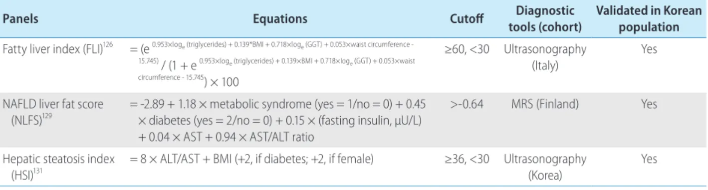
In addition to radiological examinations, various panels of sero- logical tests have been proposed to diagnose hepatic steatosis and assess its severity (Table 3). These panels can be calculated using clinical information such as age, sex, and the results from serological tests. These panels do not directly diagnose hepatic steatosis, unlike ultrasonography, but they can help physicians who suspect the presence of hepatic steatosis to decide whether further assessments are justified. Large-scale studies frequently use these noninvasive panels to test for hepatic steatosis instead of ultrasonography, which has a high cost.
The FLI was proposed by Bedogni et al.
126in an Italian study that examined 216 subjects with liver disease and 280 subjects with healthy livers. In that study, 228 patients had ultrasonogra- phy-defined NAFLD. The FLI is calculated based on triglycerides, gamma-glutamyl transpeptidase, BMI, and waist circumference. If the FLI is less than 30, NAFLD can be excluded (negative likeli- hood ratio, 0.2), and if the FLI is more than 60, NAFLD can be di-
agnosed (positive likelihood ratio, 4.3). An FLI score of more than 60 has a positive predictive value of 99% and a negative predic- tive value of 15%. The AUC of the FLI was 0.84. The FLI showed acceptable accuracy among a Korean population.
127,128The NAFLD liver fat score (NLFS) was proposed by Kotronen et al.
129in a Finnish study comprising 470 subjects. That study used MRS to diagnose NAFLD. The NLFS is calculated based on meta- bolic syndrome, T2DM, fasting insulin, aspartate aminotransferase (AST), and the AST/alanine aminotransferase (ALT) ratio. Its cutoff is -0.640 (sensitivity, 86%; specificity, 71%). If the NLFS is less than -0.640, NAFLD can be excluded, and if it is more than -0.640, NAFLD can be diagnosed. The AUC of the NLFS was 0.86–0.87. NLFS showed acceptable accuracy in a Korean popu- lation.
130The HSI was proposed by Lee et al.
131in a Korean cohort study of 10,724 subjects (5,462 with ultrasonography-defined NAFLD).
The HSI is calculated based on sex, BMI, AST, ALT, and T2DM. If the HSI is less than 30, NAFLD can be excluded (negative likeli- hood ratio of 0.2, sensitivity of 93.1%), and if it is more than 36, NAFLD can be diagnosed with high predictive accuracy (positive likelihood ratio of 6.1, specificity of 92.4%). The AUC of the HSI was 0.81. The HSI showed acceptable accuracy among a Korean population.
132The noninvasive surrogates just described show acceptable ac- curacy in most cross-section studies and have been used to trace changes in hepatic steatosis in certain studies. However, the use- fulness of those noninvasive surrogates in monitoring disease progression and evaluating treatment response should be further investigated.
Table 3. Panels for hepatic steatosis
Panels Equations Cutoff Diagnostic
tools (cohort)
Validated in Korean population Fatty liver index (FLI)
126= (e
0.953×loge (triglycerides) + 0.139*BMI + 0.718×loge (GGT) + 0.053×waist circumference -15.745)
/ (1 + e
0.953×loge (triglycerides) + 0.139×BMI + 0.718×loge (GGT) + 0.053×waist circumference - 15.745) × 100
≥60, <30 Ultrasonography (Italy)
Yes
NAFLD liver fat score (NLFS)
129= -2.89 + 1.18 × metabolic syndrome (yes = 1/no = 0) + 0.45
× diabetes (yes = 2/no = 0) + 0.15 × (fasting insulin, μU/L) + 0.04 × AST + 0.94 × AST/ALT ratio
>-0.64 MRS (Finland) Yes
Hepatic steatosis index (HSI)
131= 8 × ALT/AST + BMI (+2, if diabetes; +2, if female) ≥36, <30 Ultrasonography (Korea)
Yes
BMI, body mass index; GGT, gamma-glutamyl transpeptidase; AST, aspartate aminotransferase; ALT, alanine aminotransferase.
[Recommendations]
1. Abdominal ultrasonography, CAP, unenhanced CT, MRS, and MRI-PDFF are acceptable modalities for diagnosing hepatic steatosis. (A1)
2. If radiological examinations are infeasible, panels for hepatic steatosis can be used to assess hepatic steatosis. (B1)
Which non-invasive surrogates are available to diagnose NASH?
NASH is significantly associated with liver fibrosis progression and HCC. Non-invasive modalities to diagnose NASH are limited, but a prediction model based on liver stiffness, CAP (assessed us- ing transient elastography), and ALT levels was recently proposed by a Korean study.
133Some studies insist that NASH can be distin- guished from NAFL using cytokeratin-18 fragments (sensitivity, 66%; specificity, 82%).
134,135A few studies claim that CT or MRI can be used to differentiate NASH from NAFLD, but no clear diag- nostic criterion has been set.
101,136Based on the association be- tween the viscosity shown in shear wave dispersion imaging and necroinflammation of the liver parenchyma, a recent study showed that shear wave dispersion imaging could be helpful in diagnosing NASH.
137The AUC of magnetic resonance elastogra- phy (MRE) alone and that for the combined use of MRE and MRI- PDFF in differentiating NASH from NAFL was 0.82–0.93.
138,139Recently, multiparametric MR indices, which score the results from various MRI techniques, have been under investigation. A recent Korean study found that a multiparametric MRI index using MRE, MRS, and T1 mapping to differentiate NASH from NAFL showed sensitivity of 80%, specificity of 85.2%, and an AUC of 0.883.
140In a meta-analysis that systematically reviewed all stud- ies using MRI to differentiate NASH from NAFL, the pooled sensi- tivity and specificity were 87.4% and 74.3%, respectively.
110[Recommendations]
1. Non-invasive diagnosis of NASH remains limited, so it should be diagnosed by liver biopsy. (A1)
Which non-invasive surrogates are available to assess liver fibrosis?
The assessment of liver fibrosis is crucial in patients with NAFLD because the degree of liver fibrosis is significantly associated with long-term outcomes such as the development of HCC and liver-re- lated death.
78Furthermore, it is important to noninvasively assess
the regression or progression of liver fibrosis during the course of anti-fibrotic therapy.
Radiological surrogates
Ultrasound-based measurement techniques for liver fibrosis take advantage of shear wave elastography (SWE). They can be divided into two categories: 1) measuring the elasticity of the liver parenchyma using SWE without acquiring imaging data (transient elastography) and 2) image-based sonoelastography that acquires both elasticity and 2D image data (point SWE and 2D SWE). Tran- sient elastography is widely used in clinical practice, and many re- searchers have reported its high performance in quantifying liver fibrosis in NAFLD patients. In a recent meta-analysis, transient elastography showed high sensitivity and specificity for evaluating the degree of liver fibrosis in NAFLD patients.
48,135,141However, the accuracy of transient elastography is limited in obese patients, who commonly have NAFLD, making it unavailable in 5–20% of patients.
48,142A recent study reported that using an XL probe rath- er than an M probe can significantly lower the failure rate of tran- sient elastography.
119,143Image-based techniques have the advantage of acquiring both elasticity data and 2D images simultaneously.
The failure rate is lower than that of transient elastography be- cause operators can choose the area of the liver parenchyma to acquire elasticity data. The AUC of point SWE for quantifying liver fibrosis in NAFLD patients was higher than 0.8.
144,145The perfor- mance of point SWE for advanced liver fibrosis was excellent (100% sensitivity and 91% specificity).
146In a recent meta-analy- sis, the diagnostic performance of point SWE for liver fibrosis was similar to that of transient elastography.
1472D SWE can obtain elasticity data from a wider area than point SWE, and the report- ed failure rate of 2D SWE was lower than that of point SWE.
148,149In a prospective study, 2D SWE, MRE, and transient elastography had similar AUCs for advanced hepatic fibrosis (0.920, 0.929, and 0.915, respectively).
150MRE shows high diagnostic accuracy for liver fibrosis.
138,151,152In
contrast to transient elastography, which can examine the elastic-
ity of only a small portion of liver tissue, MRE can evaluate the
entire liver parenchyma.
152MRE also has other advantages: opera-
tor non-dependency and no limitations for obese patients. MRE is
the most accurate non-invasive test for liver fibrosis, with a diag-
nostic performance superior to that of transient elastogra-
phy.
124,141,153In a meta-analysis, MRE correlated well with each stage
of liver fibrosis, with AUCs for each stage of 0.84–0.93.
154,155The
failure rate of MRE was less than 5%, which is significantly better
than that of transient elastography. MRE was not significantly af-
fected by the MRI manufacturer or the strength of the magnet.
156In addition, MRE showed robust reproducibility in repetitive ex- aminations.
157However, the high cost of MRE limits its availability in clinical practice. In patients with iron deposition in the liver pa- renchyma, it is difficult to perform MRE, and the presence of other infiltrative diseases, such as profound hepatic steatosis, hepatic congestion, or acute inflammation, can attenuate the diagnostic accuracy of MRE.
158,159Panels
In addition to radiological examinations, various panels of sero- logical tests have been proposed to diagnose liver fibrosis (Table 4).
Well-validated panels are summarized here.
Of the noninvasive panels for liver fibrosis, the NFS has been studied the most. The NFS was proposed in a US study by Angulo et al.
160that comprised 733 subjects with biopsy-proven NAFLD.
Two cutoffs were proposed: <-1.455 (low probability, negative predictive value of 88–93%) and >0.676 (high probability, posi- tive predictive value of 82–90%).
160A meta-analysis based on 13 studies with 3,064 subjects showed that the AUC of the NFS for advanced liver fibrosis was 0.85. If the NFS is less than -1.455, advanced liver fibrosis can be excluded with a sensitivity of 90%
and a specificity of 60%, and if NFS is more than 0.676, advanced liver fibrosis can be diagnosed with a sensitivity of 67% and a specificity of 97%.
48,160-172Among 412 Korean subjects with biop- sy-proven NAFLD, an NFS of less than -1.455 allowed advanced liver fibrosis to be excluded with a high negative predictive value (86.6%), and an NFS of more than 0.676 allowed advanced liver fibrosis to be diagnosed with a positive predictive value of 50%.
173In another Korean study, which recruited 315 subjects with biop- sy-proven NAFLD, the cutoff values (<-1.455 and >0.676) showed an AUC of 0.84 in diagnosing advanced liver fibrosis (negative
predictive value of 89.3–95.7%).
174However, cases that fall be- tween the cutoff values (indeterminate probability) still require a liver biopsy.
48The fibrosis-4 index (FIB-4) was proposed by Sterling et al. in a study comprising 832 subjects with human immunodeficiency vi- rus/hepatitis C virus co-infection. FIB-4 is calculated using the platelet count, age, AST, and ALT. The AUC of FIB-4 for advanced liver fibrosis was 0.765. When the FIB-4 is less than 1.30, ad- vanced liver fibrosis can be excluded (accuracy, 90%), and when FIB-4 is more than 2.67, advanced liver fibrosis can be diagnosed (accuracy, 80%).
175A recent study of subjects with biopsy-proven NAFLD showed that the diagnostic accuracy of the NFS and FIB-4 was significantly higher than that of other noninvasive panels for liver fibrosis and similar to that of MRE in diagnosing advanced liver fibrosis.
141However, because the diagnostic accuracy of FIB-4 was inferior to that of the NFS in a Korean study, further valida- tion studies are required.
173The enhanced liver fibrosis (ELF) panel has recently been used to assess liver fibrosis in Europe. ELF was proposed by Guha et al.
163in a UK study comprising 192 subjects with biopsy-proven NAFLD. ELF is calculated based on three proteins associated with liver fibrosis: hyaluronic acid, tissue inhibitor of metalloproteinase 1, and amino terminal peptide of procollagen III. The cutoff value and AUC of ELF for advanced liver fibrosis were 0.3576 and 0.90, respectively (sensitivity, 80%; specificity, 90%; positive predictive value, 71%; negative predictive value, 94%).
163Other serological surrogates for liver fibrosis, such as M2BPGi and AsAGP, have been proposed.
176-179However, because few data for subjects with NAFLD are available, further validation studies are required. The noninvasive surrogates described above show acceptable accuracy and prognostic values in most cross- section studies. However, the usefulness of noninvasive surro-
Table 4. Panels for liver fibrosisPanels Equations Cutoff Diagnostic
tools
Validated in Korean population NAFLD fibrosis score
(NFS)
= -1.675 + 0.037 × age (years) + 0.094 × BMI (kg/m
2) + 1.13 × impaired fasting glucose or diabetes (yes=1, no=0) + 0.99 × AST/ALT ratio - 0.013 × platelet count (×10
9/L) - 0.66 × serum albumin (g/dL)
<-1.455, >0.676 Liver biopsy Yes
Fibrosis-4 index
(FIB-4)
413= Age (years) × AST [U/L]/ (platelets [10
9/L] × (ALT [U/L])
1/2<1.3, >2.67 Liver biopsy Yes Enhanced liver fibrosis
(ELF)
163,414Patented (hyaluronic acid, tissue inhibitor of metalloproteinase 1, amino terminal peptide of procollagen III as constituent variables)
0.3576 Liver biopsy Yes
NAFLD, nonalcoholic fatty liver disease; BMI, body mass index; AST, aspartate aminotransferase; ALT, alanine aminotransferase.
gates in monitoring disease progression and evaluating treatment response should be further investigated.
[Recommendations]
1. Radiological examinations such as transient elastography, point SWE, 2D SWE, and MRE are helpful in assessing hepatic fibrosis. (A1)
2. If radiological examinations are infeasible, panels such as NFS or FIB-4 can be used to diagnose liver fibrosis. (B1)
Which differential tests are available for advanced fibrosis?
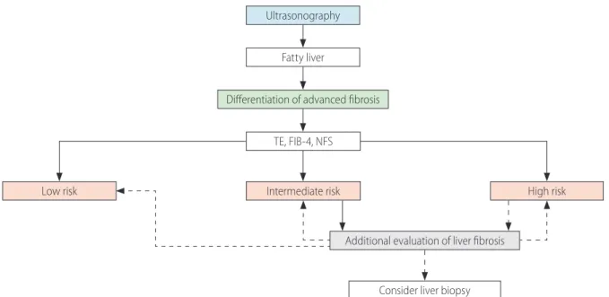
In NAFLD, liver fibrosis testing uses serologic tests, image tests, or liver biopsy. Because liver biopsy cannot be performed in all patients, an algorithm can be used to differentiate advanced fi- brosis (Fig. 2). Advanced fibrosis can be differentiated primarily using transient elastography, FIB-4, and the NFS.
180When subjects are classified as intermediate risk by transient elastography, FIB-4, or the NFS, additional tests such as M2BPGi, AsAGP, ELF, SWE, or MRE can be performed. If the algorithms suggest advanced fibrosis, additional tests to re-evaluate the liver fibrosis or a liver biopsy should be considered.
[Recommendations]
1. Non-invasive methods such as transient elastography, FIB-4, and NFS are prioritized to discriminate advanced liver fibrosis.
(A1)
2. Serological tests, imaging tests, and liver biopsies may be performed as additional liver fibrosis evaluations. (B1)
Liver biopsy
What are the indications for liver biopsy?
Non-invasive tests to replace liver biopsy have been developed and shown high accuracy.
122,181,182However, liver biopsy remains the gold standard for diagnosing NAFLD.
183In practice, it is diffi- cult to perform liver biopsy in all patients suspected of having NAFLD.
184,185However, liver biopsy can help with diagnosis, treat- ment planning, and distinguishing between NAFLD and other liver diseases (autoimmune hepatitis, drug-induced hepatitis, Wilson’s disease, etc.). Liver biopsy is necessary when NASH or advanced liver fibrosis is suspected, as well as when other liver diseases cannot be excluded.
186-188Liver biopsy has several limitations. First, sampling error is a concern because only a small portion of the liver tissue is sampled during liver biopsy. Second, intra- and inter-observer variability occurs.
189-191Third, it carries the risk of complications such as bleeding and infection, as well as increased medical costs. There-
Figure 2. Algorithm to differentiate advanced fibrosis. TE, transient elastography; FIB-4, fibrosis-4 index; NFS, nonalcoholic fatty liver disease fibrosis