Ⅰ. 서 론
타액선에는 위축성 퇴행에 의한 손상이나 폐쇄 등이 발생할 수 있고, 타액선의 실질이 염증성 세포로 대체되는 괴사의 반
응이 나타날 수도 있으며, 타액선의 세균감염이나 면역 반응 등에 의해서 변성 및 섬유화 등의 반응을 일으키게 된다. 타액 의 도관 폐쇄는 관 내관의 협착, 관 내강에 물체가 삽입되어 나 타나는 관 폐쇄, 조직 내의 점액 축적으로 인한 관 손상의 결과 로서 나타나며, 모두에서 타액이 관 내강에 축적되어 이로 인 한 압력이 선포세포에 전달되어 선포의 위축성 변성이 일어나 고 세포 변성과 괴사의 과정을 겪게 된다1-5)
.
폐쇄성 타액선염 은 임상적으로 흔한 타액선 질환 중의 하나이며, 가장 큰 조직 학적 특징은 선포세포의 소실과 선도관의 확장 및 섬유화, 염 증세포의 침윤으로 특징지어 진다6,7). 이러한 만성 폐쇄성 타액선
염에서의 다양한 선세포의 실험적 연구가 진행되어 왔으나8-12),
김 경 욱330-716
충남 천안시 신부동 산7-1
번지 단국대학교 치과대학 구강악안면외과학교실 Kyung-Wook KimDept. of Oral & Maxillofacial Surg., School of Dentistry, Dankook University, 7-1 Sinbudong, Cheonan, 330-716, Korea Tel: 82-41-550-1993 Fax: 82-41-551-8988
E-mail: [email protected]
가토 악하선 도관 절단술 및 결찰술 후 근상피세포의 변화
윤원석∙김철환∙김경욱
단국대학교 치과대학 구강악안면외과학교실
Abstract (J. Kor. Oral Maxillofac. Surg. 2006;32:81-93)
THE CHANGE OF MYOEPITHELIAL CELL AFTER LIGATION AND CUT OF SUBMANDIBULAR GLAND DUCT IN RABBIT
Weon-Suk Yun, Chul-Hwan Kim, Kyung-Wook Kim
Dept. of Oral & Maxillofacial Surgery, School of Dentistry, Dankook University
Obstructive sialadenitis is one of common disease in salivary gland, and most common histologic features are loss of acinar cell and ductal dilatation associated with fibrosis, and infiltration of inflammatory cells. Although many experimental studies has been accomplished for the salivary acinar cell change in obstructive salivary gland disease, studies for myoepithelial cell were deficient. This study is designed for salivary gland tissue change, especially myoepithelial cell when nonspecific chronic sialadenitis or salivary duct injury by duct obstruction or cut can be occurred that is common encounted clinically. After ligation and cutting of submandibular gland of rabbit, groups of aminmal were sacrificed at 1, 2, 4 weeks postoperatively, submandibular gland were removed. The histopathologic evaluation was done with light microscopy. And, with immunohistochemical staining with α-smooth muscle actin, characteristics of myoepithelial cell were examined. With transmission electron microscopy, ultrastructure of myoepithelial cell were examined for distribution and ultrastructure of myoepithelial cell.
The results were obtained as follows:
1. In the histopathologic evaluation, ligation and cutting group of 1 week, linkage of myoepithelial cell associated with acinar atrophy and degenera- tion were disappeared in both group.
2. More prominent squamous metaplasia was seen in acinar cells of ligation group of 2 weeks experimental rabbit than cutting group.
3. Acinar cells are nearly disappeared in both ligation and cutting group of 4 weeks, and myoepithelial cell also disappeared associated with acinar cell atrophy, and duct-like structure composed by squamous cells by squamous metaplasia in acinar cells were distributed.
4. In immunohistochemical study, both ligation and cutting group α-SMA distribution were diminished at 1 week experimental rabbits, but myoep- ithelial cell was more diminished in ligation group than cutting group, which were distributed around cells of squamous metaplasia.
5. Nuclear condensation, chromosome margination, and cytoplasmic vaculoation were appeared in myoepithelial cell of both cutting and ligation group after 1 week with transmission electron microscopy. But degenerative substance were seen in cytoplasm of myoepithelial cell of ligation group of 4 weeks.
From the results obtained in this study, atrophy and degeneration of myoepithelial cell was more prominent in duct ligation group than duct cutting group, and myoepithelial cells were seen around cells squamous metaplasia of acinar cell.
Key words: Obstructive sialadenitis, The change of myoepithelial cell, Submandibular gland duct
폐쇄성 만성 타액선염에서의 근상피세포의 변화에 대한 연구 는 미미하다. 근상피세포는 1898년 이하선에서 처음으로 기술 되었으며, 선포 상피층의 기저부나 개재관 혹은 선조관의 기 저부에 존재한다13)
. 이는 성상의 모양으로 존재하며 수지상의
긴 세포질을 가지는14)일명“basket cell”이라고도 명명되기도하였다13,15)
. 근상피세포는 세포질에 존재하는 근원섬유의 작용
으로 분비단위의 압력을 높혀 타액의 분비를 촉진하는 기능을 가지고 있으며, 변화가 많은 분화로 다양한 간질조직을 구성 하는 중추적 역할을 담당하고 있다16)
. 최근 면역조직화학적 검
사법 중, α-SMA(smooth muscle actin)은 타액선과 유선 조직의 근
상피세포의 표지자로 알려져 왔으며, 정상 타액선의 근상피세 포에서 강한 염색반응이 일어난다17,18).
본 연구에서는 임상적으로 많이 접할 수 있는 도관 폐쇄나 절단 등의 타액선 도관의 손상이나 비특이적 만성 악하선염 존재 시, 타액선 조직, 특히 근상피세포의 변화를 광학현미경 적, 전자현미경적으로 관찰하고, α
-SMA(smooth muscle actin)을
이용한 면역조직화학적 검사를 통한 근상피세포의 분포를 관 찰하여 임상적으로 가장 흔한 타액선 질환인 폐쇄성 타액선염 의 임상적 치료에 도움이 되고자 하였다.Ⅱ. 연구재료 및 방법 1. 연구재료
실험동물로는 일정기간동안 동일 조건에서 사육된 체중 3
kg
내외의 웅성가토 24마리를 사용하였다. 대조군으로 4마리 를 정상 악하선을 관찰하기 위하여 사용하였고, 실험군으로는 양쪽 악하선을 노출시켜, 도관 결찰군 20 부위, 도관 절단군 20 부위를 배정하였다.2. 연구방법
(1) 동물실험
실험군은 Ketamine hydrochloride(Ketalar�
, 유한양행) 10 mg/kg
및 0.15 ml/kg의 2% xylazine hydrochloride(Rumpun�, 한국바이엘)
을 근주하고, 양쪽 경부에 털을 제거하고 베타딘 용액으로 소 독한 후, 지혈을 목적으로 1:100,000의 epinephrine이 함유된0.2% 염산 리도카인(광명제약)을 침윤 주사하였다. 가토의 경
부에 수평절개를 가하고 층별 이단 후 양쪽 악하선을 노출시 킨 후 도관을 분리하여 한쪽 부위는 3-0 nylon을 이용하여 도관 의 결찰술을 시행하고, 한쪽 부위는 도관의 절단술을 시행하였다. 시행 후 양쪽 악하선을 재위치 시킨 후 층별 봉합을 시행 하였다.
(2) 희생
대조군과 실험군 모두 Ketamine hydrochloride(Ketalar�
, 유한양
행)를 이용하여 심장에 다량 주사하여 희생시킨 후 정상 가토4마리는 정상 악하선 8개를 채취하여 대조군으로 사용하였고,
실험군은 H&E염색, MT염색 및 면역조직화학 염색 및 투과전 자현미경의 관찰을 위해 1주, 2주 및 4주째에 희생하여 양쪽 악 하선을 채취하였다.(3) 표본 제작
가. H&E 염색 및 MT염색
도관을 결찰 혹은 절단한 실험군의 악하선을 채취하여 10%
중성 포르말린에 고정한 후, 4 ㎛ 파라핀 절편을 제작하고 통법 에 따라 H&E 염색 및 MT염색을 시행하였고, 광학현미경하에 서 200배로 조직병리학적 특징을 관찰하였다.
나. 면역조직화학적 검사
절취한 조직을 면역조직화학적 염색을 시행하여 조직병리 학적 특성을 관찰하였다. 먼저 절취한 조직을 고정한 후 Poly-
L-Lysine으로 처리된 슬라이드에 4-8 ㎛ 파라핀 절편을 제작하
고 면역조직화학적 염색을 시행하기 위하여 절편을 0.3% H2O
2 에 5분간 부란시키고 10 mM phosphate buffer solution(PBS)으로 세척한 후, 제 1차 항체로 처리한 후(Table 1) LSAB (labelledstreptavidine biotin)법으로 처리하고 난 후, 발색제로 Diami- nobenzidine을 이용하였다. 발삼으로 봉입한 다음 Mayer’s Hematoxylin으로 대조염색을 시행하여 광학현미경으로 200배
하에서 관찰하였다(Table 2).다. 투과전자현미경(TEM)을 이용한 관찰
도관 절단 및 결찰후 1, 2 및 4주째에 악하선 조직을 적출하여 조직을 1 ㎣의 크기로 잘라 2.5% glutaraldehyde에 넣은 후 고정 액은 acetate veronal을 사용하여 pH 7.2로 완충시키고 1% osmi-
um tetraoxide를 사용하여 30-60분 후고정한다. 고정액을 증류수
로 씻어낸 후 알코올로 탈수시키고, n-butyl methacrylate에 침윤 시킨 후, 젤라틴 캡슐에 포매하고, 60℃에서 중합하고 촉매제 는 1% benzoyl peroxide를 사용하였다. 0.2 ㎟ 정도 되는 블록에 서 초박절편을 제작하고 탄소격자상에 올려놓고 Uranyl acetate 와 Lead citrate로 염색한 후 투과전자현미경(JEOL JEA CX II,JAPAN)으로 관찰하였다.
Table 1.
Specification of primary antibody used in this studyHost/Subclass Dilution Manufacture
α
-SMA Mouse/IgM 1 : 100 Chemicom
Ⅲ. 연구결과 1. H&E 소견
가. 대조군
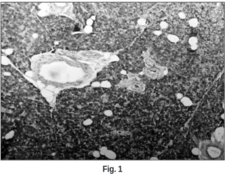
선방세포는 장액성 및 점액성 세포로 구성되어 있었고, 입방 형 및 원주형 상피세포로 이루어진 개재도관(intercalated duct)이 관찰되었다. 간질조직에 분비도관(excretory duct)이 보였고, 선조 도관(striated duct)은 윤관세포와 연결되어 있었다. 근상피세포는 선방세포를 싸고 있었으며, 방추형의 모양을 보였다(Fig. 1).
나. 실험군
① 1주 소견
도관 절단군에서는 선방세포의 퇴축이 보였으며, 간질조직 의 세포층에 만성 염증세포의 침윤이 관찰되었고, 도관은 결 체조직과 분리되어 있었다(Fig. 2). 도관 결찰군에서는 분비도 관에 호산성 물질이 저류되어 있었으며, 염증 반응으로 파괴 된 선방세포도 있었으며, 도관세포의 탈락이 보였다(Fig. 6). 부 분적으로 편평세포로의 화생(metaplasia)이 보였으나, 양 군 모 두에서 근상피세포는 선방세포의 퇴축과 파괴로 그 연결상이 소실되었다.
② 2주 소견
도관 절단군에서는 만성 염증세포의 침윤으로 선방세포의 소실이 뚜렷해졌으며 도관이 결체조직과 분리되어 있었다
(Fig. 3).
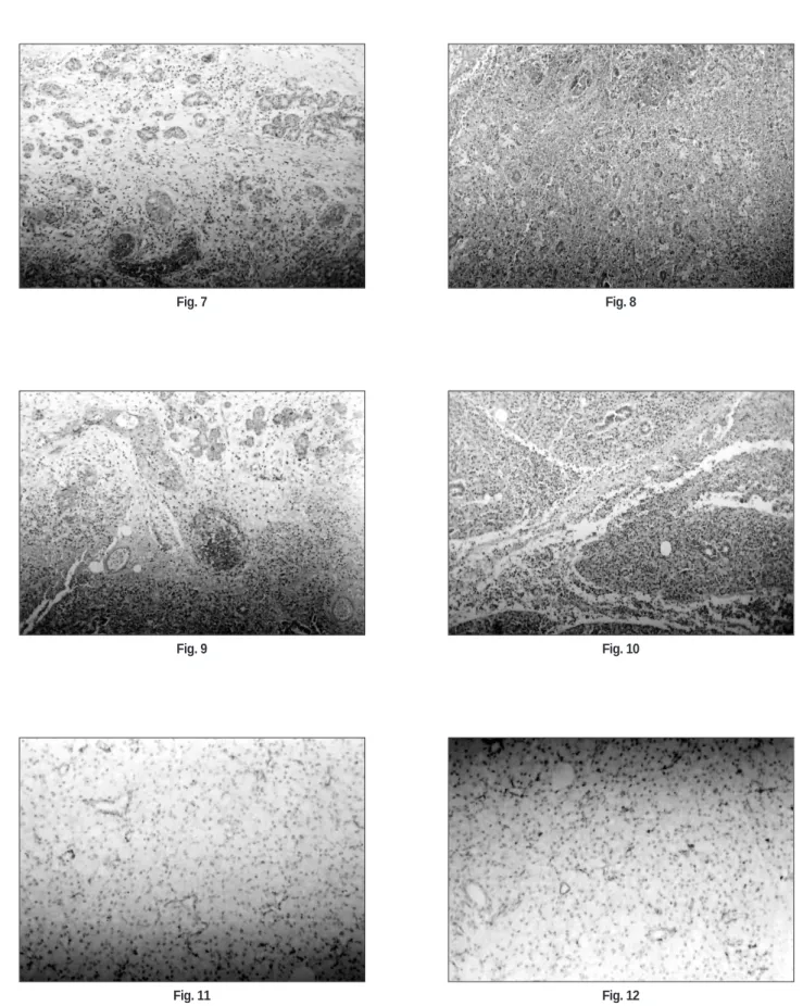
도관 결찰군에서는 분비도관에 호산성 물질이 저류되어 있 었으며, 거의 모든 도관세포의 탈락이 보였다. 편평세포로의 화생은 결찰군에서 더욱 뚜렷하게 보였으나(Fig. 7), 양 군 모두 에서 근상피세포는 선방세포의 퇴축과 파괴로 그 연결상이 소 실되었다.
③ 4주 소견
양 군 모두에서 간질조직의 세포에 만성 염증세포의 침윤으 로 선방세포의 소실이 더욱 뚜렷해 졌으며, 도관이 결체조직 과 분리되어 있었다. 선방세포의 편평세포로의 화생은 결찰군 에서 더욱 뚜렷하게 보였으며, 대부분 선방세포 주위에 근상 피세포의 소실이 관찰되었다(Fig. 5, 9 & 10).
2. 면역조직화학적 염색 결과
가. 대조군
본 연구의 면역조직화학 염색을 위하여 근상피세포에 특징 적으로 염색되는 α
-SMA(α -smooth muscle actin)을 사용하였다.
강한 양성 반응을 보이는 근상피세포는 선방세포와 개재도관 을 둘러싸고 있으며, 상호 연결되어 관찰되었다. 그러나 선조 도관에서는 면역조직화학적 염색반응이 나타나지 않아 근상 피세포는 발견되지 않았다(Fig. 11).
나. 실험군
1주 소견에서 양군 모두에서 α -SMA 양성 세포가 감소하였으
나, 도관 결찰군에서보다는 도관 절단군에서 더 많은 α
-SMA Table 2.Procedure for immunohistochemical staining
Paraffin section preparation
�
Deparaffination & hydration
�
Peroxydase inactivation (3% H
2O
2, 5 mim.)
�
Serum blocking reagent (20 min.)
�
Primary Ab (4 hrs.)
�
LASB(labelled streptavidin biotin) method (30 min.)
�
Deaminobenzidine
�
Counter staining with Mayer’ s hematoxylin
�
Balsam mounting
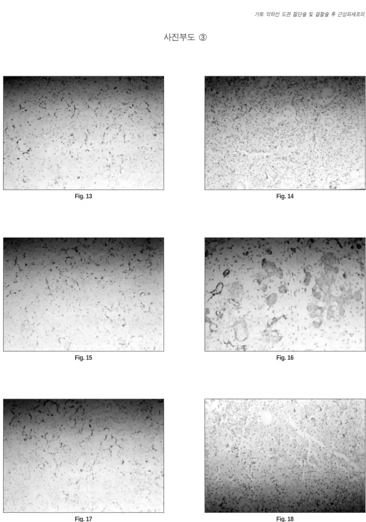
양성 세포가 관찰되었으며(Fig. 12, 17), 2주 소견에서는 근상피 세포의 퇴화로 인하여 1주 소견의 양군의 관찰 때보다 더 적은
α
-SMA 양성 세포가 관찰되었고(Fig. 13, 18), 4주에서는 근상피
세포의 심한 퇴화로 인하여 α
-SMA 양성 세포가 많이 감소된
관찰소견이 보였다. 또한 4주 군의 α-SMA 양성 세포는 결찰군
에서는 거의 관찰되지 않았다(Fig. 13-16, 19-21).3. 투과전자현미경(TEM)을 이용한 악하선 세포의 관찰
가. 대조군
윤관세포에서는 미토콘드리아와 형질세망이 뚜렷하게 관찰 되었고, 선방세포에서는 분비과립이 다양한 전자치밀상을 보 였다. 선방세포와 개재도관 주위에 방추형 세포질을 보이며 타원형의 염색질과 근세사가 풍부한 근상피세포가 보였다.
나. 실험군
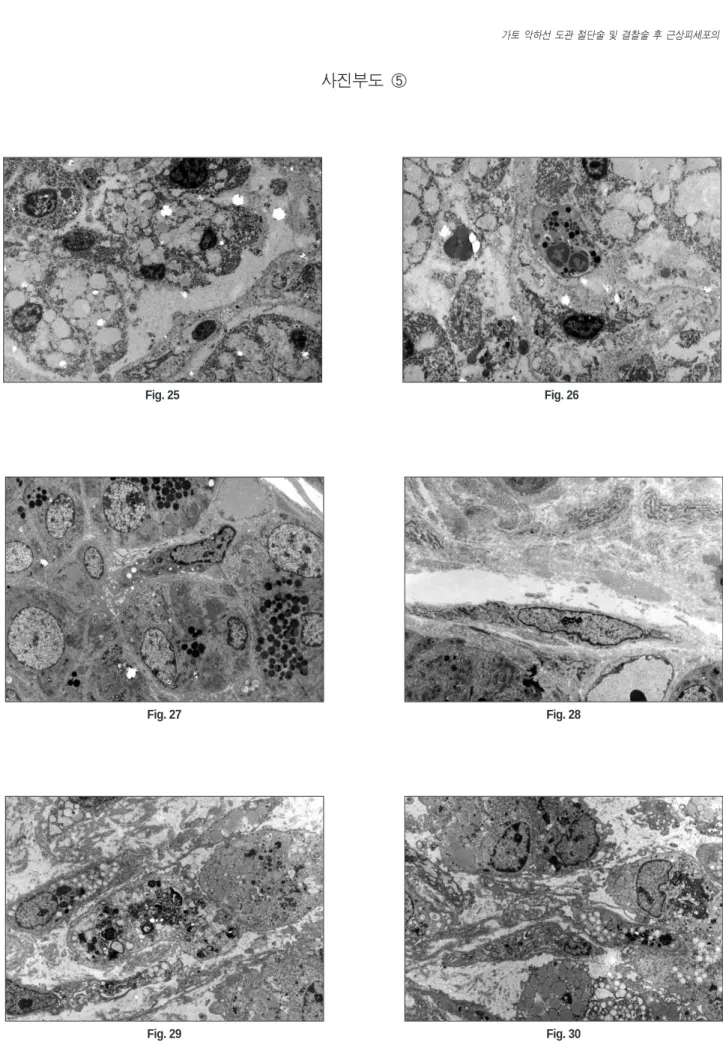
도관 절단군의 1주 소견에서 근상피세포는 핵이 농축된 형 태를 가지고 있었으며, 근상피세포 세포질은 염색체의 변연추 향(margination)과 많은 공포를 보였다(Fig. 22, 23). 도관 절단군 의 2주 및 4주 소견에서는 점액세포와 근상피세포의 퇴행성 변 화를 관찰할 수 있었다(Fig. 24, 25).
도관 결찰군에서 1주 소견에서는, 점액세포와 근상피세포의 퇴행성 변화가 나타났으나, 장액세포에서는 퇴행성 변화가 나 타나지 않았다(Fig. 26, 27). 도관 결찰군의 2주 소견에서는 염색 체의 변연추향을 동반한 근상피세포의 퇴행성 변화가 관찰되 었고(Fig. 28), 4주 소견에서는 층판형 구조를 가지며, 분절된 염 색체와 공포화를 동반하는 점액세포의 퇴행성 변화가 관찰되 었고, 근상피세포 또한 분절된 염색체와 공포화를 동반하는 퇴행성 변화를 나타내었다(Fig. 29, 30).
Ⅳ. 총괄 및 고찰
타액선은 많은 선포로 구성된 분비 단위의 집합체를 특징으 로 하는 외분비성 관상형의 구조체이다15)
. 분비 단위는 amylase
를 함유하는 장액성과 sialomucin을 분비하며 점액성과, 장액성 세포와 점액선 세포의 중간인 혼합성의 3 가지로 분류되며 그 분비단위의 구분에 따라 장액성선, 점액성선, 혼합성선으로 구분되어진다. 인체에서는 이하선은 대부분 장액선 선포세포 로 구성되어 있으며, 악하선 및 설하선은 혼합성 선포세포가 많이 분포되어 있다.선포세포는 명확한 기저막으로 둘러싸인 상피세포의 배모 양의 군락으로 구성된 장액성 선포와 이보다 좀 더 크면서 불 규칙한 형태의 점액성 선포로 그 조직학적 특성이 나누어진 다. 선포세포는 기저막에 의해 둘러싸인 수축성의 근상피세포 로 둘러싸여있다. 분비액과 단백질은 작은 입방세포로 구성된 개재관(intercalated duct)을 통과하여 미토콘드리아가 풍부한 선 조관(striated duct)으로 진행되어 가며, 이 2개의 도관은 소엽내 관(interlobular duct)의 일부이다. 많은 선포의 군락들은 다시 더 커다란 관으로 모이게 되는데 그 구조는 basilar reverse cell, 중층 상피세포와 강 내부를 둘러싸는 입방형, 또는 원주형 세포로 구성되어 소엽외분비관으로 이행된다. 즉 큰 분비관과, 소엽 사이관 또는 일명 배출관으로 구성된다19,20)
.
근상피세포는 많은 외분비선에서 관찰되는 세포로서, 선포 세포와 도관을 둘러싸고 있다21)
. 비록 근상피세포는 그 구조와
기능이 평활근세포와 유사하지만, 분비세포 또는 도관세포와 기저막 사이에 위치하여 상피세포의 특성도 함께 가지고 있으며22,23)
, 사상체를 함유하고 있다
24). 타액선에서의 근상피세포는
선포와 개재관의 기저층과 상피세포 사이에 존재하며 또한 선 조관과 개재관의 단위 내에 존재한다. 이 세포는 성상 형태를 나타내며, 수지상의 긴 세포질 돌기를 갖는다25)
. 또한 기저막
쪽으로 세포질 선조를 갖는 특징적인 소견을 보인다. 그 세포 질에 존재하는 근원섬유의 기능으로 분비단위의 압력을 높혀 타액의 분비를 촉진하는 것으로 알려져 있다. 일종의 수축성 기능세포이며, 저장과 세포 흡수 능력을 갖는다고 하였다16,26-29).
일반적으로 근상피세포는 이하선 보다는 악하선에서 더 많이 분포하며, 이하선에서는 주로 개재관에 분포하지만, 악하선에 서는 선포와 개재관 주위에 많이 분포한다21, 30-32). 또한 근상피세
포는 다형성 선종, 선양낭성암종 등의 타액성 종양의 조직발 생에도 중요한 역할을 한다고 알려져 왔으나27,33-36), 현재까지는
완전히 밝혀지지 않았다.폐쇄성 타액선염은 임상적으로 흔하게 나타나는 질환으로 써, 조직학적으로는 심한 또는 전체 선포세포의 상실과, 도관 의 확장 및 섬유화, 염증성 세포의 침윤으로 특징지어 진다7,37)
.
일반적으로 페쇄성 타액선염의 조직학적 소견은 도관 내층은 편평상의 세포와 점액세포의 화생(metaplasia) 등의 다양한 반 응성 변화를 보이지만, 도관 내층이 중층편평상피로 비후된 경우에는 내강에 위치한 세포에서 점액성 술잔세포(mucousgoblet)가 나타날 수 있고, 도관주위의 결합조직에서는 많은 염
증성 세포의 침윤을 보인다. 분비선에서는 초기에서 선포는 변성이 일어나고 일부 변성된 선포가 소엽 내에서 관찰되며,Table 3.
α-SMA expression of myoepithelial cell in submandibualr gland of rabbit after cutting and ligationControl 1 week 2 week 4 week
Cutting Group 75±9 70±4 60±4 20±3
Ligation Group 75±7 64±5 30±3 5±1
(Mean±SD, α -SMA positive cells at random 5 site under ×200 power field)
압력의 변화가 증가될수록 도관의 확장은 현저해지고 선포위 축은 진행되어 몇 개의 선포만이 남아있게 된다. 도관 폐쇄 후, 시간이 경과할수록 소엽은 콜라겐화되며, 페쇄된 분비선은 역 행성 세균성 타액성염의 소견을 나타내며, 관 내강에서 화농 성 물질과 염증성 세포가 출현하게 된다2,3,38)
. 이와 같은 조직학
적 변화는 이전에는 선포세포가 도관세포로의 분화의 억제에 의하여 일어난다고 생각되었으나39-41), 그 후 Walker 등의 실험
연구에서 이하선의 도관 결찰을 시행한 연구에서 타액선 선포 세포의 빠른 소실은 세포사멸의 과정으로 밝혀지게 되었고, 이하선의 위축과 더불어 도관세포의 증식이 동반되는 과정을 거치는 것이 밝혀지게 되었다42). 또한 이와 유사한 결과가 도관
결찰된 췌장의 변화에서도 밝혀져 왔다43-47).
일반적으로 퇴행성 악하선에서의 근상피세포의 변화는 세 포구조가 불규칙한 형태로 변화하면서 퇴행성 변화를 한다
42,48)
. 근상피세포는 세포돌기의 확장과 기저막의 불규칙적인
두께의 증가를 보이는데 이는 새로운 근상피세포의 합성과 관 련이 있다고 하였다21,49)
. 이러한 근상피세포의 변화는 분비관
의 폐쇄뿐만 아니라, 고삭신경의 손상을 통해서도 나타날 수있다50, 51)
. 성장기 동물의 실험적 연구에서 미성숙 근상피세포
또는 전구세포가 배양액이나 이식된 경우에 성공적으로 세포 의 성숙이 일어난 연구 보고는 있으나52-55)
, 성숙된 근상피세포
는 최종분화세포이기 때문에 일반적으로 증식성 변화를 하지않는다34,52,55)
. 그러나 근상피세포의 분화는 퇴행성 변화를 하는
타액선이나56)
, 호르몬에 의한 자극
57)에 의해서 나타날 수 있다.도관 결찰 후, 형태학적으로 변화된 근상피세포는 가토58)와 고 양이49)를 이용한 실험적 연구에서 alkaline phosphatase에 양성염 색반응을 보고하였고, Nagato 등은 쥐의 설하선 도관의 결찰의 연구에서 근상피세포의 조직학적 변화와 함께 actin NBD-phal-
lacidin 염색에 양성반응을 보고하면서, 세포는 형태적 변화를
나타내면서 약 6-21일경 후에는 근원섬유의 감소를 나타내었 으며, 도관 결찰 후 약 40일경에는 근상피세포가 소실되었음을 보고하였다60).
α
-SMA(smooth muscle actin)는 타액선 및 유선조직의 근상피
세포에 염색되는 근상피세포의 면역조직화학적 표지자로 잘 알려져 왔으며18)
, 일반적으로 포유 동물에서의 설하선에서는
강한 염색반응을 나타내고, 악하선에서는 약한 염색반응을 나 타낸다. 도관 결찰된 악하선과 설하선을 비교해 보면 설하선 에서 더 강한 반응을 나타낸다. 본 연구에서는 본 연구 결과의 실험군의 1주 소견에서 양군 모두에서 α-SMA 양성 세포가 감
소하였고, 2주 소견에서는 근상피세포의 퇴화로 인하여 1주 소 견의 양군의 관찰 때보다 더 적은 α-SMA 양성 세포가 관찰되
었고, 또한 4주 군의 α-SMA 양성 세포는 도관 결찰군에서는 거
의 관찰되지 않은 결과를 나타내어 상기의 Nagato 등의 연구 결 과59)와 유사하였다. 이와 같은 사실은 아마도 점액성 세포들이 도관 결찰 후 가장 먼저 퇴행성 변화가 일어나며, 이어 퇴행성 변화가 근상피세포에도 일어나 근상피세포의 기능인 수축력 을 상실하는 것으로 생각된다. 그러나 Burgess 등의 연구에 의 하면56),
도관 결찰 후, 이하선의 위축성 변화를 관찰하면서 그면역조직화학적 분석에서 근상피세포는 증식성 변화를 보였 다고 발표하였으나 악하선에서도 증식성 변화를 보이는지에 대한 연구는 시행하지는 않았으며, 투과전자현미경을 이용한 악하선 위축성 변화 시의 근상피세포의 세포사멸에 대한
Walker 등의 연구에서
42)근상피세포의 사멸을 규명할 수가 없었다. 투과전자현미경과 면역조직화학적 연구를 이용한 악하 선 위축성 변화 시의 근상피세포의 세포사멸에 관한 Takahashi 등의 연구에 의하면60)
, 악하선 결찰 후, 초기 약 1주내에서는 선
포세포의 부분적 소실과 도관 주위의 임파구의 증식을 동반하 면서 근상피세포는 위축성 변화와 증식성 변화가 동시에 나타 났음을 밝혀내었다. 본 연구의 α-SMA를 이용한 근상피세포의
변화에서도 실험군 1 주에서는 대조군과 큰 차이를 보이지 않 아 위축성 변화와 증식성 변화가 함께 나타났음을 고려할 수 있었다. 최근 연구에 의하면, 도관 결찰된 악하선 근상피세포 에서 세포외 뉴클레오타이드의 수용체 중의 하나인 P2T
2수용 체가 악하선 손상 후 반응에 대한 주된 요인으로 밝혀지게 되었다61)
. 이 연구에서 도관 결찰된 악하선에서 인사이투보합결
합법을 이용한 동물 실험에서 P2
T
2수용체 mRNA가 주로 선포 와 개재관 주위에 주로 나타났으나, 그 조절기전에 대하여서 는 알 수 없었다고 하였다. Tandler는6)만성 타액선염의 미세조 직학적 연구에서 퇴화된 선방세포외 선조도관의 기저막의 변 성, 미토콘드리아의 퇴행성 변화, lupofucin 과립의 관찰 등을 보고하였으며, 1988년 Wayne는62)선포세포의 변형이 생산능력 을 감소시키며, 조면내형질세망의 고갈과 zymogen 분비과립 의 축적을 관찰하였다. 본 연구에서도 도관 결찰군에서 개재 도관의 세포막이 불규칙하게 보였으며, 분비과립의 수가 감소 하였으며, 도관 이형성이 시작되는 증거가 보이기 시작하였으 며, 도관 절단군에서는 분비과립들이 서로 혼합되어 불규칙하 게 보이며, 핵의 염색체가 모이고 변연추향의 양상이 관찰되 었다. 14일 후 도관 결찰군에서는 염색체의 변연추향을 동반한 근상피세포의 퇴행성 변화가 관찰되었고, 세포의 첨부 쪽으로 다양하고 크기가 작은 전자 치밀상의 과립을 관찰할 수 있었 고, 핵의 변연추향과 핵막의 불규칙한 모습이 보였다. 도관 절 단군에서는 핵의 농염상과 함께 핵막이 불규칙하게 보였으며, 세포막이 뚜렷하지 않고 분비과립 수가 적으며, 세포 소기관 의 퇴축이 관찰되었다. 4주 째에는 도관 결찰군에서 핵의 변연 추향이 점점 두드러져서 퇴화되고 있었으며, 분비과립이 존재 하지 않는 세포도 나타났으며, 미세융모의 소실, 공포화를 동 반하는 점액세포의 퇴행성 변화가 관찰되었고, 도관 절단군에 서는 점액세포와 근상피세포의 퇴행성 변화를 관찰할 수 있 었다.본 연구에서는 임상적으로 많이 접할 수 있는 도관 폐쇄나 절단 등의 타액선 도관의 손상이나 비특이적 만성 악하선염의 존재 시, 타액선 조직, 특히 근상피세포의 변화를 광학현미경 적, 전자현미경적으로 관찰하고, α
-SMA(smooth muscle actin)을
이용한 면역조직화학적 검사를 통한 근상피세포의 분포를 관 찰하여 임상적으로 가장 흔한 타액선 질환인 폐쇄성 타액선염 의 임상적 치료에 도움이 되고자 하였다. 이의 결과는 근상피세포의 퇴축 및 소실이 도관 결찰군에서 더욱 뚜렷하게 나타 났으며, 근상피세포가 선방세포의 편평상피로 화생된 세포의 주위에서 관찰되었다. 그러나 근상피세포의 퇴축 및 소실에 대한 더 많은 분자유전생물학적, 생화학적, 면역전자현미경적 연구를 통하여 근상피세포의 분화 및 사멸에 관한 기전을 밝 혀야 할 것으로 사료된다.
Ⅴ. 결 론
본 연구에서는 임상적으로 많이 접할 수 있는 도관 폐쇄나 절단 등의 타액선 도관의 손상이나 비특이적 만성 악하선염의 존재 시, 타액선 조직, 특히 근상피세포의 변화를 관찰하기 위 하여, 가토의 경부 절개를 통하여 악하선 도관을 결찰, 또는 절 단한 후 1주, 2주, 4주 후에 희생시킨 후 절취된 악하선 조직을 광학현미경적, 전자현미경적으로 관찰하고, α-SMA(smooth
muscle actin)을 이용한 면역조직화학적 검사를 통한 근상피세
포의 분포를 관찰하여 다음과 같은 결과를 얻었다.1.
병리조직 1주 소견에서는 실험군 모두 근상피세포는 선방 세포의 퇴축과 파괴로 연결상이 소실되었다.2.
병리조직 2주 소견에서는 선방세포는 편평상피로 화생은 결찰군에서 더욱 뚜렷이 나타났다.3.
병리조직 4주 소견에서는 실험군 모두 선방세포가 거의 소 실되었으며, 근상피세포는 선방세포의 소실과 함께 사라진 것으로 보였고, 선방세포의 화생이 일어난 편평세포로 이 루어진 도관형태의 구조물들이 산재되어 나타났다.4.
실험군의 면역조직화학 염색에서는 양군 모두에서 실험 1 주일째부터, 감소하였으나, 도관 결찰군에서 근상피세포가 뚜렷하게 감소하였고, 화생된 편평세포 주위에서 발견되 었다.5.
실험군 모두 투과전자현미경 소견에서 1주 이후에 근상피 세포의 핵농축, 염색체 이동 및 세포질에서 공포가 나타났 으며, 도관 결찰군의 4주 소견에서만 퇴행성 구조물인 관상 조직이 세포질 내에서 관찰되었다.이상과 같은 결과에서 근상피세포의 퇴축 및 소실이 도관 결 찰군에서 더욱 뚜렷하게 나타났으며, 근상피세포가 선방세포 의 편평상피로 화생된 세포의 주위에서 관찰되었다.
참고문헌
1. Blitzer A: Inflammatory and obstructive disorders of the oral cavity. J Dent Res 1987;66:675-679.
2. Daley TD, Lovas JG: Disease of the salivary glands; a review. J Can Dent Assoc 1991;57:411-414.
3. Jensen JL, Howell FV, Rick GM, et al: Minor salivary gland calculi;
a clinicopathologic study of forty-seven new cases. Oral Surg Oral Med Oral Pathol 1979;47:44-50.
4. Chisholm DM, Waterhouse JP, Mason DK: Lymphocytic sialadenitis in the major and minor glands; a correlation in postmortem subjects. J Clin Pathol 1970;23:690-694.
5. Greenspan JS, Daniels TE, Talal N, et al: The histopathology of Sjogren’s syndrome. Rheum Dis Clin North Am 1992;18:571-589.
6. Tandler B: Ultrastructure of chronically inflammed human sub-
mandibular glands. Arch Pathol Lab Med 1977;101:425-431.
7. Matthews TW, Dardick I: Morphological alternations of salivary gland parenchyma in chronic sialadenitis. J Otolaryngol 1988;17:385- 398.
8. Bhaskar SN, Bolden TE, Weinmann JP: Experimental obstruction adenitis in mouse. J Dent Res 1956;35:852-862.
9. Standish SM, Shafer WG: Serial histologic effects of rat submaxillary and sublingual gland duct and blood vessel ligation. Exp Cell Res 1957;36:866-879.
10. Ohlin P, Perec C: Secretory responses and choline acetylase of the rat’s submaxillary gland after duct ligation. Experimentia 1967;23:248-249.
11. Junqueira LCU: Cytological, cytochemical and biochemical observa- tion on secreting and resting salivary glands. Exp Cell Res 1951;2:327-338.
12. Junqueira LCU: Rabinovitch M. Reversibility of the phenomena induced by secretory duct ligation in the rat submaxillary gland.
Texas Rep Bil Med 1954;12:94-97.
13. Hubner G, Klein HJ, Kleinsasser O, et al: Role of myoepithelial cells in the development of salivary gland tumors. Cancer 1971;27:1255- 1261.
14. Harriette JK, Reuben Baumal, Alexander Marks, et al: Myoepithelial cells in salivary gland tumors. Arch Pathol Lab Med 1987;109:190- 195.
15. Fernando MM, Christian M: Histology of the major salivary glands.
Am J Surg Pathol 1989;13(10):879-899.
16. Toto PD, Hsu DJ: Product definition of pleomorphic adenoma of minor salivary glands. J Oral Pathol 1985;14:818-831.
17. Nilsen RD, Donath K: Actin containing cells in normal human sali- vary glands. An immunohistochemical study. Virchows Arch[ Pathol Anat 1984;405: 69-83.
18. Palmer RM: The identification of myoepithelial cells in human sali- vary glands. A review and comparison of light microscopical meth- ods. J Oral Pathol 1986;15:221-229.
19. Tandler B: Ultrastructure of the human submaxillary gland. Z Zelforsch 1965;68:852-863.
20. Parks HF: On the fine structure of the parotid gland of mouse and rat.
Am J Anat 1961;108:303-329.
21. Garrett JR, Emmelin N: Activities of salivary myoepithelial cells: A review. Med. Biol 1979;57:1-5.
22. Tandler B: Ultrastructure of the human submanibular gland III:
myoepithelium. Z Zellforsch 1965;68:852-863.
23. Leeson TS, Leeson CR: Myoepithelial cells in the exorbital lacrimal and parotid glands of rat in frozen-etched replicas. Am J Anat 1971;132:133-146.
24. Franke WW, Schmid E, Freudentstein C, et al: Intermediate sized fil- aments of the prekeratin type in myoepithelial cells. J Cell Biol 1980;84:633-654.
25. Harriette JK, Reuben B, Alexander M, et al: Myoepithelial cells in salivary gland tumors. Arch Pathol Lab Med 1985;109:190-195.
26. Nagashima Y, Ono K: Myoepithelial cell ultrastructure in the sub- mandibular gland of man. Anat Embryol 1985;171:259-265.
27. Redman RS, Sweney LR, Mclaughlin ST: Differentiation of myoep- ithelial cells in the developing rat parotid gland. Am J Anat 1980;158:299-320.
28. Young JA, Van Lennep EW: Morphology and physiology of salivary myoepithelial cell. Internaltional Review of physiology 1977;12:105- 125.
29. Nagato T, Yoshida H, Uehara Y: A scanning electron microscope study of myoepithelial cells in exocrine glands. Laryngoscope 1976;80:455-466.
30. Dewey MM: A histochemical and biochemical study of the parotid gland in normal and hypophysectomized rats. Am J Anat 1958;102:243-271.
31. Bogart BI: The fine structural localization of acetylcholinesterase activity in the rat parotid and sublingual glands. Am J Anat 1971;132:259-266.
32. Redman RS: Myoepithelium of salivary glands. Microsc Res Techn
1994;27:25-45.
33. Bataski JG, Kraemer B, Sciubba JJ: The pathology of head and neck tumors: the myoepithelial cell and its participation in salivary gland neoplasia. Part 17. Head and Neck Surg 1983;5:223-233.
34. Bataski JG, Regezi JA, Luna MA: El-Naggar A. Histogenesis of sali- vary gland neoplasms: a postulate with prognostic implications J Laryngol Otol 1989;103:939-944.
35. Dardick I, Burford-Manson AP: Current status of histogenetic and morphogenetic concepts of salivary gland tumorigenesis. Crit Rev Oral Bio Med 1993;4:639-667.
37. Tandler B: Ultrastructure of chronically inflamed human sub- mandibular glands. Arch Pathol LAb Med 1977;101:425-431.
38. Bodner L: Salivary gland calculi; diagnostic imaging and surgical management. Compendium 1993;14:572.
39. Standish S, Shafer WG: Serial histologic effects of rat submaxillary and sublingual salivary gland. Duct and blood vessel ligation. J Dent Res 1957;36:866-879.
40. Bhasker SN, Lilly GE, Bhussry B: Regeneration of the salivary glands in the rabbit. J Dent Res 1966;45:37-41.
41. Tamarin A: Submaxillary gland recovery from obstruction. II.
Electron microscopic alternation of acinar cells. J Ultrastruct Res 1971;24:288-302.
42. Walker NI, Gobe GC: Cell death and cell proliferation during atrophy of the rat parotid gland induced by duct obstruction. J Pathol 1987;153:333-344.
43. Walker NI, Pound AW: An autoradiographic study of the cell prolif- eration during involution of the rat pancreas. J Pathol 1983;139:407- 418.
44. Walker NI: Ultrastructure of the rat pancreas after experimental duct ligation. I. The role of apoptosis and intraepithelial macrophages in acinar cell death. Am J Pathol 1987;126:439-451.
45. Abe K, Watanabe S: Apoptosis of mouse pancreatic acinar cells after duct ligation. Arch Histol Cytol 1995;58:221-229.
46. Doi R, Wada M, Hosotani R, et al: Role of apoptosis in duct obstruc- tion- induced pancreatic involution in rats Pancreas 1997;14:39-46.
47. Wada M, Doi R, Hosotani R, et al: Expression of bcl-2 and PCNA in duct cells after pancreatic duct ligation in rats. Pancreas 1997;15:333- 344.
48. Radnor CJP: Myoepitheium in involuting mammary glands of the rat.
J Anat 1972;112:355-365.
49. Emmelin N, Garrett JR, Ohlin P: Secretory activity and the
myoepthelial cells of salivary glands after duct ligation in cats. Arch Oral Biol 1974;19:275-283.
50. Harrison JD, Garrett JR: Mucocele formation in cats by glandular duct ligation. Arch Oral Biol 1972;17:1403-1414.
51. Harrison JD, Fouad HMA, Garrett JR: Variation in the response to ductal obstruction of feline submandibular and sublingual salivary glands and the importance of the innervation. J Oral Pathol Med 2001;30:29-34.
52. Joshi K, Smith JA, Perusinghe N, et al: Cell proliferation in the human mammary epithelium. Differential contribution by epithelial and myoepithelial cells. Am J Pathol 1986;124:199-206.
53. Lopez JM, Tolivia J, Alvarez-Uria M: An ultrastructural structure of myoepithelium maturation during postnatal development of the ham- ster Harderian gland. Anat Embryol 1992;186:573-582.
54. Taga R, Sesso A: Ultrastructural studies on developing parotid gland of the rat at early postnatal periods. Arch Histol Cytol 1979;42:427- 444.
55. Cutler LS, Chaudhry AP: Differentiation of the myoepithelial cells of the rat submandibular gland in vivo and in vitro:an ultrastructural study. J Morphol 1973;140:343-354.
56. Burgess KL, Dardick I, Gummins MM, et al: Myoepithelial cells actively proliferate during atrophy of rat parotid gland. Oral Surg Med Pathol Radiol 1996;82:674-680.
57. Sapino A, Macri L, Gugliotta P, et al: Immunohistochemical identifi- cation of proliferating cell type in mouse mammary gland. J Histochem Cytochem 1990;38:1541-1547.
58. Shiba R, Hamada T, Kawakatsh K: Histochemical and electron microscopical studies on the effect of duct ligation of rat salivary glands. Arch Oral Biol 1972;17:299-309.
59. Nagato T, Yoshida H, Yoshida A, et al: A scanning electron microscopy study of myoepithelial cells in exocrine glands. Cell Tissue Res 1980;209:1-10.
60. Takahashi S, Nakamura S, Shinzato K, et al: Apoptosis and prolifera- tion of myoepithelial cells in atrophic rat submandibular glands. J Histochem Cytochem 2001;49(12):1557-1563.
61. Ann JS, Camden JM, Schrader Am, et al: Reversible regulation of P
2T
2nucleotide receptor expression in the duct-ligated rat sub- mandibular gland. Am J Physiol 2000;279:C286-294.
62. Wayne T: Morphological alternations of salivary gland parenchyma
in chronic sialadenitis. J Otolaryngol 1988;17(7):385-394.
사진부도 설명
Fig. 1. Normal submandibular gland of rat is composed of acinar, globular convoluted, intercalated duct, striated and excretory duct cells.
Fig. 2. Acina cell degeneration, detached globular convoluted and intercalated ductal cells, Chronic inflammatory cell infiltration of mesenchymal tissue after 1 week of duct cutting.
Fig. 3. Squamous cell metaplasia from acinar cell degeneration, and chronic inflammatory cell infiltration after 2 weeks of duct cutting.
Fig. 4. Enhanced acina cell degeneration, detached globular convoluted and intercalated ductal cells infiltrated by chronic inflammatory cell infiltration after 4weeks of duct cutting.
Fig. 5. Destruction of lobular pattern by chronic inflammatory cell infiltration detached and dilated excretory ductal cells containing eosinophilc coagulum after 4 weeks of duct cutting.
Fig. 6. Acina cell degeneration, detached globular convoluted and intercalated ductal cells, chronic inflammatory cell infiltration of mesenchymal tissue after 1 week of duct ligation.
Fig. 7. Increased squamous cell metaplasia from acinar cell degeneration on the basis of chronic inflammatory cell infiltration after 2 weeks of duct ligation.
Fig. 8. Almost acina cell disappearance, and detached globular convoluted and intercalated ductal cells containing eosinophilic coagulum by chron- ic inflammatory cell infiltration after 2 weeks of duct ligation.
Fig. 9. Prominent squamous metaplasia on the basis of chronic inflammatory cell infiltration of mesenchymal tissue after 4 weeks of duct ligation.
Fig. 10. Destruction of lobualr structure and ductal structure by chronic inflammatory cell infiltration after 4 weeks of duct ligation.
Fig. 11. Myoepithelial cells surrounding acinar cell and intercalated cells of normal submandibular gland(×200, α -SMA).
Fig. 12. Decreased myoepithelial cells surrounding acinar cell and intercalated cells after 1 week of duct cutting(
×200, α-SMA).
Fig. 13. Decreased myoepithelial cells surrounding acinar cell and intercalated cells after 2weeks of duct cutting(
×200, α-SMA).
Fig. 14. Disappeared myoepithelial cells surrounding intercalated cells of normal submandibular gland after 4 weeks of duct cutting(×200, α - SMA).
Fig. 15. Decreased myoepithelial cells surrounding acinar cell and intercalated cells of normal submandibular gland after 4 weeks of duct cutting (×200, α -SMA).
Fig. 16. Expression of myoepithelial cells involved in forming squamous metaplasia from degenerated acinar cells after 4 weeks of duct cutting (×200, α -SMA).
Fig. 17. More decreased myoepithelial cells surrounding acinar cell and intercalated cells after 1 week of duct ligation(
×200, α-SMA).
Fig. 18. Almost disappeared myoepithelial cells surrounding acinar cell and intercalated cells after 2 weeks of duct ligation(×200, α -SMA).
Fig. 19. More decreased myoepithelial cells after 4 weeks of duct ligation(×200, α -SMA).
Fig. 20. More decreased myoepithelial cells after 4 weeks of duct ligation(×200, α -SMA).
Fig. 21. Active expression of myoepithelial cells involved in forming squamous metaplasia from degenerated acinar cells after 4 weeks of duct ligation(×200, α -SMA).
Fig. 22. Myoepithelial cell with pyknotic nucleus after 1 week of duct cutting(×3000, TEM).
Fig. 23. Marginated chromosome and numerous vacuolation of myoepithelial cell after 1week of duct cutting(×3000, TEM).
Fig. 24. Increased degenerative mucous and myoepithelial cell after 2 weeks of duct cutting(×3000, TEM).
Fig. 25. Degenerative mucous and myoepithelial cell after 4 weeks of duct cutting(×3000, TEM).
Fig. 26. Degenerative mucous and myoepithelial cell but intact serous cell after 1 week of duct ligation(×3000, TEM).
Fig. 27. Residual serous cell but degenerative myoepithalial cell after 1 week of duct ligation(
×3000, TEM).Fig. 28. Degenerative myoepithelial cell with chromosomal margination after 2 weeks of duct ligaiton(×3000, TEM).
Fig. 29. Degenerative mucous cell with laminated structure but degenerative myoepithelial cell with fragmental chromosome and vacuolation after 4 weeks of duct ligation(×3000, TEM).
Fig. 30. Degenerative mucous cell with fragmental chromosome and vacuolation after 4 weeks of duct ligation(
×3000, TEM).Abbreviation
TEM : Trasmission Electromicroscopy
사진부도 ①
Fig. 1 Fig. 2
Fig. 3 Fig. 4
Fig. 5 Fig. 6
사진부도 ②
Fig. 7 Fig. 8
Fig. 9 Fig. 10
Fig. 11 Fig. 12
사진부도 ③
Fig. 13 Fig. 14
Fig. 15 Fig. 16
Fig. 17 Fig. 18
사진부도 ④
Fig. 19 Fig. 20
Fig. 21 Fig. 22
Fig. 23 Fig. 24
사진부도 ⑤