J Korean Neurosurg Soc(Supplement I)/Volume 30/December, 2001 S140
KISEP Case Reports J Korean Neurosurg Soc((((Supplement ⅠⅠⅠⅠ)))) 30::::S140-S143, 2001
신경섬유종증 Ⅱ형 소아에서 발생한 거대 삼차신경초종
원광대학교 의과대학 신경외과학교실
이문영·김태영·문성근·김종문
==
== Abstract ====
A Huge Trigeminal Schwannoma in a Child with Neurofibromatosis Type Ⅱ
Mun Young Lee, M.D., Tae Young Kim, M.D., Seong Keun Moon, M.D., Jong Moon Kim, M.D.
Department of Neurosurgery, School of Medicine, Wonkwang University, Iksan, Korea
e report a case of huge trigeminal schwannoma in a 10-year-old boy with neurofibromatosis type Ⅱ, extending into the three spaces of the left middle, posterior, and infratemporal fossa. Initially we thought the child had a solitary trigeminal schwannoma, and most of the mass was resected successfully through one-stage operation, cranio-orbito-zygomatic intradural approach. However during the follow-up of the patient we found radiographically other multiple intracranial tumors of bilateral acoustic schwannomas, right trigeminal schwannoma, and foramen magnum tumor. Eventually the patient was diagnosed as neurofibromatosis type Ⅱ presenting multiple intracranial tumors. We think childhood trigeminal schwannoma, even though in the case of solitary tumor, should be considered as possible initial manifestation of neurofibromatosis type Ⅱ and that careful follow-up for the possibility of occurrence of other brain tumors such as schwannomas or meningiomas is necessary.
KEY WORDS:Trigeminal schwannoma・Child brain tumor・Neurofibromatosis type Ⅱ・Multiple intracranial tumors.
서 론
삼차신경초종은 전체 두개강내 신경초종의 0.8~8%을 차지하는 드문 두개강내 종양으로1)2) 대부분 성인에서 발 생하며 그 평균 나이는 40세 정도로 보고되어지고 있으며3) 소아에서의 발생은 매우 드물다6)9)10). 소아에서 발생된 삼 차신경초종 보고들을 보면 일부는 단독 종양으로, 일부는 다 발성 두개강내 종양의 일부로서 발생된 경우들이 있다5)6). 저자들은 소아에서 삼차신경초종을 수술하고 추적 관찰 중 양측 청신경초종, 양측 삼차신경초종 및 대후두공 종양이 발견되어 신경섬유종증 Ⅱ형으로 진단한 증례를 경험하여 문헌 고찰과 함께 보고하는 바이다.
증 례
환 자:남아, 10세.
주 소:2년 동안의 두통.
과거력:2년 전 환아는 삼차신경통과 각막염으로 소아과 에서 입원치료 받았는데 당시 뇌 전산화단층촬영 상 이상 소 견이 발견되지 않아 안면부 동통은 대상포진성 병변에 의한 것으로 추정하고 치료하였다고 함.
가족력:신경섬유종증을 비롯한 특기할 가족력은 없었음.
이학적 소견:좌안의 각막은 감각 손상으로 인하여 발생 한 신경영양성 각막염(neurotrophic keratitis)으로 혼탁하 였고 피부에는 직경 1cm 전후의 연한 갈색 반점이 3개정도 관찰되었다.
신경학적 검사:환자의 의식은 명료하였으며 좌측 안구의 외측 운동의 제한 및 좌측 각막 반사가 소실되어 있었고, 좌 측 삼차신경의 제 1 분지 영역의 이상감각 및 감각 저하 소 견이 보였으며, 좌측 교근의 위축 및 하악 반사의 감소가 관 찰되었다. 그 외 청력 감소나 운동력 저하, 보행 장애 등의 이상 소견은 보이지 않았다.
방사선학적 소견:내원 당시 시행한 뇌 전산화단층촬영
W
W
W
W
이문영·김태영·문성근·김종문
J Korean Neurosurg Soc(Supplement I)/Volume 30/December, 2001 S141
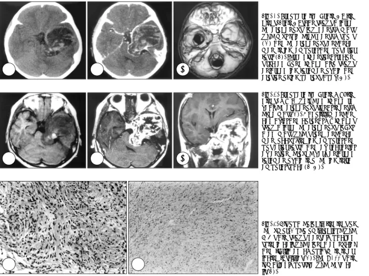
상 좌측 중두개와, 후두개와 및 측두하와에 걸친 거대한 종 괴가 저밀도의 불규칙 음영으로 나타났고 종괴의 둘레는 약한 고밀도 음영으로 주위와의 경계는 비교적 명확하였다 (Fig. 1A). 인접부 측두엽과 뇌간은 심하게 압박되어 있었 으며 종괴 내부는 고밀도 음영의 반점들이 존재하여 종양 내 출혈을 시사하였다(Fig. 1A). 조영 증강시 종양의 둘레 와 내부의 불규칙적인 증강이 보였다(Fig. 2B). 기저 두개 골의 3차원 영상에서 좌측 난원공은 매우 심하게 확장되어 있었고 좌측 추체골 첨부의 미란도 관찰되었다(Fig. 1C).
자기공명영상에서 종괴는 대체적으로 T1 강조영상에서 저 신호 강도, T2 강조영상에서는 고신호 강도로 나타났지만 내부는 불균질성과 출혈 등으로 불규칙 혼합 신호 강도를 보 였다. 조영증강 영상에서 종괴는 내부의 괴사로 생각되는 부 분을 제외하고는 강하게 조영 증강되었고 횡단, 관상, 시상 면의 사진에서 중두개와와 후두개와, 중두개와와 측두하와 에 걸친 이중 아령형의 형태를 확인할 수 있었다(Fig. 2).
치 료:수술은 두개안와관골 접근법을 이용하여 좌측 안와-관골-전두측두골에 이르는 단일 골편으로 개두술을
시행하였으며 경막을 절개하였을 때 측두엽은 중두개와 종 양에 의해 심하게 압박되어 얇아져 있었다. 측두엽을 상방 으로 견인하면서 전체적으로 관찰하였을 때 종양은 매우 두 꺼운 피막으로 둘러 쌓여 있었는데 이 피막은 종양이 두개기 저부 경막간 공간에서 발생 성장함에 따라 종양 자체의 피 막과 이에 동반된 인접 경막의 반응성 비후가 혼합되어 형 성된 것으로 생각되었다. 이 피막을 절개하고 종양의 중두 개와에 위치한 부분을 제거하였으며 측두하와에 위치한 병 변은 심하게 확장된 난원공을 통해 피막내에서 안전하게 제 거할 수 있었다. 후두개와 병변은 확장된 메켈강(Meckel’s cave)을 통한 종양의 후방 연결을 따라가면서 제거할 수 있 었으며 종양 제거 종반에 뇌간과의 지주막 경계를 확인하였다.
병리조직학적 소견:종양은 방추상핵을 갖고 있는 양극 방추형 세포가 조밀하게 결합 배열하여 있었고 S-100 단 백 면역조직 염색에 강한 양성을 보여 신경초종에 합당한 조 직 소견을 보였다(Fig. 3).
경 과:수술 직후 환아는 새로운 신경학적 결손 발생 없 이 의식은 명료하였다. 수술 3.5개월 후 촬영한 자기공명영
AAA
A BB BB
A AA
A BBBB CC CC
A AA
A BBBB CC CC
Fig. 1. Preoperative brain CT fin- gings. Brain CT scan shows a large, heterogenous, low density mass with multiple high density spots (A) and heterogenous enhance- ment in left temporal and posterior fossa(B). Three dimensional recon- structed bone image also shows enlarged left foramen ovale and erosion of left petrous apex(C).
Fig. 2. Preoperative brain MRI fin- dings. MR T1 weighted image re- veals heterogenous signal intensity huge mass(A). Gadolinium enhan- ced axial and coronal MR images show a large, heterogenous, lobul- ated mass with strong enhance- ment occupying left temporal and posterior fossa and extracranial extension through very enlarged foramen ovale into the left infrat- emporal space(B, C).
Fig. 3. Histopathologic findings of the tumor. A photomicrograph with H-E stain shows densely packed spindle cells with elongated nuclei and fibrillated cytoplasm in inter- lacing fascicles(A). The S-100 stain is markedly positive with these ce- lls(B).
신경섬유종증 Ⅱ형 소아에서 발생한 거대 삼차신경초종
J Korean Neurosurg Soc(Supplement I)/Volume 30/December, 2001 S142
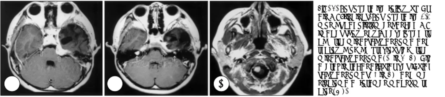
상에서 수술 부위 중 삼차신경의 신경근-신경절에 해당하 는 부위에서 잔여종양의 성장이 관찰되었다. 한편 해당 증 상은 없었으나 양측 내이도내에서도 작은 크기의 조영 증강 되는 병변이 관찰되었고, 수술 반대편의 메켈강에 해당하 는 부위에서도 조영 증강되는 병변 발생을 볼 수 있었으며 대후두공의 우측 전측방에 위치한 축외 병변도 관찰되었다 (Fig. 4). 이러한 영상 소견으로 최초 진단된 좌측의 삼차신 경초종 외에 다발성으로 우측 삼차신경초종, 양측성 청신경 초종, 대후두공 종양이 동반되어 발생했음을 알 수 있었으 며, 이를 바탕으로 다발성 두개강내 종양을 동반한 신경섬 유종증 Ⅱ형으로 진단할 수 있었다.
고 찰
삼차신경의 두개내 부위에 발생되는 삼차신경초종은 드 물며 두개강내 종양의 0.07~0.36%, 두개강내 신경초종의 0.8~8%차지하며 연령분포는 광범위하나 대부분은 중간 연령의 성인에서 발생한다고 알려져 있으며1)2)8) 10세 이하 의 소아에서는 매우 드물게 보고되고 있다6)9)10).
종양은 위치에 따라 중두개와-신경절형(middle fossa- ganglion type), 후두개와-신경근형(posterior fossa-root type), 병합-아령형(both middle & posterior fossa-du- mbbell type), 말초-신경분지형(peripheral-division type) 으로 나눌 수 있으며, 초기 증상으로 삼차신경의 손상으로 인한 통증, 무감각, 감각이상 및 두통, 복시, 청력소실 등이 발생할 수 있고, 후두개강으로의 성장으로 소뇌 및 추체로 증후도 발생할 수 있다8)9). 신경섬유종증은 말초형인 Ⅰ형 과 중심형인 Ⅱ형으로 나뉘는데 Ⅰ형은 피부의 다발성의 카 페오레 반점(cafe au lait spot)과 다발성의 홍채의 흑색소 성 과오종(Lisch nodule), 총상 또는 다발성의 신경섬유종, 액와부 반점, 시신경 교종, 골병변 등이 관찰된다. 이에 비 해 Ⅱ형은 양측성 청신경초종이 가장 흔하게 나타나며 이 밖에도 다른 두신경의 신경초종이나 수막종, 뇌실막세포종 이 동반될 수 있어 다발성의 중추신경계 종양이 특징적이
고, 피부 병변은 나타나기도 하지만 Ⅰ형보다 그 빈도는 낮 은 것으로 알려져 있다. 본 환자에서는 처음에는 피부병변 이 명확하지 않고 다른 두개강내 종양이 발견되지 않아 단 발성의 삼차신경초종으로 진단하고 수술하였으나 경과 관 찰 도중 다발성으로 두개강내 종양이 발견됨에 따라 신경 섬유종증 Ⅱ형으로 진단을 할 수 있었다.
신경섬유종증과 연관된 삼차신경초종의 보고들을 보면 대부분 증상이 없이 우연하게 발견되었거나 다발성 종양의 일부로서 발생한 것들이다5)6). 한편 삼차신경초종은 청신경 초종보다 신경섬유종증과의 병발이 더 드물고, 소아의 경 우 발생 빈도 자체가 매우 낮으며 단발성인 예들도 있어, 소 아의 삼차신경초종이라 하더라도 종양의 경과나 치료가 성 인과 유사하다고 주장하는 보고도 있다5)7)9)10). 소아에서의 삼차신경초종의 발병은 드물지만 대부분은 신경섬유종증과 병발하여 발병되고 단독으로 발병되는 경우가 매우 드묾으
로7)8)10) 소아에서 처음 발견 당시 단일 삼차신경초종일지라
도 장기적 추적으로 다른 동반 종양의 유무를 확인하여 평가 해야 할 것으로 사료된다. 저자들은 본 증례 경험으로 볼 때 소아에서 매우 드문 신경초종이 발견되었을 경우, 이것이 신 경섬유종증 Ⅱ형의 초기 임상 증상으로 표현될 수 있음을 고려해야 할 것으로 판단된다.
대부분의 경우에서 신경초종은 양성으로 성장이 매우 느 린 종양으로 생각되고 있으나, 본 환자에서와 같이 2년 간 의 기간 동안 매우 빨리 성장하여 임상적으로 악화시킬 수도 있음을 알 수 있었으며, 이러한 사실로 유추해 볼 때 신경섬 유종증에 동반된 신경초종의 경우 다른 단발성의 신경초종 에 비하여 매우 빨리 성장할 수 있음을 고려해야 할 것으로 생각되었다. 따라서 이러한 환아의 경우, 임상증상을 유발하 지 않고 있는 잠재성 다발성 종양에 대한 치료 방법에 대해 서는 종양의 성장 속도가 다른 신경초종과는 다르기 때문에 적극적인 치료 방법을 고려해야 할 것으로 판단되나, 발표 된 증례의 수가 너무 적어 가장 적합한 치료 방법의 선택에 는 논란의 여지가 있을 것으로 생각된다. 증상 없는 다발성 종양의 치료 방법 선택을 위해서는 주기적인 추적 관찰, 방
Fig. 4. Postoperative follow-up br- ain MRI findings. Post-operative 3.5 months gadolinium enhanced MR images show newly developed ri- ght trigeminal schwannoma and regrowth of the previous left trige- minal schwannoma(× in A, B), bil- ateral intracanalicular vestibular schwannomas(↑in B), and me- ningioma at foramen magnum re- gion(C).
A AA
A BB BB CCCC
이문영·김태영·문성근·김종문
J Korean Neurosurg Soc(Supplement I)/Volume 30/December, 2001 S143
사선 수술, 적극적 수술 등이 고려 될 수 있을 것이다.
결 론
저자들은 소아에서 발생한 매우 커다란 삼차 신경초종 1 예를 경험하고 문헌고찰과 함께 보고한다. 종괴는 두개안와 관골 접근법을 통하여 한번의 수술로서 성공적으로 대부분 제거되었다. 환자를 추적 관찰하던 중 양측 청신경초종, 우 측 삼차신경초종 및 대후두공 종양의 발생이 발견되어 다 발성 두개강내 종양을 병발한 신경섬유종증 Ⅱ형으로 진단 되었다. 본 증례와 같이 소아의 삼차신경초종은 신경섬유 종증 Ⅱ형의 초기 발생종양의 형태로서 발견될 수 있으며 다른 신경초종과 달리 빠른 성장을 보일 수 있기 때문에 주 의 깊은 추적 검사 및 관찰이 필요할 것으로 생각된다.
•논문접수일:2001년 6월 5일
•심사완료일:2001년 10월 21일
•책임저자:김 태 영
570-711 전북 익산시 신용동 344-2 원광대학교 의과대학 신경외과학교실
전화:063) 850-1267, 전송:063) 852-2606 E-mail:tykim@wonkwang.ac.kr
References
1) Arseni C, Dumitrescu L, Constantinescu A:Neurinomas of the
trigeminal nerve. Surg Neurol 4:497-503, 1975
2) Benedittis G, Bernasconi V, Ettorre G:Tumors of the fifth cr- anial nerve. Acta Neurochir(Wien) 38:37-64, 1977 3) Bordi L, Compton J, Symon L:Trigeminal neuroma:A report
of eleven cases. Surg Neurol 31:272-276, 1989
4) Jefferson G:Trigeminal neurinomas with some remarks on malignant invasion of the gasseran ganglion. Clin Neurosurg 1:11-54, 1955
5) Kchouk M, Aouidi L, Gouider R, Oueslati S, Touibi S, Khaldi M:Neurinomas of the trigeminal nerve in neurofibromatosis.
Apropos of two cases and review of the literature. Ann Radiol 35:533-537, 1992
6) McCormick PC, Bello JA, Post KD:Trigeminal schwan- noma:Surgical series of 14 cases with review of the literature.
J Neurosurg 69:850-860, 1988
7) Perez-Diaz CJ, Villarejo FJ, Pascual AM:Trigeminal neuri- nomas in infants:report of two cases. Childs Nerv Syst 12: 283-287, 1996
8) Pollack IF, Sekhar LN, Jannetta PJ, Janecka IP:Neurilemomas of the trigeminal nerve. J Neurosug 70:737-745, 1989 9) Ross DL, Tew JM Jr, Benton C, Eisentrout C:Trigeminal
schwannoma in a child. Neurosurgery 15:108-110, 1984 10) Tsuboi K, Fujimori H, Tomono Y, Hamano K, Nose T:Du-
mbbell-shaped trigeminal neurinoma in a Child. Acta Ne- urochir(Wien) 141:429-433, 1999