ORIGINAL ARTICLE
Korean J Intern Med 2014;29:597-602 http://dx.doi.org/10.3904/kjim.2014.29.5.597
Departments of 1Internal Medicine, 2Occupational and Environmental Medicine, and
3Biomedical Engineering, Keimyung University School of Medicine, Daegu, Korea
Received : February 27, 2014 Revised : March 31, 2014 Accepted : May 1, 2014 Correspondence to Chang-Wook Nam, M.D.
Division of Cardiology, Department of Internal Medicine, Keimyung University School of Medicine, 56
Dalseong-ro, Jung-gu, Daegu 700-712, Korea
Tel: +82-53-250-8015 Fax: +82-53-250-7034 E-mail: [email protected]
*These authors contributed equally to this work.
Background/Aims: Although complex bifurcation stenting in patients with non- left main (LM) bifurcation lesions has not yielded better clinical outcomes than simpler procedures, the utility of complex bifurcation stenting to treat LM bifur- cation lesions has not yet been adequately explored.
Methods: In the present study, patients who underwent LM-to-left anterior de- scending (LAD) coronary artery simple crossover stenting to treat significant de novo distal LM or ostial LAD disease, in the absence of angiographically sig- nificant ostial left circumflex (LCX) coronary artery disease, were consecutively enrolled. The frequencies of 3-year major adverse cardiovascular events (MACEs;
cardiac death, myocardial infarction, and target lesion revascularization), were analyzed.
Results: Of 105 eligible consecutive patients, only 12 (11.4%) required additional procedures to treat ostial LCX disease after main vessel stenting. The mean per- centage diameter of ostial LCX stenosis increased from 22.5% ± 15.2% to 32.3% ± 16.3% (p < 0.001) after LM-to-LAD simple crossover stenting. The 3-year incidence of MACEs was 9.7% (cardiac death 2.2%; myocardial infarction 2.2%; target lesion revascularization 8.6%), and that of stent thrombosis 1.1%. Of seven cases (7.5%) requiring restenosis, pure ostial LCX-related repeat revascularization was re- quired by only two.
Conclusions: Simple crossover LM-to-LAD stenting without opening of a strut on the LCX ostium was associated with acceptable long-term clinical outcomes.
Keywords: Percutaneous coronary intervention; Coronary stenosis; Stents; Out- come
INTRODUCTION
Percutaneous coronary intervention (PCI) using drug-eluting stents (DESs) is commonly used to treat bifurcation coronary artery disease [1-3]. Use of two stents (a complex strategy) was not associated with bet- ter clinical outcomes than one-stent strategies when
non-left main (LM) bifurcation PCI was performed [4- 6]. Therefore, provisional strategy is generally used to treat non-LM bifurcation. Also, the routine use of final kissing balloon dilation in patients with non-LM bi- furcation lesions, treated via main vessel stenting, did not yield clinical outcomes better than those afforded in the absence of such dilation [5,7]. However, few data
Long-term outcomes of simple crossover stenting from the left main to the left anterior descending coronary artery
Ho-Myung Lee1,*, Chang-Wook Nam1,*, Yun-Kyeong Cho1, Hyuck-Jun Yoon1, Hyoung-Seob Park1,
Hyungseop Kim1, In-Sung Chung2, Yun-Seok Heo3, Seung-Ho Hur1, Yoon-Nyun Kim1, and Kwon-Bae Kim1
on treatment of LM bifurcation lesions are available.
One pilot study showed convincingly that fraction flow reserve (FFR)-guided PCI in the left circumflex (LCX) coronary artery, performed after LM-to-left anterior descending (LAD) coronary artery simple crossover stenting, reduced the need for additional procedures [8].
Therefore, the aim of the present study was to evaluate the long-term safety and efficacy of LM-to-LAD simple crossover stenting, without opening of a strut on the LCX ostium.
METHODS
Study populationPatients with de novo distal LM or ostial LAD disease, but without significant ostial LCX disease (visually esti- mated stenosis < 50% of the arterial diameter), and with Medina classification scores of 1,1,0/1,0,0/0,1,0, were consecutively enrolled from April 2004 to June 2009 when they attended the Keimyung University Dongsan Medical Center. We excluded patients requiring addi- tional bifurcation procedures (performed at the dis- cretion of attending physicians) because of significant ostial LCX jailing evident after predilation of the main vessel or simple crossover stenting. A patient was not eligible if she/he had undergone primary or emergent PCI intervention to treat acute coronary syndrome;
had undergone previous coronary artery bypass graft surgery; exhibited severe left ventricular dysfunction (left ventricular ejection fraction < 35%); had a major life-threatening illness; or exhibited contraindications to aspirin or clopidogrel.
Procedure and outcomes
PCI was performed using standard interventional techniques. Antiplatelet and antithrombotic agents were prescribed in line with current PCI guidelines [9].
Implanted stents were commercially available DESs in all cases (sirolimus-eluting stents in 56 cases [60%];
everolimus-eluting stents in 14 [15%]; paclitaxel-eluting stents in 12 [13%]; and zotarolimus-eluting stents in 11 [12%]). All coronary angiograms were analyzed using standard definitions and measurements, following American Heart Association guidelines [10]. A guiding catheter was used for calibration and in performance
of edge-detection quantitative coronary angiography (Quantcor QCA, Pie Medical, Maastricht, the Nether- lands). An experienced operator performed QCA on three segments (the proximal main vessel, the distal main vessel, and the side branch). Variables measured included the reference diameter, the minimal lumen diameter, and the extent of stenosis (% vessel diameter).
The primary outcome was a composite of major ad- verse cardiac events (MACEs), defined as cardiac death, myocardial infarction, and any target lesion revascu- larization (TLR), by 3 years after the index procedure.
Death was defined as all-cause mortality. Myocardial infarction was defined as a 3-fold or greater elevation of the creatine kinase-MB level, or new Q-waves evident in two or more contiguous electrocardiographic leads.
Each TLR included target vessel PCI and bypass surgery of the lesion of interest, thus LM-to-LAD or ostial LCX, when symptoms and/or signs of ischemia were evident.
Clinical follow-up was performed by physicians via medical chart review or telephone interview. Angio- graphic follow-up data were gathered at the discretion of attending physicians (thus not routinely).
Statistical analysis
Data are expressed as mean ± SD for continuous vari- ables and as percentages for discrete variables. Differ- ences between continuous variables were compared us- ing Student t test or by analysis of variance. Categorical variables were compared using chi-square tests, non- parametric chi-square tests, or Fisher exact test, as ap- propriate. Cumulative incidences of MACEs were esti- mated using the Kaplan-Meier method. All calculated p values were two-sided, and a difference was considered statistically significant at p < 0.05. All statistical analyses were performed using the SPSS version 20 (IBM Co., Armonk, NY, USA).
RESULTS
Of the 105 consecutive patients with de novo distal LM or ostial LAD disease, but without significant ostial LCX disease, 12 (11.4%) underwent additional proce- dures in the LCX ostium after main vessel stenting.
Therefore, final follow-up analysis was performed on only 93 patients. Patient baseline clinical characteristics,
Lee HM, et al. Outcome of simple LM crossover stenting
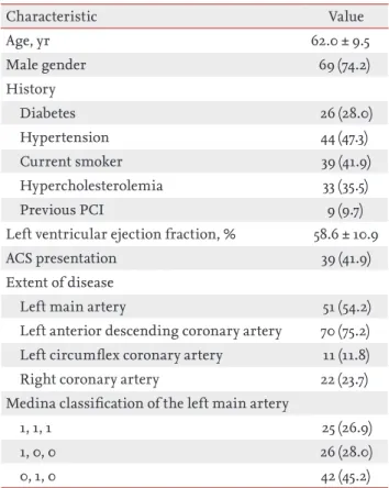
angiographic characteristics, and quantitative coronary angiographic results, are summarized in Tables 1 and 2.
Mean patient age was 62.0 ± 9.5 years (males, 75.5%), and 28.0% had diabetes. The mean left ventricular ejection fraction was 58.6% ± 10.6%. On baseline angiography, the distal reference vessel diameter of the LCX was 3.1 ± 0.5 mm. The mean stenosis of the ostial LCX (percentage of vessel diameter) increased after LM-to-LAD cross- over stenting, from 22.5% ± 15.2% to 32.3% ± 16.3% (p <
0.001).
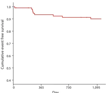
Three-year follow-up clinical data were obtained for all patients. Angiographic follow-up data were available for 73 (78.5%). During the follow-up period, the 3-year MACE rate was 9.7% (cardiac death 2.2%, myocardial in- farction 2.2%, and TLR 8.6%) (Fig. 1). One case (1.1%) of stent thrombosis was noted. Of seven restenotic cases, pure ostial LCX-associated repeat revascularization was required by only two. The relevant locations are shown in a schematic bifurcation diagram (Fig. 2). Event-free
survival is shown in Fig. 3. Upon univariate analysis, all of clinical presentation, left ventricular ejection fraction, reference vessel diameter, and stent size, were associat- ed with MACEs. However, no independent predictor of MACE was evident upon multivariate regression analy- sis.
Table 1. Baseline characteristics of all patients (n = 93)
Characteristic Value
Age, yr 62.0 ± 9.5
Male gender 69 (74.2)
History
Diabetes 26 (28.0)
Hypertension 44 (47.3)
Current smoker 39 (41.9)
Hypercholesterolemia 33 (35.5)
Previous PCI 9 (9.7)
Left ventricular ejection fraction, % 58.6 ± 10.9
ACS presentation 39 (41.9)
Extent of disease
Left main artery 51 (54.2)
Left anterior descending coronary artery 70 (75.2) Left circumflex coronary artery 11 (11.8)
Right coronary artery 22 (23.7)
Medina classification of the left main artery
1, 1, 1 25 (26.9)
1, 0, 0 26 (28.0)
0, 1, 0 42 (45.2)
Values are presented as mean ± SD or number (%).
PCI, percutaneous coronary intervention; ACS, acute coro- nary syndrome.
Table 2. Procedural results (n = 93)
Variable Value
Preprocedure LM-to-LAD
Reference vessel diameter, proximal, mm 3.7 ± 0.4 Reference vessel diameter, distal, mm 3.2 ± 0.5 Minimal lumen diameter, mm 0.6 ± 0.3
Stenosis, % diameter 82.2 ± 9.6
LM-to-LCX
Reference vessel diameter, distal, mm 3.1 ± 0.5 Minimal lumen diameter, mm 2.4 ± 0.7
Stenosis, % diameter 22.5 ± 15.2
Lesion length, mm 20.7 ± 5.8
LM procedure
Stent length, mm 22.6 ± 5.3
Stent diameter, mm 3.5 ± 0.3
Procedures for other lesions
LAD 34 (36.6)
LCX 19 (20.4)
RCA 11 (11.8)
No. of stents placed, except in LM
Single 24 (25.8)
Multiple 21 (22.6)
Postprocedure LM-to-LAD
Minimal lumen diameter, mm 3.2 ± 0.4
Stenosis, % diameter 10.2 ± 4.0
LM-to-LCX
Minimal lumen diameter, mm 2.1 ± 0.7
Stenosis, % diameter 32.3 ± 16.3
Values are presented as mean ± SD or number (%).
LM, left main coronary artery; LAD, left anterior descend- ing coronary artery; LCX, left circumflex artery; RCA, right coronary artery.
DISCUSSION
The major findings of our current study were: 1) the long-term outcomes of LM-to-LAD simple crossover stenting in patients with de novo distal LM or ostial LAD disease (without significant ostial LCX disease) were good; and 2) it was safe to not open a strut on the LCX ostium after crossover stenting. Thus, additional complex LM bifurcation strategies increasing the risk of procedural complications are not necessary. Our results should be confirmed in future large-scale ran- domized studies.
Coronary bifurcation lesions are regarded as chal- lenging by coronary intervention specialists even in the present era of DESs [11,12]. The unique characteristics of bifurcation lesions render complex procedures no more effective than simple procedures, and the reste- nosis rate at the ostium of the side branch is high after performance of complex procedures [2,5,13]. A better understanding of this lesional subset is required be- fore complex procedures are applied. A provisional approach, thus selective side-branch intervention after main vessel stenting, is currently regarded as preferred when non-LM bifurcation lesions require treatment [5,14]. Such a strategy was supported by an elegant series of investigations of FFR-guided interventional strat- egies used to treat jailed side branches [11,15,16]. The cited studies imparted two important messages. First, angiographic evaluation frequently overestimated the functional severity of a jailed side branch lesion. Sec- ond, the functional status of such lesions was stable during follow-up. However, use of such a strategy to treat a jailed LCX after LM-to-LAD simple crossover stenting was not explored. Such work was required, because a large amount of myocardium may be in jeop- ardy after LCX jailing. Coronary Bifurcation Stenting (COBIS) registry II data demonstrate the possible harm- ful effects associated with use of two-stent strategies to treat LM bifurcation lesions [17]. A small pilot study revealed discrepancies between angiographic stenosis and FFR data on jailed LCX lesions, and that use of an FFR-guided PCI strategy to treat the jailed LCX reduced the need for additional PCI [8]. However, the long-term safety of a residual strut placed on the LCX ostium was not assured. In our current study, we explored the long- term clinical outcomes of LM-to-LAD simple crossover
2.2 2.2
8.6 9.7
1.1 0
4 8 12
Cardiac death
MI TLR MACE
Stent thrombosis
%
LM
LCX
LAD
1.0
0.9
0.8
0.7
0.6
0.5
0.4
0 365 730
Day
Cumulative event free survival
1,095
Figure 1. Incidences of 3-year major adverse cardiac events and stent thrombosis. MI, myocardial infarction; TLR, target lesion revascularization; MACE, major adverse cardiovascular event.
Figure 2. Locations of restenosis. LM, left main coronary artery; LAD, left anterior descending coronary artery; LCX, left circumflex artery.
Figure 3. Three-year Kaplan-Meier event-free survival after simple left main crossover stenting.
Lee HM, et al. Outcome of simple LM crossover stenting
stenting, without opening of a strut on the LCX ostium.
The 3-year MACE rate was acceptable, being 9.7%.
Another important finding is that no additional pro- cedure was required by 88.6% of patients (93/105) who underwent successful simple crossover stenting. Only 11.4% patients required an additional kissing balloon dilation procedure (for carinal modification) after simple crossover stenting, or a two-stent procedure after LM-to-LAD stenting. Therefore, as was previously found in studies on non-LM bifurcation PCI, the use of a simple strategy reduces the need for additional pro- cedures to treat LM bifurcations. Such lesions are not only complex, but are also associated with poor progno- ses.
Although the requirement for additional procedures was lower than that of a previous study [18], a careful approach is warranted when using provisional LM bi- furcation procedures. Such lesions may have a greater plaque burden, and be at higher risk of carinal shifting, than non-LM bifurcation lesions [16,19].
Our present study had several limitations. First, although over 100 patients with a specific subset of lesions were enrolled, no single-center observational study is entirely free from selection bias. We excluded patients who received any PCI as an index procedure to treat LCX. As our study population was restricted to those exhibiting good angiographic morphology, cau- tion is required when interpreting our results, which need confirmation in a future randomized study with higher numbers of patients. Also, although we collected 3-year clinical follow-up data, the longer-term safety of our chosen strategy is not assured. Finally, we did not perform additional imaging, or gather physiological data, which would have yielded additional valuable in- formation.
In conclusion, although no strut on the ostial LCX was opened after LM-to-LAD simple crossover stenting, such a strategy was safe and effective in the long term.
Conflict of interest
No potential conflict of interest relevant to this article was reported.
Acknowledgments
This research was supported by the Scholar Research Grant of Keimyung University in 2012.
REFERENCES
1. Sharma SK, Mares AM, Kini AS. Coronary bifurcation le- sions. Minerva Cardioangiol 2009;57:667-682.
2. Colombo A, Moses JW, Morice MC, et al. Randomized study to evaluate sirolimus-eluting stents implanted at coronary bifurcation lesions. Circulation 2004;109:1244- 1249.
3. Song YB, Hahn JY, Choi SH, et al. Sirolimus- versus pacl- itaxel-eluting stents for the treatment of coronary bifur- cations results: from the COBIS (Coronary Bifurcation Stenting) Registry. J Am Coll Cardiol 2010;55:1743-1750.
4. Athappan G, Ponniah T, Jeyaseelan L. True coronary bi- furcation lesions: meta-analysis and review of literature. J Cardiovasc Med (Hagerstown) 2010;11:103-110.
5. Niemela M, Kervinen K, Erglis A, et al. Randomized com- parison of final kissing balloon dilatation versus no final kissing balloon dilatation in patients with coronary bifur- cation lesions treated with main vessel stenting: the Nor- dic-Baltic Bifurcation Study III. Circulation 2011;123:79- 86.
6. Gwon HC, Choi SH, Song YB, et al. Long-term clin- ical results and predictors of adverse outcomes after drug-eluting stent implantation for bifurcation lesions in a real-world practice: the COBIS (Coronary Bifurcation Stenting) registry. Circ J 2010;74:2322-2328.
7. Gwon HC, Hahn JY, Koo BK, et al. Final kissing balloon- ing and long-term clinical outcomes in coronary bifurca- tion lesions treated with 1-stent technique: results from the COBIS registry. Heart 2012;98:225-231.
8. Nam CW, Hur SH, Koo BK, et al. Fractional flow reserve versus angiography in left circumflex ostial interven- tion after left main crossover stenting. Korean Circ J 2011;41:304-307.
KEY MESSAGE
1. The long-term clinical outcomes of left main-to- left anterior descending coronary artery simple crossover stenting, without opening of a strut on the left circumflex coronary ostium, were accept- able.
2. Use of such a strategy reduces the need for addi- tional complex left main bifurcation procedures, which may increase the risk of complications.
9. Griswold KS, Servoss TJ, Leonard KE, et al. Connections to primary medical care after psychiatric crisis. J Am Board Fam Pract 2005;18:166-172.
10. Austen WG, Edwards JE, Frye RL, et al. A reporting system on patients evaluated for coronary artery disease: report of the Ad Hoc Committee for Grading of Coronary Artery Disease, Council on Cardiovascular Surgery, American Heart Association. Circulation 1975;51(4 Suppl):5-40.
11. Koo BK, Park KW, Kang HJ, et al. Physiological evalua- tion of the provisional side-branch intervention strategy for bifurcation lesions using fractional flow reserve. Eur Heart J 2008;29:726-732.
12. Steigen TK, Maeng M, Wiseth R, et al. Randomized study on simple versus complex stenting of coronary artery bifurcation lesions: the Nordic bifurcation study. Circula- tion 2006;114:1955-1961.
13. Hildick-Smith D, de Belder AJ, Cooter N, et al. Random- ized trial of simple versus complex drug-eluting stenting for bifurcation lesions: the British Bifurcation Coronary Study: old, new, and evolving strategies. Circulation 2010;121:1235-1243.
14. Latib A, Colombo A, Sangiorgi GM. Bifurcation stenting:
current strategies and new devices. Heart 2009;95:495-504.
15. Koo BK, Kang HJ, Youn TJ, et al. Physiologic assessment of jailed side branch lesions using fractional flow reserve.
J Am Coll Cardiol 2005;46:633-637.
16. Koo BK, De Bruyne B. FFR in bifurcation stenting: what have we learned? EuroIntervention 2010;6 Suppl J:J94-J98.
17. Song YB, Hahn JY, Yang JH, et al. Differential prognostic impact of treatment strategy among patients with left main versus non-left main bifurcation lesions undergo- ing percutaneous coronary intervention: results from the COBIS (Coronary Bifurcation Stenting) Registry II. JACC Cardiovasc Interv 2014;7:255-263.
18. Kim YH, Park SW, Hong MK, et al. Comparison of simple and complex stenting techniques in the treatment of un- protected left main coronary artery bifurcation stenosis.
Am J Cardiol 2006;97:1597-1601.
19. Lim MJ, Kern MJ. Utility of coronary physiologic hemo- dynamics for bifurcation, aorto-ostial, and ostial branch stenoses to guide treatment decisions. Catheter Cardio- vasc Interv 2005;65:461-468.