Outcomes of Percutaneous Coronary Intervention in Intermediate Coronary Artery Disease
Fractional Flow Reserve–Guided Versus Intravascular Ultrasound–Guided
Chang-Wook Nam, MD, P
HD,* Hyuck-Jun Yoon, MD,* Yun-Kyeong Cho, MD, P
HD,*
Hyoung-Seob Park, MD,* Hyungseop Kim, MD, P
HD,* Seung-Ho Hur, MD, P
HD,*
Yoon-Nyun Kim, MD, P
HD,* In-Sung Chung, MD, P
HD* Bon-Kwon Koo, MD, P
HD,†
Seung-Jae Tahk, MD, P
HD,‡ William F. Fearon, MD,§ Kwon-Bae Kim, MD, P
HD*
Daegu, Seoul, and Suwon, Korea; and Stanford, California
Objectives This study sought to evaluate the long-term clinical outcomes of a fractional flow re- serve (FFR)– guided percutaneous coronary intervention (PCI) strategy compared with intravascular ultrasound (IVUS)– guided PCI for intermediate coronary lesions.
Background Both FFR- and IVUS-guided PCI strategies have been reported to be safe and effective in intermediate coronary lesions.
Methods The study included 167 consecutive patients, with intermediate coronary lesions evalu- ated by FFR or IVUS (FFR-guided, 83 lesions vs. IVUS-guided, 94 lesions). Cutoff value of FFR in FFR- guided PCI was 0.80, whereas that for minimal lumen cross sectional area in IVUS-guided PCI was 4.0 mm
2. The primary outcome was defined as a composite of major adverse cardiac events includ- ing death, myocardial infarction, and ischemia-driven target vessel revascularization at 1 year after the index procedure.
Results Baseline percent diameter stenosis and lesion length were similar in both groups (51 ⫾ 8%
and 24 ⫾ 12 mm in the FFR group vs. 52 ⫾ 8% and 24 ⫾ 13 mm in the IVUS group, respectively).
However, the IVUS-guided group underwent revascularization therapy significantly more often (91.5% vs. 33.7%, p ⬍ 0.001). No significant difference was found in major adverse cardiac event rates between the 2 groups (3.6% in FFR-guided PCI vs. 3.2% in IVUS-guided PCI). Independent pre- dictors for performing intervention were guiding device: FFR versus IVUS (relative risk [RR]: 0.02); left anterior descending coronary artery versus non-left anterior descending coronary artery disease (RR:
5.60); and multi- versus single-vessel disease (RR: 3.28).
Conclusions Both FFR- and IVUS-guided PCI strategy for intermediate coronary artery disease were associated with favorable outcomes. The FFR-guided PCI reduces the need for revascularization of many of these lesions. (J Am Coll Cardiol Intv 2010;3:812–7) © 2010 by the American College of Cardiology Foundation
From the *Keimyung University Dongsan Medical Center, Daegu, Korea; †Seoul National University Hospital, Seoul, Korea;
‡Ajou University Hospital, Suwon, Korea; and the §Stanford University Medical Center, Cardiovascular Medicine, Stanford, California. Dr. Yoon-Nyun Kim has received research grant no. RTI04-01-01 from the Regional Technology Innovation Program of the Ministry of Knowledge Economy, Korea. All other authors report that they have no relationships to disclose.
Manuscript received January 13, 2010; revised manuscript received March 12, 2010, accepted April 17, 2010.
Because of the limitations of coronary angiography (1), adjunctive techniques to more accurately evaluate lesion severity are important in patients with intermediate coro- nary stenosis before percutaneous coronary intervention (PCI). Fractional flow reserve (FFR) has been the reference standard for the physiological assessment of coronary artery stenosis, particularly intermediate ones (2– 4). Deferring intervention of intermediate coronary lesions with a FFR ⱖ0.75 or 0.80 is associated with favorable long-term clinical outcomes (5,6). An intravascular ultrasound (IVUS)–
See page 818
derived minimal lumen area (MLA) ⱕ4.0 mm
2, or minimal lumen diameter ⱕ1.8 mm have been shown to correlate with a FFR ⬍0.75 (7), and deferring intervention in intermediate coronary lesions based on MLA ⱖ4.0 mm
2results in favorable clinical outcomes (8). However, there are few studies that compared FFR- and IVUS-guided coro- nary intervention strategies in patients with de novo coro- nary intermediate lesions. The aim of this study was to evaluate the clinical outcomes of a FFR- versus IVUS- guided PCI strategy for intermediate coronary lesions.
Methods
Patient population and study design. The patient popula- tion consisted of 167 consecutive patients (177 lesions) who underwent FFR or IVUS assessment to decide whether to perform PCI or not for de novo intermediate coronary lesions between August 2006 and June 2008. An interme- diate coronary lesion was defined as 40% to 70% diameter stenosis by visual assessment. For this study, the target vessel was a single lesion in the proximal or mid part of a major epicardial coronary artery with reference vessel diam- eter larger than 2.5 mm. The lesion had no documented evidence of ischemia by noninvasive tests (not performed, negative, inadequate, or not evaluable for a target lesion).
Patients were not eligible for enrollment if they: 1) had undergone intervention in the setting of primary or emer- gent PCI for an acute coronary syndrome; 2) had prior coronary artery bypass graft surgery; 3) had multiple lesions in the same epicardial artery; 4) had left main disease, primary myocardial disease, or a major life threatening illness; or 5) had contraindications to adenosine, aspirin, or clopidogrel.
The use of FFR or IVUS was made based on operator preference. The cutoff value of FFR in the FFR-guided PCI group was 0.80 (6,9,10) and that of MLA in the IVUS- guided PCI was 4.0 mm
2(7,8). Implanted stents were commercially available drug-eluting stents (DES) in all cases.
Procedural details. Coronary angiography was performed in multiple views after the intracoronary injection of 0.2 mg
nitroglycerin. Percutaneous coronary intervention was per- formed following standard interventional techniques. Anti- platelet and antithrombotic agents were prescribed accord- ing to current PCI guidelines (3). All coronary angiograms were analyzed using standard definitions and measurements by quantitative coronary angiography (Quantcor QCA, version 4.0, Pie Medical Imaging, Maastricht, the Nether- lands) by an experienced physician who was blinded to the type of PCI guidance.
Fractional flow reserve was defined as the ratio between mean distal coronary pressure and mean aortic pressure, both measured simultaneously at maximal hyperemia. Cor- onary pressure was measured using a 0.014-inch sensor- tipped PCI guidewire (Pressure Wire, Radi Medical Sys- tems, Uppsala, Sweden). The wire was introduced through a 6- or 7-F guiding catheter, equalized, and advanced distal to the stenosis as previously described (9). The FFR value was checked after administration of adenosine to induce maximal hyperemia, either in-
travenously (140 g/kg/min) or intracoronarily (40 g in the right, 80 g in the left coronary artery).
Intravascular ultrasound guid- ance was performed using con- ventional 6- or 7-F guiding cath- eters and a 0.014-mm guidewire positioned distally, and IVUS catheters of 30 or 40 MHz (Bos- ton Scientific Corp., Natick, Mas- sachusetts) pulled back automati- cally at a constant speed of 0.5 mm/s. The lesion site selected for analysis was the image slice with MLA and minimal stent area, which were measured following
the guidelines for IVUS measurements by the American College of Cardiology (11).
Definitions and study outcomes. The primary outcome was defined as a composite of major adverse cardiac events (MACE), defined as death, myocardial infarction, and ischemia-driven target vessel revascularization (TVR) at 12 months after the index procedure. Death was defined as all-cause mortality. The diagnosis of myocardial infarction was based on either the development of new pathological Q waves in ⱖ2 contiguous electrocardiogram leads and/or cardiac enzyme level elevation ⬎3 times the upper limit of normal value. TVR included target lesion PCI and bypass surgery of the target lesion. TVR was performed only in the presence of symptoms and/or signs of ischemia. Stent thrombosis was defined according to the Academic Re- search Consortium guidelines (12).
Statistical analyses. Data are expressed as mean ⫾ SD for continuous variables and as percentages for discrete vari-
Abbreviations and Acronyms
DESⴝ drug-eluting stent(s) FFRⴝ fractional flow reserve
IVUSⴝ intravascular ultrasound LADⴝ left anterior descending coronary artery MACEⴝ major adverse cardiac event
MLAⴝ minimal lumen area PCIⴝ percutaneous coronary intervention TVRⴝ target vessel revascularization
ables. Continuous variables were compared using Student t test. Categorical variables were compared using chi-square tests or Fisher exact test, as appropriate. All calculated p values were 2-sided and differences were considered to be statistically significant when the respective p values were
⬍0.05. Multivariate logistic regression analysis was used to assess independent predictors of performing PCI and of MACE. The parameters analyzed in multivariate analysis were selected when p value was lesser than 0.5 in univariate analysis. All statistical analyses were performed using SPSS version 15.0 for Windows (SPSS Inc., Chicago, Illinois).
Results
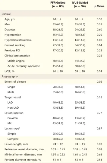
Baseline clinical, angiographic, and procedural characteris- tics are summarized in Tables 1 and 2. Among 167 consecutive patients (177 lesions), 83 lesions were assessed
by FFR and 94 lesions by IVUS. Intracoronary adenosine was used in 79 lesions to induce maximal hyperemia. The resulting groups were well matched without significant differences in the frequency of clinical cardiovascular risk factors. The incidence of multivessel disease was significantly higher in the FFR group (66.3% vs. 48.9%, p ⫽ 0.02). The most common target lesion location was the left anterior descending coronary artery (LAD) in both groups (48.2% in the FFR group vs. 58.8% in the IVUS group, p ⫽ 0.18).
There was no significant difference in clinical presentation.
Both groups had similar percent diameter stenosis (51 ⫾ 8% vs. 52 ⫾ 8% in the FFR and IVUS groups respec- tively, p ⫽ 0.53). However, reference vessel diameter was larger in the IVUS-guided group (3.39 ⫾ 0.49 mm vs.
3.23 ⫾ 0.43 mm, p ⫽ 0.03). Although angiographic lesion length was similar between 2 groups, IVUS lesion length was slightly longer than angiographic lesion length in IVUS group (23.5 ⫾ 12.7 mm in angiographic lesion length vs. 25.3 ⫾ 11.0 mm in IVUS lesion length, p ⫽ 0.008).
The incidence of performing PCI was much lower in the FFR-guided group (33.7% vs. 91.5%, p ⬍0.001) as shown in Figure 1. The mean FFR value of deferred lesions was 0.87 ⫾ 0.06 whereas that of revascularized lesions was 0.72 ⫾ 0.07 in the FFR-guided group. The mean MLA of deferred lesions was 5.1 ⫾ 1.5 mm
2and that of lesions treated by PCI was 2.9 ⫾ 0.9 mm
2in the IVUS-guided group (Table 2). When evaluating LAD lesions alone, the frequency of PCI con- tinued to be greater with IVUS guidance compared with FFR guidance (52.5% in FFR group vs. 90.9% in IVUS group, p ⬍ 0.001).
The 12-month clinical outcomes are summarized in Figure 2. There were no significant differences in TVR (3.6% in FFR group vs. 2.1% in the IVUS group, p ⫽ 0.67)
Table 1.Baseline Patient and Lesion Characteristics FFR-Guided
(nⴝ 83)
IVUS-Guided
(nⴝ 94) p Value Clinical
Age, yrs 63⫾ 9 62⫾ 9 0.50
Men 55 (66.3) 55 (58.5) 0.35
Diabetes 18 (21.7) 24 (25.5) 0.60
Hypertension 35 (42.2) 48 (51.1) 0.29
Hypercholesterolemia 13 (15.7) 14 (14.9) 1.00
Current smoking 27 (32.5) 34 (36.2) 0.64
Previous PCI 17 (20.5) 12 (12.8) 0.22
Clinical presentation 0.22
Stable angina 38 (45.8) 34 (36.2)
Acute coronary syndrome 45 (54.2) 60 (63.8)
LVEF, % 61⫾ 10 59⫾ 10 0.14
Angiography
Extent of disease 0.02
Single 28 (33.7) 48 (51.1)
Multi 55 (66.3) 46 (48.9)
Target vessel 0.18
LAD 40 (48.2) 55 (58.5)
Non-LAD 43 (51.8) 39 (41.5)
Lesion location 0.77
Proximal 40 (48.2) 43 (45.7)
Mid 43 (51.8) 51 (54.3)
Lesion type* 0.87
Simple 25 (30.1) 30 (31.9)
Complex 58 (69.9) 64 (68.1)
Lesion length, mm 24⫾ 12 24⫾ 13 0.92
Reference vessel diameter, mm 3.23⫾ 0.43 3.39⫾ 0.49 0.03 Minimal lumen diameter, mm 1.59⫾ 0.32 1.61⫾ 0.45 0.68
Percent diameter stenosis, % 51⫾ 8 52⫾ 8 0.53
Values are mean⫾ SD or n (%). *According to the American College of Cardiology/American Heart Association classification, type A and B1 lesions are simple, whereas type B2 and C lesions are complex.
FFR⫽ fractional flow reserve; IVUS ⫽ intravascular ultrasound; LAD ⫽ left anterior descending coronary artery; LVEF⫽ left ventricular ejection fraction; PCI ⫽ percutaneous coronary intervention.
Table 2.Procedural Results
FFR-Guided IVUS-Guided
p Value Defer
(nⴝ 55) PCI (nⴝ 28)
Defer (nⴝ 8)
PCI (nⴝ 86) FFR
Pre-intervention 0.87⫾ 0.06 0.72 ⫾ 0.07
Post-intervention 0.91⫾ 0.05
IVUS, mm2
Pre-interventional MLA 5.1⫾ 1.5 2.9 ⫾ 0.9
Post-interventional MSA 7.3⫾ 2.8
Stent number 1.1⫾ 0.5 1.1⫾ 0.5 0.97
Stent length, mm 31⫾ 13 28⫾ 14 0.42
Stent size, mm 3.2⫾ 0.4 3.3⫾ 0.5 0.16
Post-intervention
MLD, mm 2.89⫾ 0.42 3.03⫾ 0.47 0.19
DS, % 11⫾ 4 11⫾ 3 0.45
Values are mean⫾ SD.
DS⫽ percent diameter stenosis; MLA ⫽ minimal lumen diameter; MLD ⫽ minimal lumen diameter; MSA⫽ minimal stent area; other abbreviations as inTable 1.
and MACE (3.6% in FFR group vs. 3.2% in IVUS group, p ⫽ 1.00) between the 2 groups. The 3 TVR cases in the FFR-guided group were due to in-stent restenosis, deferred lesion aggravation, and progression of a nontarget lesion.
The 2 TVR cases in the IVUS-guided group were secondary to in-stent restenosis. One noncardiac death was observed in the IVUS group, which was related to a respiratory infection. No myocardial infarction or stent thrombosis was observed in either group. Kaplan-Meier estimates of cumu- lative freedom from MACE during 12-month follow-up are shown in Figure 3.
In multivariate logistic regression analysis, performing intervention was significantly affected by FFR versus IVUS
guidance (relative risk [RR]: 0.02, 95% confidence interval [CI]: 0.01 to 0.07), LAD versus non-LAD lesion (RR:
5.60, 95% CI: 1.98 to 15.80), and multi- versus single-vessel disease (RR: 3.28, 95% CI: 1.02 to 10.60) (Table 3). No variable was related to MACE in univariate analysis.
Discussion
The major findings in the current study are: 1) FFR- or IVUS-guided PCI in patients with intermediate coronary lesions was associated with favorable clinical outcomes; and 2) the rate of performed PCI in intermediate coronary
Figure 1.The Rate of Performing PCI According to Type of Guiding Device
The fractional flow reserve (FFR)– guided group showed significantly lower rates of performing percutaneous coronary intervention (PCI) compared to the intravascular ultrasound (IVUS)– guided group.
Figure 2.1-Year Clinical Outcomes According to the Type of Guiding Device
The FFR- and IVUS-guided groups demonstrated excellent 12-month clinical outcomes without significant between-group differences. All p values were⬎0.05.
MACE⫽ major adverse cardiac event; MI ⫽ myocardial infarction; TVR ⫽ target vessel revascularization; other abbreviations as inFigure 1.
Figure 3.Kaplan-Meier Survival Curves for Freedom From Adverse Cardiac Events During 12 Months of Follow-Up
Kaplan-Meier survival curves for freedom from adverse cardiac events dur- ing 12 months of follow-up for both groups. p⬎ 0.05. Abbreviations as in Figure 1.
lesions was significantly lower in the FFR-guided compared with the IVUS-guided group without any increase of adverse event rates according to established criteria.
Assessment of a coronary lesion with intermediate sever- ity remains challenging for interventional cardiologists. The clinical importance of a mild-to-moderate coronary stenosis has been increased by some who propose that acute myo- cardial infarctions originate from such lesions (13,14). Some argue that for this reason, PCI with DES might be beneficial for the patients with an intermediate coronary lesion (15,16). However, recent studies showed that stent- ing intermediate stenoses, without demonstrating their physiologic significance, does not improve outcome (5,6), and optimal medical treatment might result in similar outcomes (17). In this study, the overall incidence of MACE at 1 year was very low, namely 3.4% (6 of 177 lesions). Although 36% (63 of 177 lesions) of cases were deferred, the MACE rate was similar or lower than the results of recent DES clinical trials. The MACE rate of the deferral group was 3.2% (2 of 63) and similar to that of the PCI group, which was 3.5% (4 of 114). In a pooled analysis in which outcomes after DES were evaluated from 4 randomized trials (16), the MACE rate of DES is similar or even higher than that of FFR- or IVUS-guided deferral group in the current study. Therefore, FFR or IVUS measurements can be helpful in guiding the clinical decision process in patients with doubtful angiographic severity by reducing unnecessary stenting without increasing adverse clinical events.
By multivariate regression analysis, performing interven- tion was more frequent in the IVUS-guided group, in an LAD lesion, or in patients with multivessel disease. The last 2 factors seem intuitive, because the LAD territory has a large burden of myocardium compared with that of other
major coronary arteries, and multivessel disease patients have a higher chance of having a significant lesion, whether assessed by IVUS or FFR. In the current study, the rate of PCI was 3 times higher in the IVUS-guided group than in the FFR-guided group. However, the MACE rate was not different between the 2 groups. Although there have been several studies reporting a good correlation between FFR- and IVUS-derived MLA for assessing coronary lesions (7,18), the cutoff point based on MLA ⬍4.0 mm
2does not reflect the location of a lesion or the amount of myocardium subtended by the vessel. When the rate of PCI was reanalyzed according to reference vessel diameter (ⱖ3.0 mm or ⬍3.0 mm) or lesion location (proximal or mid), the results did not change. Therefore, IVUS can overestimate the clinical significance of intermediate coronary lesions (19) and likely explains the increased rate of PCI in this group. If MLA ⬍3.0 mm
2was applied for the cutoff point in the current study, as previously reported (20), the incidence of performing PCI might be decreased to 42.6%, similar to that of the FFR-guided group. When a cut-point was used that resulted in equal PCI rates of FFR group, MLA 2.5 mm
2was the most indicated value. Unfortu- nately, this retrospective pilot study was not powered to detect a difference in clinical outcome. With larger numbers, one might anticipate that the excess PCI in the IVUS- guided group would translate into an increase in the adverse event rate. This study further confirms the safety of defer- ring PCI in nonischemia-producing lesions (21).
Study limitations. First, it was a retrospective study. There were several different baseline characteristics such as a higher incidence of single-vessel disease and larger reference vessel diameter in the IVUS group. Second, the decision to select IVUS or FFR was left at the discretion of the operators. Although there was no statistically significant
Table 3.Predictors of Performing PCI in the Intermediate Coronary Lesions
Univariate Variables Multivariate Variables
Relative Risk 95% CI p Value Relative Risk 95% CI p Value
Age, yrs 0.98 0.95–1.02 0.30 0.97 0.92–1.03 0.30
Men 1.30 0.69–2.45 0.41 2.41 0.85–6.82 0.10
Diabetes 0.74 0.36–1.45 0.40 0.42 0.14–1.28 0.13
Current smoking 1.46 0.75–2.84 0.27 0.79 0.28–2.25 0.66
ACS 1.81 0.96–3.38 0.07 2.17 0.83–5.66 0.12
LVEF 0.98 0.94–1.01 0.12 0.98 0.94–1.03 0.49
FFR (vs. IVUS) 0.02 0.01–0.07 ⬍0.001 0.02 0.01–0.07 ⬍0.001
Multi (vs. single) VD 0.77. 0.41–1.44 0.41 3.28 1.02–10.60 0.047
LAD (vs. non-LAD) lesion 2.56 1.36–4.82 0.004 5.60 1.98–15.80 0.001
Lesion type 1.71 0.88–3.30 0.11 2.56 0.83–7.86 0.10
Lesion length 1.01 0.99–1.04 0.36 1.01 0.96– 1.05 0.85
Reference vessel diameter 1.77 0.87–3,61 0.11 1.80 0.61–5.32 0.29
All parameters described in this table did not have significant interactions with each other.
ACS⫽ acute coronary syndrome; CI ⫽ confidence interval; VD ⫽ vessel disease; other abbreviations as inTable 1.
difference of selection of guided method between operators, this study’s results are subject to selection bias. It is not a direct comparison of IVUS and FFR within the same patients, attempting to see which patients would be accept- able for PCI using each technology. Third, the number of patients included in the study was small and the duration of the follow-up was relatively short considering the low event rate at 1-year follow-up. Therefore, these results should be confirmed by larger randomized studies with a longer follow-up period. Fourth, the consequences of the multi- variate test could be overfitted, because the parameters were selected when p value was ⬍0.5 in univariate analysis.
Conclusions
Both FFR- and IVUS-guided PCI for intermediate coro- nary artery disease were associated with favorable outcomes.
The FFR-guided PCI reduces the need for revascularization of many of these lesions.
Reprint requests and correspondence: Dr. Kwon-Bae Kim, Divi- sion of Cardiology, Department of Internal Medicine, Keimyung University Dongsan Medical Center, 194 Dongsan-dong, Jung-gu, Daegu 700-712, South Korea. E-mail: [email protected]. Dr. Wil- liam F. Fearon, Division of Cardiovascular Medicine, Stanford University Medical Center, 300 Pasteur Drive, H3554, Stanford, California 94305. E-mail: [email protected].
REFERENCES